Hirschsprung's disease

Last updated
Hirschsprung's disease
Other namesAganglionic megacolon, congenital megacolon, congenital intestinal aganglionosis [1]
Hirschsprung acetylcholine.jpg
Histopathology of Hirschsprung disease showing abnormal acetylcholine esterase (AchE)-positive nerve fibers (brown) in the mucosa
Specialty Medical genetics
Symptoms Constipation, vomiting, abdominal pain, diarrhea, slow growth [1]
Complications Enterocolitis, megacolon, bowel obstruction, intestinal perforation [1] [2]
Usual onsetFirst 2 months of life [1]
TypesShort-segment, long-segment [1]
Causes Genetic [1]
Risk factors Family history [1]
Diagnostic method Based on symptoms, biopsy [3]
Differential diagnosis Chronic intestinal pseudo-obstruction, meconium ileus [2]
TreatmentSurgery [2]
Frequency1 in 5,000 newborns [1]

Hirschsprung's disease (HD or HSCR) is a birth defect in which nerves are missing from parts of the intestine. [1] [3] The most prominent symptom is constipation. [1] Other symptoms may include vomiting, abdominal pain, diarrhea and slow growth. [1] Most children develop signs and symptoms shortly after birth. However, others may be diagnosed later in infancy or early childhood. [4] [5] About half of all children with Hirschsprung's disease are diagnosed in the first year of life. [4] Complications may include enterocolitis, megacolon, bowel obstruction and intestinal perforation. [1] [2]

Contents

The disorder may occur by itself or in association with other genetic disorders such as Down syndrome or Waardenburg syndrome. [1] [2] About half of isolated cases are linked to a specific genetic mutation, and about 20% occur within families. [1] Some of these occur in an autosomal dominant manner. [1] The cause of the remaining cases is unclear. [1] If otherwise normal parents have one child with the condition, the next child has a 4% risk of being affected. [2] The condition is divided into two main types, short-segment and long-segment, depending on how much of the bowel is affected. [1] Rarely, the small bowel may be affected, as well. [2] Diagnosis is based on symptoms and confirmed by biopsy. [3]

Treatment is generally by surgery to remove the affected section of bowel. [2] The surgical procedure most often carried out is known as a "pull through". [3] Occasionally, an intestinal transplantation may be recommended. [2] Hirschsprung's disease occurs in about one in 5,000 of newborns. [1] Males are more often affected than females. [1] The condition is believed to have first been described in 1691 by Dutch anatomist Frederik Ruysch [6] and is named after Danish physician Harald Hirschsprung following his description in 1888. [7] [8]

Signs and symptoms

Typically, Hirschsprung disease is diagnosed shortly after birth, although it may develop well into adulthood, because of the presence of megacolon, or because the baby fails to pass the first stool (meconium) [9] within 48 hours of delivery. Normally, 90% of babies pass their first meconium within 24 hours, and 99% within 48 hours. [10]

Some other signs and symptoms in newborns include a swollen belly, vomiting (green or brown vomit), and flatulence. In older children, some other signs and symptoms include chronic constipation, flatulence, swollen belly, fatigue, and failure to thrive. [11]

Other symptoms include symptoms of bowel perforation such as vomiting, constipation, poor feeding, lethargy, and diarrhea. Symptoms of bowel obstruction would include vomiting of bile and abdominal distension. Those who do not pass stools after 36 to 48 hours after birth should raise suspicion of Hirschsprung disease. Such suspicion can also be risen if there is only passage of stools after suppository, rectal exam, or enema. Children who do not respond to constipation treatment for six months should also raise suspicion of such disease. Enterocolitis, an acute complication of Hirschsprung disease, is characterised by sudden onset of fever, abdominal distension, vomiting, passage of bloody stools or release of explosive gas or stools after rectal examination. [12]

Some cases are diagnosed later, into childhood, but usually before age 10. [9] The child may experience fecal retention, constipation, or abdominal distention. [9]

Associated syndromes

Hirschsprung's disease can also present as part of multi system disorders, such as: [13]

Cause

The disorder may occur by itself or in association with other genetic disorders such as Down syndrome. [2] About half of isolated cases are linked to a specific genetic mutation and about 20% occur within families. [1] Some of these occur in an autosomal dominant manner. [1] The cause of the remaining cases is unclear. [1] If otherwise normal parents have one child with the condition, the next child has a 4% risk of being affected. [2]

Genetics

Type OMIM GeneLocus
HSCR1 142623 RET 10q11.2
HSCR2 600155 EDNRB 13q22
HSCR3 600837 GDNF 5p13.1-p12
HSCR4 131242 EDN3 20q13.2-q13.3
HSCR5 600156  ?21q22
HSCR6 606874  ?3p21
HSCR7 606875  ?19q12
HSCR8 608462  ?16q23
HSCR9 611644  ?4q31-32
602229 SOX10 22q13
600423 ECE1 1p36.1
602018 NRTN 19p13.3
602595 GEMIN2 (Gem-associated protein 2)14q13-q21
191315 NTRK1 1q23.1
605802 ZEB2 2q22.3

Several genes and specific regions on chromosomes (loci) have been shown or suggested to be associated with Hirschsprung's disease:

The RET proto-oncogene accounts for the highest proportion of both familial and sporadic cases, with a wide range of mutations scattered along its entire coding region. [20] A proto-oncogene can cause cancer if it is mutated or overexpressed. [21]

RET proto-oncogene

RET is a gene that codes for proteins that assist cells of the neural crest in their movement through the digestive tract during the development of the embryo. Those neural crest cells eventually form bundles of nerve cells called ganglions. EDNRB codes for proteins that connect these nerve cells to the digestive tract. Thus, mutations in these two genes could directly lead to the absence of certain nerve fibers in the colon. Research suggests that several genes are associated with Hirschsprung's disease. [22] Also, new research suggests that mutations in genomic sequences involved in regulating EDNRB have a bigger impact on Hirschsprung's disease than previously thought.[ citation needed ]

RET can mutate in many ways and is associated with Down syndrome. Since Down syndrome is comorbid in 2% of Hirschsprung's cases, a likelihood exists that RET is involved heavily in both Hirschsprung's disease and Down syndrome. RET is also associated with medullary thyroid cancer and neuroblastoma, which is a type of cancer common in children. Both of these disorders are more common in Hirschsprung's patients than in the general population. One function that RET controls is the travel of the neural crest cells through the intestines in the developing fetus. The earlier the RET mutation occurs in Hirschsprung's disease, the more severe the disorder becomes.[ citation needed ]

Other genes

Common and rare DNA variations in the neuregulin 1 (NRG1) and NRG3 (NRG3) were first shown to be associated with the disease in Chinese patients through a Genome Wide Association Study by the Hong Kong team in 2009 [23] and 2012, respectively [24] Subsequent studies in both Asian and Caucasian patients confirmed the initial findings by the University of Hong Kong. Both rare and common variants in these two genes have been identified in additional Chinese, [25] Thai, Korean, Indonesian, and Spanish patients. These two genes are known to play a role in the formation of the enteric nervous system; thus, they are likely to be involved in the pathology of Hirschsprung's disease, at least in some cases.[ citation needed ]

Another gene associated with this condition is NADPH oxidase, EF-hand calcium binding domain 5 (NOX5). [26] This gene is located on the long arm of chromosome 15 (15q23).[ citation needed ]

Pathophysiology

During normal prenatal development, cells from the neural crest migrate into the large intestine (colon) to form the networks of nerves called the myenteric plexus (Auerbach plexus) (between the smooth muscle layers of the gastrointestinal tract wall) and the submucosal plexus (Meissner plexus) (within the submucosa of the gastrointestinal tract wall). In Hirschsprung disease, the migration is not complete and part of the colon lacks these nerve bodies that regulate the activity of the colon. The affected segment of the colon cannot relax and pass stool through the colon, creating an obstruction. [27]

The most accepted theory of the cause of Hirschsprung is a defect in the craniocaudal migration of neuroblasts originating from the neural crest that occurs during the first 12 weeks of gestation. Defects in the differentiation of neuroblasts into ganglion cells and accelerated ganglion cell destruction within the intestine may also contribute to the disorder. [28]

This lack of ganglion cells in the myenteric and submucosal plexus is well documented in Hirschsprung's disease. [9] With Hirschsprung's disease, the segment lacking neurons (aganglionic) becomes constricted, causing the normal, proximal section of bowel to become distended with feces. This narrowing of the distal colon and the failure of relaxation in the aganglionic segment are thought to be caused by the lack of neurons containing nitric oxide synthase. [9]

The most cited feature is absence of ganglion cells: notably in males, 75% have none in the end of the colon (rectosigmoid) and 8% lack ganglion cells in the entire colon. The enlarged section of the bowel is found proximally, while the narrowed, aganglionic section is found distally, closer to the end of the bowel. The absence of ganglion cells results in a persistent overstimulation of nerves in the affected region, resulting in contraction.[ citation needed ]

The equivalent disease in horses is lethal white syndrome. [29]

Diagnosis

A: Plain abdominal radiograph showing a PARTZ at rectosigmoid, arrow. B: Plain abdominal radiograph showing a PARTZ at midsigmoid, arrow. C: Plain abdominal radiograph showing a PARTZ at descending colon, arrow. D: Contrast enema showing a CETZ at rectosigmoid, arrow. E: Contrast enema showing a CETZ at midsigmoid, arrow. F: Contrast enema showing a CETZ at descending colon, arrow. Hirschsprung.jpg
A: Plain abdominal radiograph showing a PARTZ at rectosigmoid, arrow. B: Plain abdominal radiograph showing a PARTZ at midsigmoid, arrow. C: Plain abdominal radiograph showing a PARTZ at descending colon, arrow. D: Contrast enema showing a CETZ at rectosigmoid, arrow. E: Contrast enema showing a CETZ at midsigmoid, arrow. F: Contrast enema showing a CETZ at descending colon, arrow.

Definitive diagnosis is made by suction biopsy of the distally narrowed segment. [30] A histologic examination of the tissue would show a lack of ganglionic nerve cells. Diagnostic techniques involve anorectal manometry, [31] barium enema, and rectal biopsy. The suction rectal biopsy is considered the current international gold standard in the diagnosis of Hirschsprung's disease. [32]

Radiologic findings may also assist with diagnosis. [33] An abdominal x-ray can reveal a lack of stool in the large intestine or a bulging caused by blocked stool. [34] Cineanography (fluoroscopy of contrast medium passing anorectal region) assists in determining the level of the affected intestines. [35]

Treatment

Treatment of Hirschsprung's disease consists of surgical removal (resection) of the abnormal section of the colon, followed by reanastomosis.[ citation needed ]

Colostomy

The first stage of treatment used to be a reversible colostomy. In this approach, the healthy end of the large intestine is cut and attached to an opening created on the front of the abdomen. The contents of the bowel are discharged through the hole in the abdomen and into a bag. Later, when the patient's weight, age, and condition are right, the "new" functional end of the bowel is connected with the anus. The first surgical treatment involving surgical resection followed by reanastomosis without a colostomy occurred as early as 1933 by Doctor Baird in Birmingham on a one-year-old boy.[ citation needed ]

Other procedures

The Swedish-American surgeon Orvar Swenson (1909–2012), who discovered the cause of Hirschsprung's, first performed its surgical treatment, the pull-through surgery, in 1948. [36] The pull-through procedure repairs the colon by connecting the functioning portion of the bowel to the anus. The pull-through procedure is the typical method for treating Hirschsprung's in younger patients. Swenson devised the original procedure, and the pull-through surgery has been modified many times. [37] '

Currently, several different surgical approaches are used, which include the Swenson, Soave, Duhamel, and Boley procedures. [37] The Swenson procedure leaves a small portion of the diseased bowel. The Soave procedure, named after the Italian pediatric surgeon, Franco Soave  [ it ] (1917–1984), leaves the outer wall of the colon unaltered. The Boley procedure, pioneered by the American surgeon, Scott Boley (born 1941), is a small modification of the Soave procedure, so the term "Soave-Boley" procedure is sometimes used. [38] [39] The Duhamel procedure, named for the French pediatric surgeon Bernard Duhamel (1917–1996), uses a surgical stapler to connect the good and bad bowel.[ citation needed ]

For the 15% of children who do not obtain full bowel control, other treatments are available. Constipation may be remedied by laxatives or a high-fiber diet. In those patients, serious dehydration can play a major factor in their lifestyles. A lack of bowel control may be addressed by an ileostomy – similar to a colostomy, but uses the end of the small intestine rather than the colon. The Malone antegrade colonic enema (ACE) is also an option. [40] In a Malone ACE, a tube goes through the abdominal wall to the appendix, or if available, to the colon. The bowel is then flushed daily. [41] Children as young as 6 years of age may administer this daily flush on their own.[ citation needed ]

If the affected portion of the lower intestine is restricted to the lower portion of the rectum, other surgical procedures may be performed, such as a posterior rectal myectomy. The prognosis is good in 70% of cases. Chronic postoperative constipation is present in 7 to 8% of the operated cases. Postoperative enterocolitis, a severe manifestation, is present in the 10–20% of operated patients.[ citation needed ]

Epidemiology

According to a 1984 study conducted in Maryland, Hirschsprung's disease appears in 18.6 per 100,000 live births. [42] In Japan, it occurs at a similar rate of about one in 5,000 births (20 per 100,000). [43] It is more common in male than female (4.32:1) and in white rather than nonwhite. [44] Nine percent of the Hirschsprung cases were also diagnosed as having Down syndrome. [42] Most cases are diagnosed before the patient is 10 years of age. [9]

History

The first report of Hirschsprung's disease dates to 1691, [45] when it was described by Dutch anatomist Frederik Ruysch. [46] However, the disease is named after Harald Hirschsprung, the Danish physician who first described two infants who died of this disorder in 1888. [7] [8]

Hirschsprung's disease is a congenital disorder of the colon in which certain nerve cells, known as ganglion cells, are absent, causing chronic constipation. [47] In patients with Hirschsprung disease, both myenteric and submucosal plexuses are absent. [48] A barium enema is the mainstay of diagnosis of Hirschsprung's, though a rectal biopsy showing the lack of ganglion cells is the only certain method of diagnosis.[ citation needed ]

The first publication on an important genetic discovery of the disease was from Martucciello Giuseppe et al. in 1992. The authors described a case of a patient with total colonic aganglionosis associated with a 46, XX, del 10 (q11.21 q21.2) karyotype. [49] The major gene of Hirschsprung disease was identified in this chromosomal 10 region, it was the RET proto-oncogene. [50]

The usual treatment is "pull-through" surgery where the portion of the colon that does have nerve cells is pulled through and sewn over the part that lacks nerve cells. [51] For a long time, Hirschsprung's was considered a multifactorial disorder, where a combination of nature and nurture was considered to be the cause. However, in August 1993, two articles by independent groups in Nature Genetics said that Hirschsprung's disease could be mapped to a stretch of chromosome 10. [52] [53]

This research also suggested that a single gene was responsible for the disorder. However, the researchers were unable to isolate it.[ citation needed ]

See also

Related Research Articles

<span class="mw-page-title-main">Constipation</span> Bowel dysfunction

Constipation is a bowel dysfunction that makes bowel movements infrequent or hard to pass. The stool is often hard and dry. Other symptoms may include abdominal pain, bloating, and feeling as if one has not completely passed the bowel movement. Complications from constipation may include hemorrhoids, anal fissure or fecal impaction. The normal frequency of bowel movements in adults is between three per day and three per week. Babies often have three to four bowel movements per day while young children typically have two to three per day.

<span class="mw-page-title-main">Rectal prolapse</span> Medical condition

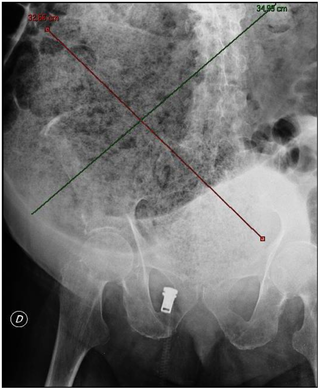
A rectal prolapse occurs when walls of the rectum have prolapsed to such a degree that they protrude out of the anus and are visible outside the body. However, most researchers agree that there are 3 to 5 different types of rectal prolapse, depending on whether the prolapsed section is visible externally, and whether the full or only partial thickness of the rectal wall is involved.

<span class="mw-page-title-main">Diverticulosis</span> Condition of the wall of the intestine

Diverticulosis is the condition of having multiple pouches (diverticula) in the colon that are not inflamed. These are outpockets of the colonic mucosa and submucosa through weaknesses of muscle layers in the colon wall. Diverticula do not cause symptoms in most people. Diverticular disease occurs when diverticula become clinically inflamed, a condition known as diverticulitis.

<span class="mw-page-title-main">Gastrointestinal disease</span> Medical condition

Gastrointestinal diseases refer to diseases involving the gastrointestinal tract, namely the esophagus, stomach, small intestine, large intestine and rectum, and the accessory organs of digestion, the liver, gallbladder, and pancreas.

<span class="mw-page-title-main">Volvulus</span> Twisting of part of the intestine, causing a bowel obstruction

A volvulus is when a loop of intestine twists around itself and the mesentery that supports it, resulting in a bowel obstruction. Symptoms include abdominal pain, abdominal bloating, vomiting, constipation, and bloody stool. Onset of symptoms may be rapid or more gradual. The mesentery may become so tightly twisted that blood flow to part of the intestine is cut off, resulting in ischemic bowel. In this situation there may be fever or significant pain when the abdomen is touched.

<span class="mw-page-title-main">Fecal impaction</span> Medical condition

A fecal impaction or an impacted bowel is a solid, immobile bulk of feces that can develop in the rectum as a result of chronic constipation. Fecal impaction is a common result of neurogenic bowel dysfunction and causes immense discomfort and pain. Its treatment includes laxatives, enemas, and pulsed irrigation evacuation (PIE) as well as digital removal. It is not a condition that resolves without direct treatment.

Intestinal malrotation is a congenital anomaly of rotation of the midgut. It occurs during the first trimester as the fetal gut undergoes a complex series of growth and development. Malrotation can lead to a dangerous complication called volvulus, in which cases emergency surgery is indicated. Malrotation can refer to a spectrum of abnormal intestinal positioning, often including:

Intestinal neuronal dysplasia (IND) is an inherited disease of the intestine that affects one in 3000 children and adults. The intestine uses peristalsis to push its contents toward the anus; people with IND have a problem with the motor neurons that lead to the intestine, inhibiting this process and thus preventing digestion.

<span class="mw-page-title-main">Megacolon</span> Medical condition

Megacolon is an abnormal dilation of the colon. This leads to hypertrophy of the colon. The dilation is often accompanied by a paralysis of the peristaltic movements of the bowel. In more extreme cases, the feces consolidate into hard masses inside the colon, called fecalomas, which can require surgery to be removed.

<span class="mw-page-title-main">Intestinal pseudo-obstruction</span> Medical condition

Intestinal pseudo-obstruction (IPO) is a clinical syndrome caused by severe impairment in the ability of the intestines to push food through. It is characterized by the signs and symptoms of intestinal obstruction without any lesion in the intestinal lumen. Clinical features mimic those seen with mechanical intestinal obstructions and can include abdominal pain, nausea, abdominal distension, vomiting, dysphagia and constipation depending upon the part of the gastrointestinal tract involved.

Stercoral ulcer is an ulcer of the colon due to pressure and irritation resulting from severe, prolonged constipation due to a large bowel obstruction, damage to the autonomic nervous system, or stercoral colitis. It is most commonly located in the sigmoid colon and rectum. Prolonged constipation leads to production of fecaliths, leading to possible progression into a fecaloma. These hard lumps irritate the rectum and lead to the formation of these ulcers. It results in fresh bleeding per rectum. These ulcers may be seen on imaging, such as a CT scan but are more commonly identified using endoscopy, usually a colonoscopy. Treatment modalities can include both surgical and non-surgical techniques.

A lower anterior resection, formally known as anterior resection of the rectum and colon and anterior excision of the rectum or simply anterior resection, is a common surgery for rectal cancer and occasionally is performed to remove a diseased or ruptured portion of the intestine in cases of diverticulitis. It is commonly abbreviated as LAR.

Solitary rectal ulcer syndrome or SRUS is a chronic, benign disorder of the rectal mucosa. It commonly occurs with varying degrees of rectal prolapse. The condition is thought to be caused by different factors, such as long term constipation, straining during defecation, and dyssynergic defecation. Treatment is by normalization of bowel habits, biofeedback, and other conservative measures. In more severe cases various surgical procedures may be indicated. The condition is relatively rare, affecting approximately 1 in 100,000 people per year. It affects mainly adults aged 30–50. Females are affected slightly more often than males. The disorder can be confused clinically with rectal cancer or other conditions such as inflammatory bowel disease, even when a biopsy is done.

<span class="mw-page-title-main">Anismus</span> Medical condition

Anismus or dyssynergic defecation is the failure of normal relaxation of pelvic floor muscles during attempted defecation. It can occur in both children and adults, and in both men and women. It can be caused by physical defects or it can occur for other reasons or unknown reasons. Anismus that has a behavioral cause could be viewed as having similarities with parcopresis, or psychogenic fecal retention.

Obstructed defecation syndrome is a major cause of functional constipation, of which it is considered a subtype. It is characterized by difficult and/or incomplete emptying of the rectum with or without an actual reduction in the number of bowel movements per week. Normal definitions of functional constipation include infrequent bowel movements and hard stools. In contrast, ODS may occur with frequent bowel movements and even with soft stools, and the colonic transit time may be normal, but delayed in the rectum and sigmoid colon.

Constipation in children refers to the medical condition of constipation in children. It is a functional gastrointestinal disorder.

<span class="mw-page-title-main">Neurogenic bowel dysfunction</span> Human disease involving inability to control defecation

Neurogenic bowel dysfunction (NBD) is the inability to control defecation due to a deterioration of or injury to the nervous system, resulting in faecal incontinence or constipation. It is common in people with spinal cord injury (SCI), multiple sclerosis (MS) or spina bifida.

<span class="mw-page-title-main">Orvar Swenson</span>

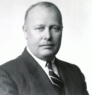
Orvar Swenson was a Swedish-born American pediatric surgeon. He discovered the cause of Hirschsprung's disease and in 1948, with Alexander Bill, performed the first pull-through operation in a child with megacolon, which then became a treatment for the disease.

A pull-through procedure is the definitive operation for Hirschsprung disease, involving the removal of the abnormal segment of bowel that has no nerves, pulling through the normal bowel and connecting it to the anus. Several types of pull-through procedures exist including the Soave, Swenson and Duhamel. It can be performed using an open or minimally invasive approach.

Waardenburg syndrome type 4A is an extremely rare congenital disorder caused by a mutation in an endothelin receptor gene. It results in common Waardenburg syndrome symptoms such as abnormal hair and skin pigmentation and heterochromia, but also present with symptoms of Hirschsprung's disease. Symptoms include abdominal pain and bowel obstruction. Waardenburg syndrome type 4A is the rarest among the types, appearing only once in about every 1,000,000 individuals. There have only been a total of 50 cases reported in total as of 2016.

References

  1. 1 2 3 4 5 6 7 8 9 10 11 12 13 14 15 16 17 18 19 20 21 22 "Hirschsprung disease". Genetics Home Reference. August 2012. Retrieved 14 December 2017.
  2. 1 2 3 4 5 6 7 8 9 10 11 "Hirschsprung's disease". Genetic and Rare Diseases Information Center (GARD) – an NCATS Program. 2017. Archived from the original on 24 November 2018. Retrieved 14 December 2017.
  3. 1 2 3 4 "Hirschsprung Disease". NORD (National Organization for Rare Disorders). 2017. Retrieved 14 December 2017.
  4. 1 2 "Definition & Facts for Hirschsprung Disease - NIDDK". National Institute of Diabetes and Digestive and Kidney Diseases. Retrieved 2023-03-24.
  5. "Hirschprung's Disease (HSCR) Diagnosis & Treatment". Children's Hospital of Pittsburgh. Retrieved 2023-03-27.
  6. Holschneider, Alexander Matthias; Puri, Prem (2007). Hirschsprung's Disease and Allied Disorders. Springer Science & Business Media. p. 1. ISBN   9783540339359.
  7. 1 2 "Hirschsprung's disease". www.whonamedit.com. Retrieved 8 October 2019.
  8. 1 2 Hirschsprung, H. (1888). "Stuhlträgheit Neugeborener in Folge von Dilatation und Hypertrophie des Colons". Jahrbuch für Kinderheilkunde und physische Erziehung. 27. Berlin: 1–7.
  9. 1 2 3 4 5 6 Goldman, Lee (2012). Goldman's Cecil Medicine (24th ed.). Philadelphia: Elsevier Saunders. p. 867. ISBN   978-1437727883.
  10. Kimura, Ken; Loening-Baucke, Vera (1999-11-01). "Failure to Pass Meconium: Diagnosing Neonatal Intestinal Obstruction". American Family Physician. 60 (7): 2043–50. ISSN   0002-838X. PMID   10569507.
  11. "Hirschsprung's disease". Mayo Clinic. Retrieved 30 January 2023.
  12. Heuckeroth RO (March 2018). "Hirschsprung disease - integrating basic science and clinical medicine to improve outcomes". Nature Reviews. Gastroenterology & Hepatology. 15 (3): 152–167. doi:10.1038/nrgastro.2017.149. PMID   29300049. S2CID   3303746.
  13. Online Mendelian Inheritance in Man (OMIM): 142623
  14. Mäkitie O, Heikkinen M, Kaitila I, Rintala R (2002). "Hirschsprung's disease in cartilage-hair hypoplasia has poor prognosis". J Pediatr Surg. 37 (11): 1585–8. doi:10.1053/jpsu.2002.36189. PMID   12407544.
  15. de Pontual L, Pelet A, Clement-Ziza M, Trochet D, Antonarakis SE, Attie-Bitach T, Beales PL, Blouin JL, Dastot-Le Moal F, Dollfus H, Goossens M, Katsanis N, Touraine R, Feingold J, Munnich A, Lyonnet S, Amiel J (2007). "Epistatic interactions with a common hypomorphic RET allele in syndromic Hirschsprung disease". Human Mutation . 28 (8): 790–6. doi:10.1002/humu.20517. PMID   17397038. S2CID   25686389.
  16. Saunders CJ, Zhao W, Ardinger HH (2009). "Comprehensive ZEB2 gene analysis for Mowat-Wilson syndrome in a North American cohort: a suggested approach to molecular diagnostics". American Journal of Medical Genetics . 149A (11): 2527–31. doi:10.1002/ajmg.a.33067. PMID   19842203. S2CID   22472646.
  17. Bonnard A, Zeidan S, Degas V, Viala J, Baumann C, Berrebi D, Perrusson O, El Ghoneimi A (2009). "Outcomes of Hirschsprung's disease associated with Mowat-Wilson syndrome". Journal of Pediatric Surgery . 44 (3): 587–91. doi:10.1016/j.jpedsurg.2008.10.066. PMID   19302864.
  18. Mueller C, Patel S, Irons M, Antshel K, Salen G, Tint GS, Bay C (2003). "Normal cognition and behavior in a Smith-Lemli-Opitz syndrome patient who presented with Hirschsprung disease". American Journal of Medical Genetics . 123A (1): 100–6. doi:10.1002/ajmg.a.20491. PMC   1201564 . PMID   14556255.
  19. Flori E, Girodon E, Samama B, Becmeur F, Viville B, Girard-Lemaire F, Doray B, Schluth C, Marcellin L, Boehm N, Goossens M, Pingault V (2005). "Trisomy 7 mosaicism, maternal uniparental heterodisomy 7 and Hirschsprung's disease in a child with Silver-Russell syndrome". European Journal of Human Genetics . 13 (9): 1013–8. doi: 10.1038/sj.ejhg.5201442 . PMID   15915162.
  20. Martucciello G, Ceccherini I, Lerone M, Jasonni V (2000). "Pathogenesis of Hirschsprung's disease". Journal of Pediatric Surgery. 35 (7): 1017–1025. doi:10.1053/jpsu.2000.7763. PMID   10917288.
  21. Chial, H (2008). "Proto-oncogenes to Oncogenes to Cancer". Nature Education . 1: 33.
  22. Puri P, Shinkai T (2004). "Pathogenesis of Hirschsprung's disease and its variants: recent progress". Semin. Pediatr. Surg. 13 (1): 18–24. doi:10.1053/j.sempedsurg.2003.09.004. PMID   14765367. S2CID   11395791.
  23. Garcia-Barcelo, Maria-Merce (2009). "Genome-wide association study identifies NRG1 as a susceptibility locus for Hirschsprung's disease". Proc. Natl. Acad. Sci. USA. 106 (8): 2694–2699. Bibcode:2009PNAS..106.2694G. doi: 10.1073/pnas.0809630105 . PMC   2650328 . PMID   19196962.
  24. Tang, Clara (May 10, 2012). "Genome-wide copy number analysis uncovers a new HSCR gene: NRG3". PLOS Genet. 8 (5): e1002687. doi: 10.1371/journal.pgen.1002687 . PMC   3349728 . PMID   22589734.
  25. Yang J, Duan S, Zhong R, Yin J, Pu J, Ke J, Lu X, Zou L, Zhang H, Zhu Z, Wang D, Xiao H, Guo A, Xia J, Miao X, Tang S, Wang G (2013). "Exome sequencing identified NRG3 as a novel susceptible gene of Hirschsprung's disease in a Chinese population". Mol. Neurobiol. 47 (3): 957–66. doi:10.1007/s12035-012-8392-4. PMID   23315268. S2CID   16842806.
  26. Shin JG, Seo JY, Seo JM, Kim DY, Oh JT, Park KW, Kim HY, Kim JH, Shin HD (2019) Association analysis of NOX5 polymorphisms with Hirschsprung disease. J Pediatr Surg
  27. Parisi MA, Pagon RA, Bird TD, Dolan CR, Stephens K, Adam MP (2002). Pagon RA, Bird TC, Dolan CR, Stephens K (eds.). "Hirschsprung Disease Overview". GeneReviews. PMID   20301612.
  28. Kays DW (1996). "Surgical conditions of the neonatal intestinal tract". Clinics in Perinatology. 23 (2): 353–75. doi:10.1016/S0095-5108(18)30246-X. PMID   8780909.
  29. Metallinos DL, Bowling AT, Rine J (Jun 1998). "A missense mutation in the endothelin-B receptor gene is associated with Lethal White Foal Syndrome: an equine version of Hirschsprung disease". Mamm. Genome. 9 (6): 426–31. doi:10.1007/s003359900790. PMID   9585428. S2CID   19536624. Archived from the original on 2000-09-16.
  30. Dobbins WO, Bill AH (1965). "Diagnosis of Hirschsprung's Disease Excluded by Rectal Suction Biopsy". New England Journal of Medicine. 272 (19): 990–993. doi:10.1056/NEJM196505132721903. PMID   14279253.
  31. Eli Ehrenpreis (Oct 2003). Anal and rectal diseases explained. Remedica. pp. 15–. ISBN   978-1-901346-67-1 . Retrieved 2010-11-12.
  32. Martucciello G, Pini Prato A, Puri P, Holschneider AM, Meier-Ruge W, Jasonni V, Tovar JA, Grosfeld JL (2005). "Controversies concerning diagnostic guidelines for anomalies of the enteric nervous system: a report from the fourth International Symposium on Hirschsprung's disease and related neurocristopathies". J Pediatr Surg. 40 (10): 1527–31. doi: 10.1016/j.jpedsurg.2005.07.053 . PMID   16226977.
  33. Kim HJ, Kim AY, Lee CW, Yu CS, Kim JS, Kim PN, Lee MG, Ha HK (2008). "Hirschsprung disease and hypoganglionosis in adults: radiologic findings and differentiation". Radiology. 247 (2): 428–34. doi:10.1148/radiol.2472070182. PMID   18430875.
  34. "Pediatric Hirschsprung's Disease | Children's National Hospital".
  35. Hope, John W.; Borns, Patricia Flint; Berg, Paul K. (September 1965). "Roentgenologic Manifestations of Hirschsprung's Disease in Infancy". American Journal of Roentgenology. 95 (1): 217–229. doi:10.2214/ajr.95.1.217. PMID   14344366.
  36. Swenson O (1989). "My early experience with Hirschsprung's disease". J. Pediatr. Surg. 24 (8): 839–44, discussion 844–5. doi:10.1016/S0022-3468(89)80549-4. PMID   2671336.
  37. 1 2 "Hirschsprung disease". American Pediatric Surgical Association. Retrieved 11 June 2019.
  38. W. Allan Walker (2004-07-01). Pediatric gastrointestinal disease: pathophysiology, diagnosis, management. PMPH-USA. pp. 2120–. ISBN   978-1-55009-240-0 . Retrieved 2010-11-12.
  39. Timothy R. Koch (2003). Colonic diseases. Humana Press. pp. 387–. ISBN   978-0-89603-961-2 . Retrieved 2010-11-12.[ permanent dead link ]
  40. Malone PS, Ransley PG, Kiely EM (1990). "Preliminary report: the antegrade continence enema". Lancet. 336 (8725): 1217–1218. doi:10.1016/0140-6736(90)92834-5. PMID   1978072. S2CID   9203632.
  41. Walsh RA, Koyle MA, Waxman SW (2000). "The Malone ACE Procedure for Fecal Incontinence". Infections in Urology. 13 (4). Archived from the original on 2011-04-07. Retrieved 2012-01-04.
  42. 1 2 Goldberg EL (1984). "An epidemiological study of Hirschsprung's disease". Int J Epidemiol. 13 (4): 479–85. doi:10.1093/ije/13.4.479. PMID   6240474.
  43. Suita S, Taguchi T, Ieiri S, Nakatsuji T (2005). "Hirschsprung's disease in Japan: analysis of 3852 patients based on a nationwide survey in 30 years". Journal of Pediatric Surgery . 40 (1): 197–201, discussion 201–2. doi:10.1016/j.jpedsurg.2004.09.052. PMID   15868585.
  44. Colwell, Janice (2004). Fecal and Urinary Diversion Management. Mosby. p. 264. ISBN   978-0-323-02248-4.
  45. Hirschsprung's Disease and Allied Disorders. Berlin: Springer. 2007. ISBN   978-3-540-33934-2.
  46. "Hirschsprung Disease: Background, Pathophysiology, Epidemiology". 2017-01-08.{{cite journal}}: Cite journal requires |journal= (help)
  47. Worman S, Ganiats TG (1995). "Hirschsprung's disease: a cause of chronic constipation in children". Am Fam Physician. 51 (2): 487–94. PMID   7840044.
  48. "What is the pathophysiology of Hirschsprung disease?". 2 August 2021.
  49. Martucciello G; Bicocchi MP; Dodero P.; Lerone M.; Silengo Cirillo M; Puliti A; Gimelli G; Romeo G. (1992). "Total colonic aganglionosis associated with interstitial deletion of the long arm of chromosome 10". Pediatric Surgery International. 7 (4): 308–310. doi:10.1007/BF00183991. S2CID   40123658.
  50. Romeo G, Ronchetto P, Luo Y, Barone V, Seri M, Ceccherini I, Pasini B, Bocciardi R, Lerone M, Kääriäinen H (1994). "Point mutations affecting the tyrosine kinase domain of the RET proto-oncogene in Hirschsprung's disease". Nature. 367 (6461): 377–378. Bibcode:1994Natur.367..377R. doi:10.1038/367377a0. PMID   8114938. S2CID   157274.
  51. (National Digestive Diseases Information Clearinghouse).
  52. Angrist M, Kauffman E, Slaugenhaupt SA, Matise TC, Puffenberger EG, Washington SS, Lipson A, Cass DT, Reyna T, Weeks DE (1993). "A gene for Hirschsprung disease (megacolon) in the pericentromeric region of human chromosome 10". Nat. Genet. 4 (4): 351–6. doi:10.1038/ng0893-351. PMID   8401581. S2CID   22031571.
  53. Lyonnet S, Bolino A, Pelet A, Abel L, Nihoul-Fékété C, Briard ML, Mok-Siu V, Kaariainen H, Martucciello G, Lerone M, Puliti A, Luo Y, Weissenbach J, Devoto M, Munnich A, Romeo G (1993). "A gene for Hirschsprung disease maps to the proximal long arm of chromosome 10". Nat. Genet. 4 (4): 346–50. doi:10.1038/ng0893-346. PMID   8401580. S2CID   29089707.