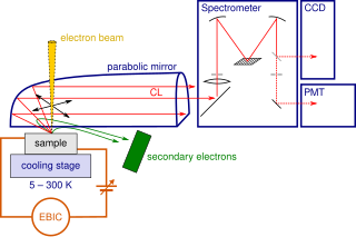
Cathodoluminescence is an optical and electromagnetic phenomenon in which electrons impacting on a luminescent material such as a phosphor, cause the emission of photons which may have wavelengths in the visible spectrum. A familiar example is the generation of light by an electron beam scanning the phosphor-coated inner surface of the screen of a television that uses a cathode ray tube. Cathodoluminescence is the inverse of the photoelectric effect, in which electron emission is induced by irradiation with photons.
Force spectroscopy is a set of techniques for the study of the interactions and the binding forces between individual molecules. These methods can be used to measure the mechanical properties of single polymer molecules or proteins, or individual chemical bonds. The name "force spectroscopy", although widely used in the scientific community, is somewhat misleading, because there is no true matter-radiation interaction.
The term biophotonics denotes a combination of biology and photonics, with photonics being the science and technology of generation, manipulation, and detection of photons, quantum units of light. Photonics is related to electronics and photons. Photons play a central role in information technologies, such as fiber optics, the way electrons do in electronics.

Near-field scanning optical microscopy (NSOM) or scanning near-field optical microscopy (SNOM) is a microscopy technique for nanostructure investigation that breaks the far field resolution limit by exploiting the properties of evanescent waves. In SNOM, the excitation laser light is focused through an aperture with a diameter smaller than the excitation wavelength, resulting in an evanescent field on the far side of the aperture. When the sample is scanned at a small distance below the aperture, the optical resolution of transmitted or reflected light is limited only by the diameter of the aperture. In particular, lateral resolution of 20 nm and vertical resolution of 2–5 nm have been demonstrated.
Nanophotonics or nano-optics is the study of the behavior of light on the nanometer scale, and of the interaction of nanometer-scale objects with light. It is a branch of optics, optical engineering, electrical engineering, and nanotechnology. It often involves metallic components, which can transport and focus light via surface plasmon polaritons.
Extraordinary optical transmission (EOT) is the phenomenon of greatly enhanced transmission of light through a subwavelength aperture in an otherwise opaque metallic film which has been patterned with a regularly repeating periodic structure. Generally when light of a certain wavelength falls on a subwavelength aperture, it is diffracted isotropically in all directions evenly, with minimal far-field transmission. This is the understanding from classical aperture theory as described by Bethe. In EOT however, the regularly repeating structure enables much higher transmission efficiency to occur, up to several orders of magnitude greater than that predicted by classical aperture theory. It was first described in 1998.

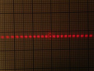
A Bessel beam is a field of electromagnetic, acoustic or even gravitational radiation whose amplitude is described by a Bessel function of the first kind. A true Bessel beam is non-diffractive. This means that as it propagates, it does not diffract and spread out; this is in contrast to the usual behavior of light, which spreads out after being focused down to a small spot. Bessel beams are also self-healing, meaning that the beam can be partially obstructed at one point, but will re-form at a point further down the beam axis.
Magnetic tweezers (MT) are scientific instruments for the manipulation and characterization of biomolecules or polymers. These apparatus exert forces and torques to individual molecules or groups of molecules. It can be used to measure the tensile strength or the force generated by molecules.

Supercritical angle fluorescence microscopy (SAF) is a technique to detect and characterize fluorescent species and their behaviour close or even adsorbed or linked at surfaces. The method is able to observe molecules in a distance of less than 100 to 0 nanometer from the surface even in presence of high concentrations of fluorescent species around. Using an aspheric lens for excitation of a sample with laser light, fluorescence emitted by the specimen is collected above the critical angle of total internal reflection selectively and directed by a parabolic optics onto a detector. The method was invented in 1998 in the laboratories of Stefan Seeger at University of Regensburg/Germany and later at University of Zurich/Switzerland.

Tethered particle motion (TPM) is a biophysical method that is used for studying various polymers such as DNA and their interaction with other entities such as proteins.
Optoelectrofluidics, also known as optically induced electrohydrodynamics, refers to the study of the motions of particles or molecules and their interactions with optically-induced electric field and the surrounding fluid.
Microrheology is a technique used to measure the rheological properties of a medium, such as microviscosity, via the measurement of the trajectory of a flow tracer. It is a new way of doing rheology, traditionally done using a rheometer. There are two types of microrheology: passive microrheology and active microrheology. Passive microrheology uses inherent thermal energy to move the tracers, whereas active microrheology uses externally applied forces, such as from a magnetic field or an optical tweezer, to do so. Microrheology can be further differentiated into 1- and 2-particle methods.

The superconducting nanowire single-photon detector (SNSPD) is a type of near-infrared and optical single-photon detector based on a current-biased superconducting nanowire. It was first developed by scientists at Moscow State Pedagogical University and at the University of Rochester in 2001.
Through-Focus Scanning Optical Microscopy (TSOM) is an imaging method that produces nanometer-scale three-dimensional measurement sensitivity using a conventional bright-field optical microscope. TSOM has been introduced and maintained by Ravikiran Attota at NIST. It was given an R&D 100 Award in 2010. In the TSOM method a target is scanned through the focus of an optical microscope, acquiring conventional optical images at different focal positions. The TSOM images are constructed using the through-focus optical images. A TSOM image is unique under given experimental conditions and is sensitive to changes in the dimensions of a target in a distinct way, which is very well applicable in nanoscale dimensional metrology. The TSOM method is alleged to have several nanometrology applications ranging from nanoparticles to through-silicon-vias (TSV).
Differential dynamic microscopy (DDM) is an optical technique that allows performing light scattering experiments by means of a simple optical microscope. DDM is suitable for typical soft materials such as for instance liquids or gels made of colloids, polymers and liquid crystals but also for biological materials like bacteria and cells.
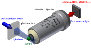
Light sheet fluorescence microscopy (LSFM) is a fluorescence microscopy technique with an intermediate-to-high optical resolution, but good optical sectioning capabilities and high speed. In contrast to epifluorescence microscopy only a thin slice of the sample is illuminated perpendicularly to the direction of observation. For illumination, a laser light-sheet is used, i.e. a laser beam which is focused only in one direction. A second method uses a circular beam scanned in one direction to create the lightsheet. As only the actually observed section is illuminated, this method reduces the photodamage and stress induced on a living sample. Also the good optical sectioning capability reduces the background signal and thus creates images with higher contrast, comparable to confocal microscopy. Because LSFM scans samples by using a plane of light instead of a point, it can acquire images at speeds 100 to 1000 times faster than those offered by point-scanning methods.
The Optical Stretcher is a dual-beam optical trap that is used for trapping and deforming ("stretching") micrometer-sized soft matter particles, such as biological cells in suspension. The forces used for trapping and deforming objects arise from photon momentum transfer on the surface of the objects, making the Optical Stretcher - unlike atomic force microscopy or micropipette aspiration - a tool for contact-free rheology measurements.
David G. Grier is an American physicist whose research focuses on experimental soft condensed matter physics—an interdisciplinary field that includes physics, chemistry, biology, and nanotechnology, aiming to understand how objects interacting in simple ways manage to organize into sophisticated hierarchies of structure and function.
Interferometric scattering microscopy (iSCAT) is an optical microscopy technique that relies on collecting light scattered by an object together with a reference light field, provided by the reflection at an interface. In the presence of the scattering object, the total intensity of light returning in the direction of the incident beam is different than in the absence of that object due to interference of the scattered and reflected light. Thus, the object appears as a small dip on top of a large background.