3D cell culture

Last updated

A 3D cell culture is an artificially created environment in which biological cells are permitted to grow or interact with their surroundings in all three dimensions. Unlike 2D environments (e.g. a Petri dish), a 3D cell culture allows cells in vitro to grow in all directions, similar to how they would in vivo. [1] These three-dimensional cultures are usually grown in bioreactors, small capsules in which the cells can grow into spheroids, or 3D cell colonies. Approximately 300 spheroids are usually cultured per bioreactor. [1]

Contents

Background

3D cell cultures have been used in research for several decades. [2] One of the first recorded approaches for their development was at the beginning of the 20th century, with the efforts of Alexis Carrel to develop methods for prolonged in vitro tissue cultures. [3] Early studies in the 80's, led by Mina Bissell from the Lawrence Berkeley National Laboratory, highlighted the importance of 3D techniques for creating accurate in vitro culturing models. This work focused on the importance of the extracellular matrix and the ability of cultures in artificial 3D matrices to produce physiologically relevant multicellular structures, such as acinar structures in healthy and cancerous breast tissue models. These techniques have been applied to in vitro disease models used to evaluate cellular responses to pharmaceutical compounds. [4]

Eric Simon, in a 1988 NIH SBIR grant report, showed that electrospinning could be used to produce nano- and submicron-scale polystyrene and polycarbonate fibrous mats (now known as scaffolds) specifically intended for use as in vitro cell substrates. This early use of electrospun fibrous lattices for cell culture and tissue engineering showed that various cell types including Human Foreskin Fibroblasts (HFF), transformed Human Carcinoma (HEp-2), and Mink Lung Epithelium (MLE) would adhere to and proliferate upon the fibers. It was noted that as opposed to the flattened morphology typically seen in 2D culture, cells grown on the electrospun fibers exhibited a more histotypic rounded 3-dimensional morphology generally observed in vivo. [5]

3D cell culture, by emulating essential aspects of the in vivo environment, including interactions between cells and the extracellular matrix, allows for the faithful recreation of structural architecture and specialized functions in normal tissues or tumors in a laboratory setting. This approach authentically models the conditions and processes of living tissues, producing responses akin to those observed in vivo. Since its inception in the 1970s, 3D cell culture has provided significant insights into the mechanisms regulating tissue homeostasis and cancer. [6] Moreover, it has expedited translational research in the realms of cancer biology and tissue engineering. [7]

Properties

In living tissue, cells exist in 3D microenvironments with intricate cell-cell and cell-matrix interactions and complex transport dynamics for nutrients and cells. [8] [9] [10] [11] [12] [13] [14] [15] [16] Standard 2D, or monolayer, cell cultures are inadequate representations of this environment, which often makes them unreliable predictors of in vivo drug efficacy and toxicity. [17] [14] 3D spheroids more closely resemble in vivo tissue in terms of cellular communication and the development of extracellular matrices. [1] These matrices help the cells to be able to move within their spheroid similar to the way cells would move in living tissue. [10] The spheroids are thus improved models for cell migration, differentiation, survival, and growth. [15] Furthermore, 3D cell cultures provide more accurate depiction of cell polarization, since in 2D, the cells can only be partially polarized. [10] Moreover, cells grown in 3D exhibit different gene expression than those grown in 2D. [10]

The third dimension of cell growth provides more contact space for mechanical inputs and for cell adhesion, which is necessary for integrin ligation, cell contraction and even intracellular signalling. [18] [19] Normal solute diffusion and binding to effector proteins (like growth factors and enzymes) is also reliant on the 3D cellular matrix, so it is critical for the establishment of tissue scale solute concentration gradients [20] [21]

For the purposes of drug toxicology screening, it is much more useful to test gene expression of in vitro cells grown in 3D than 2D, since the gene expression of the 3D spheroids will more closely resemble gene expression in vivo. Lastly, 3D cell cultures have greater stability and longer lifespans than cell cultures in 2D. [22] This means that they are more suitable for long-term studies and for demonstrating long-term effects of the drug. 3D environments also allow the cells to grow undisturbed. In 2D, the cells must undergo regular trypsinization to provide them with sufficient nutrients for normal cell growth. [23] 3D spheroids have been cultured in a lab setting for up to 302 days while still maintaining healthy, non-cancerous growth. [22]

In the interdisciplinary research of biology and aerospace, the 3D printed-scaffolds are also being used for protecting cells from the effect of gravity during the launching. [24]

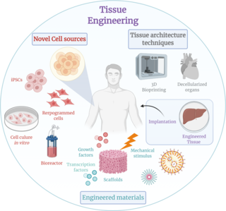
Classification of 3D culture methods

There are a large number of commercially available culturing tools that claim to provide the advantages of 3D cell culture. In general, the platforms can be classified in two types of 3D culturing methods: scaffold techniques and scaffold-free techniques.

A model showing three examples of techniques used for culturing cells in a 3D environment. 3d cell culture (1).svg
A model showing three examples of techniques used for culturing cells in a 3D environment.

Scaffold techniques

Scaffold techniques include the use of solid scaffolds, hydrogels and other materials. In a recent study potentiality of human CD34+ stem cells explored by generating in vitro agarose gel 3D model to understand the bone ossification process. [25] Scaffolds can be used to generate microtissue 3D model by culturing fibroblasts outside of tumour cells, mimicking the tumor stroma interaction. [26]

The effectiveness of scaffolds in various applications, particularly in tissue engineering, is significantly impacted by factors such as pore distribution, exposed surface area, and porosity. The quantity and arrangement of these elements influence both the depth and rate at which cells penetrate the scaffold volume, the structure of the resulting extracellular matrix, and ultimately, the success of the regenerative process. [27] Scaffolds can be produced with diverse architectures depending on the manufacturing method, leading to either random or precisely designed pore distribution. [28] Recently, advanced computer-controlled rapid prototyping techniques have been employed to create scaffolds with well-organized geometries. [29]

Hydrogels

As the natural extracellular matrix (ECM) is important in the survival, proliferation, differentiation and migration of the cells, different hydrogel matrices mimicking natural ECM structure are considered as potential approaches towards in vivo –like cell culturing. [30] [31] [32] Hydrogels are composed of interconnected pores with high water retention, which enables efficient transport of e.g. nutrients and gases. Several different types of hydrogels from natural and synthetic materials are available for 3D cell culture, including e.g. animal ECM extract hydrogels, protein hydrogels, peptide hydrogels, polymer hydrogels, and wood-based nanocellulose hydrogel.

The approach to crafting the optimal ECM replica relies on the specific characteristics of the culture in question and typically involves employing diverse and independent chemical processes. [33] For example, the utilization of photolabile chemistries can lead to the erosion of specific regions within a gel, and subsequently exposing these areas allows for the application of adhesive ligands, promoting cell adhesion and migration. [34] The development of more intricate frameworks is anticipated, comprising interwoven networks of chemistries under the control of both cells and users. In essence, there is no singular network capable of faithfully emulating the intricate ECM of every tissue type. However, a thoughtful integration of bioinspired cues into synthetic gels holds the potential to yield resilient and versatile scaffolds applicable across various cell culture systems. [35]

Scaffold-free techniques

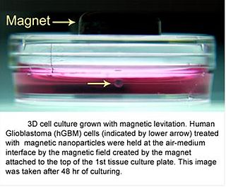
Scaffold free techniques employ another approach independent from the use scaffold. Scaffold-free methods include e.g. the use of low adhesion plates, hanging drop plates, micropatterned surfaces, and rotating bioreactors, magnetic levitation, and magnetic 3D bioprinting.

Spheroids

Electron microscopy of a mesothelioma spheroid (NCI-H226). Scale bars, 200 mm. Electron microscopy (Ho).tif
Electron microscopy of a mesothelioma spheroid (NCI-H226). Scale bars, 200 μm.

Spheroids are a type of three-dimensional cell modeling that better simulate a live cell's environmental conditions compared to a two-dimensional cell model, specifically with the reactions between cells and the reactions between cells and the matrix. [37] Spheroids are useful in the study of changing physiological characteristics of cells, [38] the difference in the structure of healthy cells and tumor cells, and the changes cells undergo when forming a tumor. [39] Spheroids co-cultured with tumor and healthy cells were used to simulate how cancerous cells interact with normal cells. [40] Spheroids can also be co-cultured with fibroblasts to mimic tumor-stroma interaction. [41] Spheroids can be grown with a few different methods. One common method is to use low cell adhesion plates, typically a 96 well plate, to mass-produce spheroid cultures, where the aggregates form in the rounded bottom of the cell plates. [36] [42] Spheroids can also be cultured using the hanging drop method [43] involving forming cell aggregates in drops that hang from the surface of a cell plate. [37] Other methods under investigation include the use of rotating wall vessel bioreactors, which spins and cultures the cells when they are constantly in free fall and forms aggregates in layers [44] Recently, some protocols have been standardized to produce uniform and reliable spheroids. [45] Researchers had also explored standardized, economical and reproducible methods for 3D cell culture. [46] To improve reproducibility and transparency in spheroid experiments, an international consortium developed MISpheroID (Minimal Information in Spheroid Identity). [47]

Clusteroids

clusteroids are a type of three-dimensional cell modeling similar to spheroids but are distinguished by their creation method; grown as clusters of cells in an aqueous two-phase system of water-in-water Pickering emulsion using interfacial tension and osmotic shrinkage to pack the cells into dense clusters which are then cultured in a hydrogel into tissues or organoids. [48] [49]

In the absence of blood vessels, oxygen permeability is impaired during necrotic nucleus formation and this prevents the ex vivo use of 3D cell culture. There is an emulsion template that can overcome this problem. This approach allowed researchers to adjust the cell composition to attain the ideal conditions for promoting the synthesis of diverse angiogenic protein markers within the co-cultured clusteroids. [50] HUVEC cells exhibit a reaction to the presence of Hep-G2 cells and their derivatives by generating endothelial cell sprouts in Matrigel, all without the external introduction of vascular endothelial growth factor (VEGF) or other agents that induce angiogenesis. [51] [52] The replication of this cultivation technique is straightforward for generating various cell co-culture spheroids. [53] The w/w Pickering emulsion template greatly aids in constructing 3D co-culture models, offering significant potential for applications in drug testing and tissue engineering. [54]

Bioreactors

The bioreactors used for 3D cell cultures are small plastic cylindrical chambers that are specifically engineered for the purpose of growing cells in three dimensions. The bioreactor uses bioactive synthetic materials such as polyethylene terephthalate membranes to surround the spheroid cells in an environment that maintains high levels of nutrients. [55] [56] They are easy to open and close, so that cell spheroids can be removed for testing, yet the chamber is able to maintain 100% humidity throughout. [1] This humidity is important to achieve maximum cell growth and function. The bioreactor chamber is part of a larger device that rotates to ensure equal cell growth in each direction across three dimensions. [1]
MC2 Biotek has developed a bioreactor to incubate proto-tissue that uses gas exchange to maintain high oxygen levels within the cell chamber. [57] This is an improvement over previous bioreactors because the higher oxygen levels help the cell grow and undergo normal cell respiration. [15]

Collaborative efforts between tissue engineering (TE) firms, academic institutions, and industrial partners can enhance the transformation of research-oriented bioreactors into efficient commercial manufacturing systems. [58] Academic collaborators contribute foundational aspects, while industrial partners provide essential automation elements, ensuring compliance with regulatory standards and user-friendliness. [59] Established consortia in Europe, such as REMEDI, AUTOBONE, and STEPS, focus on developing automated systems to streamline the engineering of autologous cell-based grafts. [60] The aim is to meet regulatory criteria and ensure cost-effectiveness, making tissue-engineered products more clinically accessible and advancing the translational paradigm of TE from research to a competitive commercial field. [61]

Microfluidics

The utilization of microfluidic technology facilitates the generation of intricate micro-scale structures and the precise manipulation of parameters, thereby emulating the in vivo cellular milieu. The integration of microfluidic technology with 3D cell culture holds considerable potential for applications that seek to replicate in vivo tissue characteristics, notably exemplified by the evolving organ-on-a-chip system. [62] The various cell structures in the human body must be vascularized to receive the nutrients and gas exchange in order to survive. Similarly, 3D cell cultures in vitro require certain levels of fluid circulation, which can be problematic for dense, 3D cultures where cells may not all have adequate exposure to nutrients. This is particularly important in hepatocyte cultures because the liver is a highly vascularized organ. One study cultured hepatocytes and vascular cells together on a collagen gel scaffold between microfluidic channels, and compared growth of cells in static and flowing environments, and showed the need for models with tissues and a microvascular network. [63] Another study showed that hanging-drop based spheroid co-culture device can be useful, generating two different cell spheroids on adjacent channels of microfluidic hanging drop device, and co-culturing spheroids with merging droplets, to monitor tumor-induced angiogenesis. [64]

Microfluidic 3D cell culture, with its potential applications in biomedical research and tissue engineering, is an area of growing interest. However, its advancement is accompanied by several formidable challenges. [65] One such challenge pertains to the difficulty in accessing cultured cells within microsystems, coupled with the intricate nature of sample extraction for subsequent assays. [66] Additionally, the development of methodologies and devices dedicated to in vivo-like cell metabolism and functions study, as well as drug discovery, represents a significant hurdle for microfluidic 3D cell culture devices. [67] Another noteworthy impediment is the limited availability of microfabrication instrumentation in conventional biology laboratories. Moreover, the commercialization of mature and user-friendly microfluidic devices poses a substantial challenge, hindering their accessibility to biologists. [68] Lastly, while biologists often seek high-throughput assay tools with optimal reproducibility, microfluidics encounters technical limitations in meeting these demands, despite the potential feasibility of parallel assays. [69]

High-throughput screening

Advanced development of 3D models for high-throughput screening in high density formats has recently been achievable due to technological achievements related to increased microplate density. These can be found in 384 and 1536-well formats that are cell repellent, cost effective, and amenable to fully automated screening platforms. [70] Two options that afford 1536-well formats are available from either Greiner Bio-One using the m3D Magnetic 3D bioprinting [71] and Corning Life Sciences which incorporates a ultra-low attachment surface coating, along with a microcavity geometry and gravity to create 3D models. [72] [73] Due to the rapid and affordable methods and technologies that have been developed for 3D screening, parallel high-throughput screening approaches to test isogenic pairs of oncogene related mutants versus wildtype have been enabled. [74] Moreover, High-throughput screening techniques play a pivotal role in connecting the realms of pharmacology and toxicology within the framework of 3D cell culture.

Pharmacology and toxicology

A primary purpose of growing cells in 3D scaffolds and as 3D cell spheroids in vitro is to test pharmacokinetic and pharmacodynamic effects of drugs and nanomaterials in preclinical trials. [15] [75] [76] [77] [78] Toxicology studies have shown 3D cell cultures to be nearly on par with in vivo studies for the purposes of testing toxicity of drug compounds. When comparing LD50 values for 6 common drugs: acetaminophen, amiodarone, diclofenac, metformin, phenformin, and valproic acid, the 3D spheroid values correlated directly with those from in vivo studies. [79] Although 2D cell cultures have previously been used to test for toxicity along with in vivo studies, the 3D spheroids are better at testing chronic exposure toxicity because of their longer life spans. [80] The matrix in 3D Spheroids causes cells to maintain actin filaments and is more relevant physiologically in cytoskeletal organization and cell polarity and shape of human cells. [81] The three-dimensional arrangement allows the cultures to provide a model that more accurately resembles human tissue in vivo without using animal test subjects. [82]

The current protocols for evaluating drug candidates and assessing toxicity heavily depend on outcomes derived from early-stage in vitro cell-based assays, with the expectation that these assays faithfully capture critical aspects of in vivo pharmacology and toxicology. [83] Various in vitro designs have been fine-tuned for high throughput to enhance screening efficiency, allowing exhaustive libraries of potential pharmacologically relevant or potentially toxic molecules to undergo scrutiny for cell signals indicative of tissue damage or aligned with therapeutic objectives. [84] Innovative approaches to multiplexed cell-based assay designs, involving the selection of specific cell types, signaling pathways, and reporters, have become standard practice. [85]

Despite these advancements, a considerable percentage of new chemical and biological entities (NCEs/NBEs) encounter setbacks in late-stage human drug testing. Some receive regulatory "black box" warnings, while others are withdrawn from the market due to safety concerns post-regulatory approval. [86] This recurrent pattern underscores the inadequacy of in vitro cell-based assays and subsequent preclinical in vivo studies in furnishing comprehensive pharmacological and toxicity data or reliable predictive capacity for comprehending the in vivo performance of drug candidates. [87]

The absence of a dependable translational assay toolkit for pharmacology and toxicology contributes to the high cost and inefficiency of transitioning from initial in vitro cell-based screens to in vivo testing and subsequent clinical approvals. [88] Particular emphasis is placed on their capacity to retain essential cell and molecular interactions, as well as physiological parameters influencing cell phenotypes and responses to bioactive agents. The distinctive advantages and challenges associated with these models are scrutinized, with a specific focus on their suitability for cell-based assays and their predictive capabilities, crucial for establishing accurate correlations with in vivo mechanisms of drug toxicity. [89]

While assessing safety and efficacy, these models are well equipped to model a wide range of disease states. Each of these models has advantages and limitations that require model development and data interpretation. Public-private partnerships are critical to advance and stimulate research in this area. [90]

Criticisms

Existing 3D methods are not without limitations, including scalability, reproducibility, sensitivity, and compatibility with high-throughput screening (HTS) instruments. Cell-based HTS relies on rapid determination of cellular response to drug interaction, such as dose dependent cell viability, cell-cell/cell-matrix interaction, and/or cell migration, but the available assays are not optimized for 3D cell culturing. Another challenge faced by 3D cell culturing is the limited amount of data and publications that address mechanisms and correlations of drug interaction, cell differentiation, and cell-signalling in these 3D environments. None of the 3D methods have yet replaced 2D culturing on a large scale, including in the drug development process; although the number of 3D cell culturing publications is increasing rapidly, the current limited biochemical characterization of 3D tissue diminishes the adoption of new methods.

Drug-induced liver injury (DILI) stands as a primary cause of compound attrition in the pharmaceutical realm during the course of drug development. [91] To preemptively assess the toxicity of compounds before embarking on laboratory animal testing, a range of in-vitro cell culture toxicity assays has been employed over the years. [92] While two-dimensional (2D) in-vitro cell culture models are commonly utilized and have contributed significantly to our understanding, they frequently exhibit limitations in faithfully replicating the natural structures of in-vivo tissues. [93] Although the most logical testing method involves humans, ethical constraints associated with human trials pose significant challenges. [94] Consequently, there is a pressing need for enhanced human-relevant and predictive models to overcome these limitations. [95]

The past decade has witnessed substantial endeavors aimed at advancing three-dimensional (3D) in-vitro cell culture models to better emulate in-vivo physiological conditions. The intrinsic advantages of 3D cell culture lie in its ability to represent cellular interactions akin to those in-vivo. When appropriately validated, 3D cell culture models can serve as a pivotal intermediary, bridging the gap between conventional 2D cell culture models and in-vivo animal models. This review endeavors to offer a comprehensive overview of the challenges associated with the sensitivity of biomarkers employed in detecting DILI during drug development. [96] Additionally, it explores the potential of 3D cell culture models to address the existing gaps in the current paradigm, offering a promising avenue for more accurate toxicity assessments. [97]

There are also problems using spheroids as a model for cancerous tissue. Although beneficial for 3D tissue culture, tumor spheroids have been criticized for being challenging or impossible to "manipulate gradients of soluble molecules in [3D spheroid] constructs, and to characterize cells in these complex gradients", unlike the paper-supported 3D cell culture for tissue-based bioassays explored by Ratmir et al. [56] Further challenges associated with complex 3D cell culture techniques include: imaging due to large scaffold sizes and incompatibility with many fluorescence microscopes, flow cytometry because it requires the dissociation of spheroids into a single-cell suspension, and the automation of liquid handling. [98]

2D models cannot study cell-cell and cell-matrix interactions. As a result of the scarcity of preclinical models relevant to 2D cultures, [99] [100] [101] 3D culture provides a pathophysiological microenvironment and has the potential to play a role in cancer drug discovery. [102] [103] [104] [105] [106]

Tissue engineering requires 3D cellular scaffolds. As biomaterials, various natural and synthetic polymer hydrogels have been used by scientists to design 3D scaffolds. Since this barrier is a structure that mimics the natural ECM microenvironment, synthetic scaffolds may be more useful for studying specific tumorigenic steps. [35] Finally, it is suggested that the most suitable three-dimensional models should be carefully selected according to specific targets. [106]

See also

Related Research Articles

<i>In vitro</i> Latin term meaning outside a natural biological environment

In vitro studies are performed with microorganisms, cells, or biological molecules outside their normal biological context. Colloquially called "test-tube experiments", these studies in biology and its subdisciplines are traditionally done in labware such as test tubes, flasks, Petri dishes, and microtiter plates. Studies conducted using components of an organism that have been isolated from their usual biological surroundings permit a more detailed or more convenient analysis than can be done with whole organisms; however, results obtained from in vitro experiments may not fully or accurately predict the effects on a whole organism. In contrast to in vitro experiments, in vivo studies are those conducted in living organisms, including humans, known as clinical trials, and whole plants.

<span class="mw-page-title-main">Tissue engineering</span> Biomedical engineering discipline

Tissue engineering is a biomedical engineering discipline that uses a combination of cells, engineering, materials methods, and suitable biochemical and physicochemical factors to restore, maintain, improve, or replace different types of biological tissues. Tissue engineering often involves the use of cells placed on tissue scaffolds in the formation of new viable tissue for a medical purpose, but is not limited to applications involving cells and tissue scaffolds. While it was once categorized as a sub-field of biomaterials, having grown in scope and importance, it can is considered as a field of its own.

In vitro toxicity testing is the scientific analysis of the toxic effects of chemical substances on cultured bacteria or mammalian cells. In vitro testing methods are employed primarily to identify potentially hazardous chemicals and/or to confirm the lack of certain toxic properties in the early stages of the development of potentially useful new substances such as therapeutic drugs, agricultural chemicals and food additives.

<span class="mw-page-title-main">Cell culture</span> Process by which cells are grown under controlled conditions

Cell culture or tissue culture is the process by which cells are grown under controlled conditions, generally outside of their natural environment. After cells of interest have been isolated from living tissue, they can subsequently be maintained under carefully controlled conditions. They need to be kept at body temperature (37 °C) in an incubator. These conditions vary for each cell type, but generally consist of a suitable vessel with a substrate or rich medium that supplies the essential nutrients (amino acids, carbohydrates, vitamins, minerals), growth factors, hormones, and gases (CO2, O2), and regulates the physio-chemical environment (pH buffer, osmotic pressure, temperature). Most cells require a surface or an artificial substrate to form an adherent culture as a monolayer (one single-cell thick), whereas others can be grown free floating in a medium as a suspension culture. This is typically facilitated via use of a liquid, semi-solid, or solid growth medium, such as broth or agar. Tissue culture commonly refers to the culture of animal cells and tissues, with the more specific term plant tissue culture being used for plants. The lifespan of most cells is genetically determined, but some cell-culturing cells have been 'transformed' into immortal cells which will reproduce indefinitely if the optimal conditions are provided.

<span class="mw-page-title-main">Organ printing</span> Method of creating artificial organs

Organ printing utilizes techniques similar to conventional 3D printing where a computer model is fed into a printer that lays down successive layers of plastics or wax until a 3D object is produced. In the case of organ printing, the material being used by the printer is a biocompatible plastic. The biocompatible plastic forms a scaffold that acts as the skeleton for the organ that is being printed. As the plastic is being laid down, it is also seeded with human cells from the patient's organ that is being printed for. After printing, the organ is transferred to an incubation chamber to give the cells time to grow. After a sufficient amount of time, the organ is implanted into the patient.

<span class="mw-page-title-main">Organoid</span> Miniaturized and simplified version of an organ

An organoid is a miniaturised and simplified version of an organ produced in vitro in three dimensions that mimics the key functional, structural and biological complexity of that organ. They are derived from one or a few cells from a tissue, embryonic stem cells or induced pluripotent stem cells, which can self-organize in three-dimensional culture owing to their self-renewal and differentiation capacities. The technique for growing organoids has rapidly improved since the early 2010s, and The Scientist names it as one of the biggest scientific advancements of 2013. Scientists and engineers use organoids to study development and disease in the laboratory, drug discovery and development in industry, personalized diagnostics and medicine, gene and cell therapies, tissue engineering and regenerative medicine.

An organ-on-a-chip (OOC) is a multi-channel 3-D microfluidic cell culture, integrated circuit (chip) that simulates the activities, mechanics and physiological response of an entire organ or an organ system. It constitutes the subject matter of significant biomedical engineering research, more precisely in bio-MEMS. The convergence of labs-on-chips (LOCs) and cell biology has permitted the study of human physiology in an organ-specific context. By acting as a more sophisticated in vitro approximation of complex tissues than standard cell culture, they provide the potential as an alternative to animal models for drug development and toxin testing.

<span class="mw-page-title-main">3D bioprinting</span> Utilization of 3D printing to fabricate biomedical parts

Three dimensional (3D) bioprinting is the utilization of 3D printing–like techniques to combine cells, growth factors, bio-inks, and biomaterials to fabricate functional structures that were traditionally used for tissue engineering applications but in recent times have seen increased interest in other applications such as biosensing, and environmental remediation. Generally, 3D bioprinting utilizes a layer-by-layer method to deposit materials known as bio-inks to create tissue-like structures that are later used in various medical and tissue engineering fields. 3D bioprinting covers a broad range of bioprinting techniques and biomaterials. Currently, bioprinting can be used to print tissue and organ models to help research drugs and potential treatments. Nonetheless, translation of bioprinted living cellular constructs into clinical application is met with several issues due to the complexity and cell number necessary to create functional organs. However, innovations span from bioprinting of extracellular matrix to mixing cells with hydrogels deposited layer by layer to produce the desired tissue. In addition, 3D bioprinting has begun to incorporate the printing of scaffolds which can be used to regenerate joints and ligaments. Apart from these, 3D bioprinting has recently been used in environmental remediation applications, including the fabrication of functional biofilms that host functional microorganisms that can facilitate pollutant removal.

<span class="mw-page-title-main">3D cell culturing by magnetic levitation</span> Application of growing 3D tissue

3D cell culturing by Magnetic LevitationMethod (MLM) is the application of growing 3D tissue by inducing cells treated with magnetic nanoparticle assemblies in spatially varying magnetic fields, using neodymium magnetic drivers and promoting cell-to-cell interactions by levitating the cells up to the air/liquid interface of a standard petri dish. The magnetic nanoparticle assemblies consist of magnetic iron oxide nanoparticles, gold nanoparticles, and the polymer polylysine. 3D cell culturing is scalable, with the capability of culturing as few as 500 cells up to millions of cells, or from a single dish to high-throughput low volume systems. Once magnetized cultures are generated, they can also be used as the building block material, or the "ink" for the magnetic 3D bioprinting process.

MC2 Biotek is a biotechnology company established in 2006, with offices in Denmark and external labs in the United Kingdom. MC2 is a holding company, comprising three smaller Biotechnology companies with their own biotechnology solutions, they are: DrugMode (DK), Zadec (DK), and Drug Delivery Solutions (UK). DrugMode specializes in 3D cell culture, and is based out of the University of Southern Denmark at Odense. Zadec focuses on Diabetes and nutrition, and has developed an oral anti-diabetes drug, RX-1, which is currently in clinical trials. Drug Delivery Solutions works in the field of dermatology and ophthalmology, developing topical drugs such as a cream to treat psoriasis.

Magnetic 3D bio printing is a methodology that employs biocompatible magnetic nanoparticles to print cells into 3D structures or 3D cell cultures. In this process, cells are tagged with magnetic nanoparticles that are used to render them magnetic. Once magnetic, these cells can be rapidly printed into specific 3D patterns using external magnetic forces that mimic tissue structure and function.

The in vivo bioreactor is a tissue engineering paradigm that uses bioreactor methodology to grow neotissue in vivo that augments or replaces malfunctioning native tissue. Tissue engineering principles are used to construct a confined, artificial bioreactor space in vivo that hosts a tissue scaffold and key biomolecules necessary for neotissue growth. Said space often requires inoculation with pluripotent or specific stem cells to encourage initial growth, and access to a blood source. A blood source allows for recruitment of stem cells from the body alongside nutrient delivery for continual growth. This delivery of cells and nutrients to the bioreactor eventually results in the formation of a neotissue product. 

<span class="mw-page-title-main">3D cell culture in wood-based nanocellulose hydrogel</span>

Hydrogel from wood-based nanofibrillated cellulose (NFC) is used as a matrix for 3D cell culture, providing a three-dimensional environment that more closely resembles the conditions found in living tissue. As plant based material, it does not contain any human- or animal-derived components. Nanocellulose is instead derived from wood pulp that has been processed to create extremely small, nanoscale fibers. These fibers can be used to create a hydrogel, which is a type of material that is made up of a network of cross-linked polymer chains and is able to hold large amounts of water.

Microfluidic cell culture integrates knowledge from biology, biochemistry, engineering, and physics to develop devices and techniques for culturing, maintaining, analyzing, and experimenting with cells at the microscale. It merges microfluidics, a set of technologies used for the manipulation of small fluid volumes within artificially fabricated microsystems, and cell culture, which involves the maintenance and growth of cells in a controlled laboratory environment. Microfluidics has been used for cell biology studies as the dimensions of the microfluidic channels are well suited for the physical scale of cells. For example, eukaryotic cells have linear dimensions between 10 and 100 μm which falls within the range of microfluidic dimensions. A key component of microfluidic cell culture is being able to mimic the cell microenvironment which includes soluble factors that regulate cell structure, function, behavior, and growth. Another important component for the devices is the ability to produce stable gradients that are present in vivo as these gradients play a significant role in understanding chemotactic, durotactic, and haptotactic effects on cells.

<span class="mw-page-title-main">Antonios Mikos</span> Greek-American biomedical engineer

Antonios Georgios Mikos is a Greek-American biomedical engineer who is the Louis Calder Professor of Bioengineering and Chemical and Biomolecular Engineering at Rice University. He specialises in biomaterials, drug delivery, and tissue engineering.

Multicellular tumor spheroids are scaffold-free spherical self-assembled aggregates of cancer cells. It is a 3 dimensional culture model which closely models oxygen gradients in small avascular tumors. They are cellular model used in cancer research to assess drug response.

Cultrex Basement Membrane Extract (BME) is the trade name for a extracellular protein mixture secreted by Engelbreth-Holm-Swarm (EHS) mouse sarcoma cells and manufactured into a hydrogel by R&D Systems, a brand of Bio-Techne. Similar to Matrigel, this hydrogel is a natural extracellular matrix that mimics the complex extracellular environment within complex tissues. It is used as a general cell culture substrate across a wide variety of research applications.

Intestines-on-a-chip are microfluidic bioengineered 3D-models of the real organ, which better mimic physiological features than conventional 3D intestinal organoid culture. A variety of different intestine-on-a-chip models systems have been developed and refined, all holding their individual strengths and weaknesses and collectively holding great promise to the ultimate goal of establishing these systems as reliable high-throughput platforms for drug testing and personalised medicine. The intestine is a highly complex organ system performing a diverse set of vital tasks, from nutrient digestion and absorption, hormone secretion, and immunological processes to neuronal activity, which makes it particularly challenging to model in vitro.

<span class="mw-page-title-main">HepaRG</span> Hepatic cell line model

HepaRG cell line is a human hepatic in vitro line used in liver biology research and for assessing liver pathology, hepatotoxicity, and drug-induced injury. The HepaRG model is considered a surrogate for Primary Human Hepatocytes, which are the most pertinent model to reproduce the human liver functioning as they express 99% of the same genes.

A clusteroid is a method of producing 3D cell cultures that was first developed in 2019. Clusteroids are grown as not true spheroids but as dense clusters of cells in an aqueous two-phase system of water-in-water Pickering emulsion. The cells are incapsulated by mixing two aqueous solutions containing the incompatible polymers: Polyethylene oxide (PEO) solution as a continuous phase and dextran solution (DEX) as a dispersed phase, using whey protein as a stabiliser. Clusteoids as an in vitro model are more accurate to the complexities of in vivo, and aren't as susceptible to some of the problems 2D cultures present, for example; A large problem in culturing cells as a 2D monolayer is confluence as most cell lines used in research tend to decline in growth and health above 80% due to competition between cells for nutrients and oxygen in their growth media. A unique problem in non-vascularised clusteroids is necrotic core formation; as nutrients and oxygen cannot diffuse into the centre of the clusteroid without other cells taking it up, the cells within become starved and subsequently die. This necrotic core formation is similar to that of poorly-vascularised solid tumours.

References

  1. 1 2 3 4 5 Fey S, Wrzesinski K (2013). "Determination of Acute Lethal and Chronic Lethal Thresholds of Valproic Acid Using 3D Spheroids Constructed From the Immortal Human Hepatocyte Cell Line HEPG2/C3A" (PDF). In Boucher A (ed.). Valproic Acid. Nova Science Publishers, Inc. pp. 141–165. ISBN   978-1-62417-952-5. Archived from the original (PDF) on 2 December 2013.
  2. Mapanao AK, Voliani V (June 2020). "Three-dimensional tumor models: Promoting breakthroughs in nanotheranostics translational research". Applied Materials Today. 19: 100552. doi:10.1016/j.apmt.2019.100552. S2CID   213634060.
  3. Carrel A (May 1912). "On the Permannet Life of Tissues Outside of the Organisms". The Journal of Experimental Medicine. 15 (5): 516–28. doi:10.1084/jem.15.5.516. PMC   2124948 . PMID   19867545.
  4. MERIT Award Recipient: Mina J. Bissell, Ph.D. (n.d.). Retrieved 16 June 2016, from http://www.cancer.gov/research/nci-role/spotlight/merit/Bissell Archived 17 September 2020 at the Wayback Machine
  5. Simon, Eric M. (1988). "NIH Phase I Final Report: Fibrous Substrates for Cell Culture (R3RR03544A) (PDF Download Available)". ResearchGate. Retrieved 22 May 2017.
  6. Xu Q, Yan M, Tang Y. 3D-Autologous Culture Method for Precision Oncology. Methods Mol Biol. 2023;2660:61-68. doi: 10.1007/978-1-0716-3163-8_5. PMID 37191790.
  7. Koledova Z. 3D Cell Culture: An Introduction. Methods Mol Biol. 2017;1612:1-11. doi: 10.1007/978-1-4939-7021-6_1. PMID 28634931.
  8. Marx, Vivien (11 April 2013). "A Better Brew" (PDF). Nature. Retrieved 9 July 2013.
  9. Souza GR, Molina JR, Raphael RM, Ozawa MG, Stark DJ, Levin CS, et al. (April 2010). "Three-dimensional tissue culture based on magnetic cell levitation". Nature Nanotechnology. 5 (4): 291–6. Bibcode:2010NatNa...5..291S. doi:10.1038/nnano.2010.23. PMC   4487889 . PMID   20228788.
  10. 1 2 3 4 Pampaloni F, Reynaud EG, Stelzer EH (October 2007). "The third dimension bridges the gap between cell culture and live tissue". Nature Reviews Molecular Cell Biology. 8 (10): 839–45. doi:10.1038/nrm2236. PMID   17684528. S2CID   23837249.
  11. Chun TH, Hotary KB, Sabeh F, Saltiel AR, Allen ED, Weiss SJ (May 2006). "A pericellular collagenase directs the 3-dimensional development of white adipose tissue". Cell. 125 (3): 577–91. doi: 10.1016/j.cell.2006.02.050 . PMID   16678100. S2CID   15822397.
  12. Yamada KM, Cukierman E (August 2007). "Modeling tissue morphogenesis and cancer in 3D". Cell. 130 (4): 601–10. doi: 10.1016/j.cell.2007.08.006 . PMID   17719539. S2CID   9233152.
  13. Friedrich J, Seidel C, Ebner R, Kunz-Schughart LA (12 February 2009). "Spheroid-based drug screen: considerations and practical approach". Nature Protocols. 4 (3): 309–24. doi:10.1038/nprot.2008.226. PMID   19214182. S2CID   21783074.
  14. 1 2 Prestwich GD (August 2007). "Simplifying the extracellular matrix for 3-D cell culture and tissue engineering: a pragmatic approach". Journal of Cellular Biochemistry. 101 (6): 1370–83. doi:10.1002/jcb.21386. PMID   17492655. S2CID   45152239.
  15. 1 2 3 4 Griffith LG, Swartz MA (March 2006). "Capturing complex 3D tissue physiology in vitro". Nature Reviews Molecular Cell Biology. 7 (3): 211–24. doi:10.1038/nrm1858. PMID   16496023. S2CID   34783641.
  16. Lee J, Cuddihy MJ, Kotov NA (March 2008). "Three-dimensional cell culture matrices: state of the art" (PDF). Tissue Engineering. Part B, Reviews. 14 (1): 61–86. doi:10.1089/teb.2007.0150. hdl: 2027.42/63369 . PMID   18454635.
  17. Haycock JW (2011). "3D Cell Culture: A Review of Current Approaches and Techniques". 3D Cell Culture. Methods in Molecular Biology. Vol. 695. pp. 1–15. doi:10.1007/978-1-60761-984-0_1. ISBN   978-1-60761-983-3. PMID   21042962.
  18. Suuronen EJ, Sheardown H, Newman KD, McLaughlin CR, Griffith M (2005). "Building in vitro models of organs". A Survey of Cell Biology. International Review of Cytology. Vol. 244. pp. 137–73. doi:10.1016/s0074-7696(05)44004-8. ISBN   9780123646484. PMID   16157180.
  19. Louekari K (October 2004). "Status and prospects of in vitro tests in risk assessment". Alternatives to Laboratory Animals. 32 (4): 431–5. doi: 10.1177/026119290403200416 . PMID   15651929. S2CID   25708371.
  20. Knight B, Laukaitis C, Akhtar N, Hotchin NA, Edlund M, Horwitz AR (May 2000). "Visualizing muscle cell migration in situ". Current Biology. 10 (10): 576–85. doi: 10.1016/s0960-9822(00)00486-3 . PMID   10837222. S2CID   5830501.
  21. Roskelley CD, Desprez PY, Bissell MJ (December 1994). "Extracellular matrix-dependent tissue-specific gene expression in mammary epithelial cells requires both physical and biochemical signal transduction". Proceedings of the National Academy of Sciences of the United States of America. 91 (26): 12378–82. Bibcode:1994PNAS...9112378R. doi: 10.1073/pnas.91.26.12378 . PMC   45441 . PMID   7528920.
  22. 1 2 Wrzesinski K, Magnone MC, Hansen LV, Kruse ME, Bergauer T, Bobadilla M, Gubler M, Mizrahi J, Zhang K, Andreasen CM, Joensen KE (2013). "HepG2/C3A spheroids exhibit stable physiological functionality for at least 24 days after recovering from trypsinisation". Toxicol. Res. 2 (3): 163–172. doi:10.1039/C3TX20086H.
  23. "After trypsinisation, 3D spheroids of C3A hepatocytes need 18 days to re-establish similar levels of key physiological functions to those seen in the liver" (PDF). Archived from the original (PDF) on 2 April 2015. Retrieved 25 November 2013.
  24. Han, Y; Zeger, L; Tripathi, R; Egli, M; Ille, F; Lockowandt, C; Florin, G; Atic, E; Redwan, IN; Fredriksson, R; Kozlova, EN (October 2021). "Molecular genetic analysis of neural stem cells after space flight and simulated microgravity on earth". Biotechnology and Bioengineering. 118 (10): 3832–3846. doi: 10.1002/bit.27858 . PMID   34125436. S2CID   235425528.
  25. Srikanth L, Sunitha MM, Kumar PS, Chandrasekhar C, Vengamma B, Sarma PV (November 2016). "+ stem cells". Molecular Biology Reports. 43 (11): 1233–1242. doi:10.1007/s11033-016-4053-4. PMID   27497820. S2CID   13230517.
  26. Pednekar, Kunal P.; Heinrich, Marcel A.; van Baarlen, Joop; Prakash, Jai (6 October 2021). "Novel 3D µtissues Mimicking the Fibrotic Stroma in Pancreatic Cancer to Study Cellular Interactions and Stroma-Modulating Therapeutics". Cancers. 13 (19): 5006. doi: 10.3390/cancers13195006 . ISSN   2072-6694. PMC   8508009 . PMID   34638490.
  27. Wang H, van Blitterswijk CA. The role of three-dimensional polymeric scaffold configuration on the uniformity of connective tissue formation by adipose stromal cells. Biomaterials. 2010 May;31(15):4322-9. doi: 10.1016/j.biomaterials.2010.02.008. Epub 2010 Mar 2. PMID 20199809.
  28. Melchels FP, Barradas AM, van Blitterswijk CA, de Boer J, Feijen J, Grijpma DW. Effects of the architecture of tissue engineering scaffolds on cell seeding and culturing. Acta Biomater. 2010 Nov;6(11):4208-17. doi: 10.1016/j.actbio.2010.06.012. Epub 2010 Jun 16. PMID 20561602.
  29. Carletti E, Motta A, Migliaresi C. Scaffolds for tissue engineering and 3D cell culture. Methods Mol Biol. 2011;695:17-39. doi: 10.1007/978-1-60761-984-0_2. PMID 21042963.
  30. Sadat-Shojai M (2018). "Controlled pattern of cell growth in modulated protein nanocomplexes: Regulating cells spreading in three dimensions". Materials Today. 21 (6): 686–688. doi:10.1016/j.mattod.2018.06.003. S2CID   139837561.
  31. Tibbitt MW, Anseth KS (July 2009). "Hydrogels as extracellular matrix mimics for 3D cell culture". Biotechnology and Bioengineering. 103 (4): 655–63. doi:10.1002/bit.22361. PMC   2997742 . PMID   19472329.
  32. Geckil H, Xu F, Zhang X, Moon S, Demirci U (April 2010). "Engineering hydrogels as extracellular matrix mimics". Nanomedicine. 5 (3). London, England: 469–84. doi:10.2217/nnm.10.12. PMC   2892416 . PMID   20394538.
  33. Birgersdotter A, Sandberg R, Ernberg I. Gene expression perturbation in vitro--a growing case for three-dimensional (3D) culture systems. Semin Cancer Biol. 2005 Oct;15(5):405-12. doi: 10.1016/j.semcancer.2005.06.009. PMID 16055341.
  34. Barralet JE, Wang L, Lawson M, Triffitt JT, Cooper PR, Shelton RM. Comparison of bone marrow cell growth on 2D and 3D alginate hydrogels. J Mater Sci Mater Med. 2005 Jun;16(6):515-9. doi: 10.1007/s10856-005-0526-z. PMID 15928866.
  35. 1 2 Tibbitt MW, Anseth KS. Hydrogels as extracellular matrix mimics for 3D cell culture. Biotechnol Bioeng. 2009 Jul 1;103(4):655-63. doi: 10.1002/bit.22361. PMID 19472329; PMCID: PMC2997742.
  36. 1 2 Xiang X, Phung Y, Feng M, Nagashima K, Zhang J, Broaddus VC, et al. (January 2011). "The development and characterization of a human mesothelioma in vitro 3D model to investigate immunotoxin therapy". PLOS ONE. 6 (1): e14640. Bibcode:2011PLoSO...614640X. doi: 10.1371/journal.pone.0014640 . PMC   3031536 . PMID   21305058.
  37. 1 2 Fennema E, Rivron N, Rouwkema J, van Blitterswijk C, de Boer J (February 2013). "Spheroid culture as a tool for creating 3D complex tissues" (PDF). Trends in Biotechnology. 31 (2): 108–15. doi:10.1016/j.tibtech.2012.12.003. PMID   23336996.
  38. Jiang Y, Pjesivac-Grbovic J, Cantrell C, Freyer JP (December 2005). "A multiscale model for avascular tumor growth". Biophysical Journal. 89 (6): 3884–94. Bibcode:2005BpJ....89.3884J. doi:10.1529/biophysj.105.060640. PMC   1366955 . PMID   16199495.
  39. Guttilla IK, Phoenix KN, Hong X, Tirnauer JS, Claffey KP, White BA (February 2012). "Prolonged mammosphere culture of MCF-7 cells induces an EMT and repression of the estrogen receptor by microRNAs". Breast Cancer Research and Treatment. 132 (1): 75–85. doi:10.1007/s10549-011-1534-y. PMID   21553120. S2CID   6930899.
  40. Kunz-Schughart LA, Heyder P, Schroeder J, Knuechel R (May 2001). "A heterologous 3-D coculture model of breast tumor cells and fibroblasts to study tumor-associated fibroblast differentiation". Experimental Cell Research. 266 (1): 74–86. doi:10.1006/excr.2001.5210. PMID   11339826.
  41. Priwitaningrum, Dwi L.; Blondé, Jean-Baptiste G.; Sridhar, Adithya; van Baarlen, Joop; Hennink, Wim E.; Storm, Gert; Le Gac, Séverine; Prakash, Jai (December 2016). "Tumor stroma-containing 3D spheroid arrays: A tool to study nanoparticle penetration". Journal of Controlled Release. 244 (Pt B): 257–268. doi:10.1016/j.jconrel.2016.09.004. PMID   27616660.
  42. Phung YT, Barbone D, Broaddus VC, Ho M (2011). "Rapid generation of in vitro multicellular spheroids for the study of monoclonal antibody therapy". Journal of Cancer. 2: 507–14. doi:10.7150/jca.2.507. PMC   3204399 . PMID   22043235.
  43. Tung YC, Hsiao AY, Allen SG, Torisawa YS, Ho M, Takayama S (February 2011). "High-throughput 3D spheroid culture and drug testing using a 384 hanging drop array". The Analyst. 136 (3): 473–8. Bibcode:2011Ana...136..473T. doi:10.1039/c0an00609b. PMC   7454010 . PMID   20967331.
  44. Xu X, Farach-Carson MC, Jia X (November 2014). "Three-dimensional in vitro tumor models for cancer research and drug evaluation". Biotechnology Advances. 32 (7): 1256–1268. doi:10.1016/j.biotechadv.2014.07.009. PMC   4171250 . PMID   25116894.
  45. Santi, Melissa; Mapanao, Ana Katrina; Cappello, Valentina; Voliani, Valerio (1 July 2020). "Production of 3D tumor models of head and neck squamous cell carcinomas for nanotheranostics assessment". ACS Biomaterials Science & Engineering. 6 (9): 4862–4869. doi: 10.1021/acsbiomaterials.0c00617 . ISSN   2373-9878. PMC   7735655 . PMID   33395269.
  46. Tan, Loh Teng Hern; Low, Liang Ee; Tang, Siah Ying; Yap, Wei Hsum; Chuah, Lay Hong; Chan, Chim Kei; Lee, Learn Han; Goh, Bey Hing (2019). "A reliable and affordable 3D tumor spheroid model for natural product drug discovery: A case study of curcumin". Progress in Drug Discovery & Biomedical Science. 2. doi: 10.36877/pddbs.a0000017 .
  47. Peirsman, Arne; Blondeel, Eva; Ahmed, Tasdiq; Anckaert, Jasper; Audenaert, Dominique; Boterberg, Tom; Buzas, Krisztina; Carragher, Neil; Castellani, Gastone; Castro, Flávia; Dangles-Marie, Virginie (1 November 2021). "MISpheroID: a knowledgebase and transparency tool for minimum information in spheroid identity". Nature Methods. 18 (11): 1294–1303. doi:10.1038/s41592-021-01291-4. ISSN   1548-7105. PMC   8566242 . PMID   34725485.
  48. Celik; Dominici; Filby; Das; Madden; Paunov (11 July 2019). "Fabrication of Human Keratinocyte Cell Clusters for Skin Graft Applications by Templating Water-in-Water Pickering Emulsions". Biomimetics. 4 (3): 50. doi: 10.3390/biomimetics4030050 . ISSN   2313-7673. PMC   6784416 . PMID   31336810.
  49. Wang, Anheng; Madden, Leigh A.; Paunov, Vesselin N. (2020). "High-throughput fabrication of hepatic cell clusteroids with enhanced growth and functionality for tissue engineering applications". Materials Advances. 1 (8): 3022–3032. doi: 10.1039/D0MA00635A . ISSN   2633-5409. S2CID   229265148.
  50. Wang, A.; Madden, L.A.; Paunov, V.N. High-throughput fabrication of hepatic cell clusteroids with enhanced growth and functionality for tissue engineering applications. Mater. Adv. 2020, 1, 3022–3032.
  51. Chiew, G.G.Y.; Fu, A.; Perng Low, K.; Qian Luo, K. Physical supports from liver cancer cells are essential for differentiation and remodeling of endothelial cells in a HepG2-HUVEC co-culture model. Sci. Rep. 2015, 5, 10801.
  52. Lasli, S.; Kim, H.-J.; Lee, K.; Suurmond, C.-A.E.; Goudie, M.; Bandaru, P.; Sun, W.; Zhang, S.; Zhang, N.; Ahadian, S.; et al. A human liver-on-a-chip platform for modeling nonalcoholic fatty liver disease. Adv. Biosyst. 2019, 3, e1900104.
  53. Wang, A.; Weldrick, P.J.; Madden, L.A.; Paunov, V.N. Biofilm-infected human clusteroid three-dimensional coculture platform to replace animal models in testing antimicrobial nanotechnologies. ACS Appl. Mater. Interfaces 2021, 13, 22182–22194.
  54. Wang A, Madden LA, Paunov VN. Vascularized Co-Culture Clusteroids of Primary Endothelial and Hep-G2 Cells Based on Aqueous Two-Phase Pickering Emulsions. Bioengineering (Basel). 2022 Mar 21;9(3):126. doi: 10.3390/bioengineering9030126. PMID 35324815; PMCID: PMC8945860.
  55. Du Y, Han R, Wen F, Ng San San S, Xia L, Wohland T, et al. (January 2008). "Synthetic sandwich culture of 3D hepatocyte monolayer". Biomaterials. 29 (3): 290–301. doi:10.1016/j.biomaterials.2007.09.016. PMID   17964646.
  56. 1 2 Derda R, Laromaine A, Mammoto A, Tang SK, Mammoto T, Ingber DE, Whitesides GM (November 2009). "Paper-supported 3D cell culture for tissue-based bioassays". Proceedings of the National Academy of Sciences of the United States of America. 106 (44): 18457–62. Bibcode:2009PNAS..10618457D. doi: 10.1073/pnas.0910666106 . PMC   2773961 . PMID   19846768.
  57. Fey, Stephen J. "WO2012022351". European Patent Register.
  58. Freed LE, Vunjak-Novakovic G. Spaceflight bioreactor studies of cells and tissues. Adv Space Biol Med. 2002;8:177-95. doi: 10.1016/s1569-2574(02)08019-x. PMID 12951697.
  59. Wendt D, Riboldi SA, Cioffi M, Martin I. Bioreactors in tissue engineering: scientific challenges and clinical perspectives. Adv Biochem Eng Biotechnol. 2009;112:1-27. doi: 10.1007/978-3-540-69357-4_1. PMID 19290495.
  60. Schmid J, Schwarz S, Meier-Staude R, Sudhop S, Clausen-Schaumann H, Schieker M, Huber R. A Perfusion Bioreactor System for Cell Seeding and Oxygen-Controlled Cultivation of Three-Dimensional Cell Cultures. Tissue Eng Part C Methods. 2018 Oct;24(10):585-595. doi: 10.1089/ten.TEC.2018.0204. PMID 30234443; PMCID: PMC6208160.
  61. Wendt D, Riboldi SA, Cioffi M, Martin I. Potential and bottlenecks of bioreactors in 3D cell culture and tissue manufacturing. Adv Mater. 2009 Sep 4;21(32-33):3352-67. doi: 10.1002/adma.200802748. PMID 20882502.
  62. Li XJ, Valadez AV, Zuo P, Nie Z. Microfluidic 3D cell culture: potential application for tissue-based bioassays. Bioanalysis. 2012 Jun;4(12):1509-25. doi: 10.4155/bio.12.133. PMID 22793034; PMCID: PMC3909686.
  63. Sudo R, Chung S, Zervantonakis IK, Vickerman V, Toshimitsu Y, Griffith LG, Kamm RD (July 2009). "Transport-mediated angiogenesis in 3D epithelial coculture". FASEB Journal. 23 (7): 2155–64. doi: 10.1096/fj.08-122820 . PMC   2718841 . PMID   19246488.
  64. Rodoplu, Didem; Matahum, Jefunnie Sierra; Hsu, Chia-Hsien (29 March 2022). "A microfluidic hanging drop-based spheroid co-culture platform for probing tumor angiogenesis". Lab on a Chip. 22 (7): 1275–1285. doi:10.1039/D1LC01177D. ISSN   1473-0189. PMID   35191460. S2CID   247024765.
  65. Marimuthu M, Kim S. Microfluidic cell coculture methods for understanding cell biology, analyzing bio/pharmaceuticals, and developing tissue constructs. Anal Biochem. 2011 Jun 15;413(2):81-9. doi: 10.1016/j.ab.2011.02.027. Epub 2011 Feb 24. PMID 21354094.
  66. Elliott NT, Yuan F. A review of three-dimensional in vitro tissue models for drug discovery and transport studies. J Pharm Sci. 2011 Jan;100(1):59-74. doi: 10.1002/jps.22257. Epub 2010 Jun 8. PMID 20533556.
  67. Chen SY, Hung PJ, Lee PJ. Microfluidic array for three-dimensional perfusion culture of human mammary epithelial cells. Biomed Microdevices. 2011 Aug;13(4):753-8. doi: 10.1007/s10544-011-9545-3. PMID 21556741.
  68. Musick K, Khatami D, Wheeler BC. Three-dimensional micro-electrode array for recording dissociated neuronal cultures. Lab Chip. 2009 Jul 21;9(14):2036-42. doi: 10.1039/b820596e. Epub 2009 Apr 8. PMID 19568672; PMCID: PMC2818679.
  69. Malik M, Yang Y, Fathi P, Mahler GJ, Esch MB. Critical Considerations for the Design of Multi-Organ Microphysiological Systems (MPS). Front Cell Dev Biol. 2021 Sep 9;9:721338. doi: 10.3389/fcell.2021.721338. PMID 34568333; PMCID: PMC8459628.
  70. Baillargeon, P; Shumate, J; Hou, S; Fernandez-Vega, V; Marques, N; Souza, G; et al. (2019). "Automating a Magnetic 3D Spheroid Model Technology for High-Throughput Screening". SLAS Technol. 24 (4): 420–428. doi: 10.1177/2472630319854337 . PMC   7704036 . PMID   31225974.
  71. Hou, S; Tiriac, H; Sridharan, BP; Scampavia, L; Madoux, F; Seldin, J; et al. (2018). "Advanced Development of Primary Pancreatic Organoid Tumor Models for High-Throughput Phenotypic Drug Screening". SLAS Discov. 23 (6): 574–584. doi:10.1177/2472555218766842. PMC   6013403 . PMID   29673279.
  72. Madoux, F; Tanner, A; Vessels, M; Willetts, L; Hou, S; Scampavia, L; et al. (2017). "A 1536-Well 3D Viability Assay to Assess the Cytotoxic Effect of Drugs on Spheroids". SLAS Discov. 22 (5): 516–524. doi: 10.1177/2472555216686308 . PMID   28346088.
  73. Quereda, V; Hou, S; Madoux, F; Scampavia, L; Spicer, TP; Duckett, D (2018). "A Cytotoxic Three-Dimensional-Spheroid, High-Throughput Assay Using Patient-Derived Glioma Stem Cells". SLAS Discov. 23 (8): 842–849. doi:10.1177/2472555218775055. PMC   6102052 . PMID   29750582.
  74. Kota, S; Hou, S; Guerrant, W; Madoux, F; Troutman, S; Fernandez-Vega, V; et al. (2018). "A novel three-dimensional high-throughput screening approach identifies inducers of a mutant KRAS selective lethal phenotype". Oncogene. 37 (32): 4372–4384. doi:10.1038/s41388-018-0257-5. PMC   6138545 . PMID   29743592.
  75. Cassano D, Santi M, D'Autilia F, Mapanao AK, Luin S, Voliani V (2019). "Photothermal effect by NIR-responsive excretable ultrasmall-in-nano architectures". Materials Horizons. 6 (3): 531–537. doi: 10.1039/C9MH00096H . ISSN   2051-6347.
  76. Mapanao AK, Santi M, Faraci P, Cappello V, Cassano D, Voliani V (September 2018). "Endogenously Triggerable Ultrasmall-in-Nano Architectures: Targeting Assessment on 3D Pancreatic Carcinoma Spheroids". ACS Omega. 3 (9): 11796–11801. doi:10.1021/acsomega.8b01719. PMC   6173554 . PMID   30320273.
  77. Zustiak, Silviya Petrova; Dadhwal, Smritee; Medina, Carlos; Steczina, Sonette; Chehreghanianzabi, Yasaman; Ashraf, Anisa; Asuri, Prashanth (February 2016). "Three-dimensional matrix stiffness and adhesive ligands affect cancer cell response to toxins". Biotechnology and Bioengineering. 113 (2): 443–452. doi: 10.1002/bit.25709 . ISSN   1097-0290. PMID   26184715. S2CID   38031281.
  78. Otieno, Monicah A.; Gan, Jinping; Proctor, William (2018), Chen, Minjun; Will, Yvonne (eds.), "Status and Future of 3D Cell Culture in Toxicity Testing", Drug-Induced Liver Toxicity, Methods in Pharmacology and Toxicology, New York, NY: Springer, pp. 249–261, doi:10.1007/978-1-4939-7677-5_12, ISBN   978-1-4939-7677-5
  79. Fey SJ, Wrzesinski K (June 2012). "Determination of drug toxicity using 3D spheroids constructed from an immortal human hepatocyte cell line". Toxicological Sciences. 127 (2): 403–11. doi:10.1093/toxsci/kfs122. PMC   3355318 . PMID   22454432.
  80. Messner S, Agarkova I, Moritz W, Kelm JM (January 2013). "Multi-cell type human liver microtissues for hepatotoxicity testing". Archives of Toxicology. 87 (1): 209–13. doi:10.1007/s00204-012-0968-2. PMC   3535351 . PMID   23143619.
  81. Jensen J, Hyllner J, Björquist P (June 2009). "Human embryonic stem cell technologies and drug discovery". Journal of Cellular Physiology. 219 (3): 513–9. doi:10.1002/jcp.21732. PMID   19277978. S2CID   36354049.
  82. Alexander F, Eggert S, Wiest J (February 2018). "A novel lab-on-a-chip platform for spheroid metabolism monitoring". Cytotechnology. 70 (1): 375–386. doi:10.1007/s10616-017-0152-x. PMC   5809666 . PMID   29032507.
  83. Amacher DE. The discovery and development of proteomic safety biomarkers for the detection of drug-induced liver toxicity. Toxicol Appl Pharmacol. 2010 May 15;245(1):134-42. doi: 10.1016/j.taap.2010.02.011. Epub 2010 Feb 26. PMID 20219512.
  84. Zhang M, Chen M, Tong W. Is toxicogenomics a more reliable and sensitive biomarker than conventional indicators from rats to predict drug-induced liver injury in humans? Chem Res Toxicol. 2012 Jan 13;25(1):122-9. doi: 10.1021/tx200320e. Epub 2011 Dec 13. PMID 22122743.
  85. Stevens JL. Future of toxicology--mechanisms of toxicity and drug safety: where do we go from here? Chem Res Toxicol. 2006 Nov;19(11):1393-401. doi: 10.1021/tx060213n. PMID 17112225.
  86. Lee MY, Dordick JS. High-throughput human metabolism and toxicity analysis. Curr Opin Biotechnol. 2006 Dec;17(6):619-27. doi: 10.1016/j.copbio.2006.09.003. Epub 2006 Oct 12. PMID 17046235.
  87. Houck KA, Kavlock RJ. Understanding mechanisms of toxicity: insights from drug discovery research. Toxicol Appl Pharmacol. 2008 Mar 1;227(2):163-78. doi: 10.1016/j.taap.2007.10.022. Epub 2007 Nov 4. PMID 18063003.
  88. Prestwich GD. Evaluating drug efficacy and toxicology in three dimensions: using synthetic extracellular matrices in drug discovery. Acc Chem Res. 2008 Jan;41(1):139-48. doi: 10.1021/ar7000827. Epub 2007 Jul 27. PMID 17655274.
  89. Astashkina A, Mann B, Grainger DW. A critical evaluation of in vitro cell culture models for high-throughput drug screening and toxicity. Pharmacol Ther. 2012 Apr;134(1):82-106. doi: 10.1016/j.pharmthera.2012.01.001. Epub 2012 Jan 10. PMID 22252140.
  90. Wang H, Brown PC, Chow ECY, Ewart L, Ferguson SS, Fitzpatrick S, Freedman BS, Guo GL, Hedrich W, Heyward S, Hickman J, Isoherranen N, Li AP, Liu Q, Mumenthaler SM, Polli J, Proctor WR, Ribeiro A, Wang JY, Wange RL, Huang SM. 3D cell culture models: Drug pharmacokinetics, safety assessment, and regulatory consideration. Clin Transl Sci. 2021 Sep;14(5):1659-1680. doi: 10.1111/cts.13066. Epub 2021 Jun 16. PMID 33982436; PMCID: PMC8504835
  91. Chipangura JK, Ntamo Y, Mohr B, Chellan N. A review of challenges and prospects of 3D cell-based culture models used for studying drug induced liver injury during early phases of drug development. Hum Exp Toxicol. 2023 Jan-Dec;42:9603271221147884. doi: 10.1177/09603271221147884. PMID 36879529.
  92. Jiang J, Wolters JE, van Breda SG, Kleinjans JC, de Kok TM. Development of novel tools for the in vitro investigation of drug-induced liver injury. Expert Opin Drug Metab Toxicol. 2015;11(10):1523-37. doi: 10.1517/17425255.2015.1065814. Epub 2015 Jul 9. PMID 26155718.
  93. Funk C, Roth A. Current limitations and future opportunities for prediction of DILI from in vitro. Arch Toxicol. 2017 Jan;91(1):131-142. doi: 10.1007/s00204-016-1874-9. Epub 2016 Oct 20. PMID 27766365.
  94. Tutty MA, Movia D, Prina-Mello A. Three-dimensional (3D) liver cell models - a tool for bridging the gap between animal studies and clinical trials when screening liver accumulation and toxicity of nanobiomaterials. Drug Deliv Transl Res. 2022 Sep;12(9):2048-2074. doi: 10.1007/s13346-022-01147-0. Epub 2022 May 4. PMID 35507131; PMCID: PMC9066991.
  95. Nguyen DG, Funk J, Robbins JB, Crogan-Grundy C, Presnell SC, Singer T, Roth AB. Bioprinted 3D Primary Liver Tissues Allow Assessment of Organ-Level Response to Clinical Drug Induced Toxicity In Vitro. PLoS One. 2016 Jul 7;11(7):e0158674. doi: 10.1371/journal.pone.0158674. PMID 27387377; PMCID: PMC4936711.
  96. Fang Y, Eglen RM. Three-Dimensional Cell Cultures in Drug Discovery and Development. SLAS Discov. 2017 Jun;22(5):456-472. doi: 10.1177/1087057117696795. Erratum in: SLAS Discov. 2021 Oct;26(9):NP1. PMID 28520521; PMCID: PMC5448717.
  97. İpek S, Üstündağ A, Can Eke B. Three-dimensional (3D) cell culture studies: a review of the field of toxicology. Drug Chem Toxicol. 2023 May;46(3):523-533. doi: 10.1080/01480545.2022.2066114. Epub 2022 Apr 21. PMID 35450503.
  98. Jensen C, Teng Y (2020). "Is It Time to Start Transitioning From 2D to 3D Cell Culture?". Frontiers in Molecular Biosciences. 7: 33. doi: 10.3389/fmolb.2020.00033 . PMC   7067892 . PMID   32211418.
  99. Szot, C.S.; Buchanan, C.F.; Freeman, J.W.; Rylander, M.N. 3D in Vitro Bioengineered Tumors Based on Collagen I Hydrogels. Biomaterials 2011, 32, 7905–7912.
  100. Yamada, K.M.; Cukierman, E. Modeling Tissue Morphogenesis and Cancer in 3D. Cell 2007, 130, 601–610,26]
  101. ], Kim, J.B. Three-Dimensional Tissue Culture Models in Cancer Biology. In Seminars in Cancer Biology; Academic Press: Cambridge, MA, USA, 2005; Volume 15, pp. 365–377
  102. Horning, J.L.; Sahoo, S.K.; Vijayaraghavalu, S.; Dimitrijevic, S.; Vasir, J.K.; Jain, T.K.; Panda, A.K.; Labhasetwar, V. 3-D Tumor Model for in Vitro Evaluation of Anticancer Drugs. Mol. Pharm. 2008, 5, 849–862Horning, J.L.; Sahoo, S.K.; Vijayaraghavalu, S.; Dimitrijevic, S.; Vasir, J.K.; Jain, T.K.; Panda, A.K.; Labhasetwar, V. 3-D Tumor Model for in Vitro Evaluation of Anticancer Drugs. Mol. Pharm. 2008, 5, 849–862
  103. Soares, C.P.; Midlej, V.; de Oliveira, M.E.W.; Benchimol, M.; Costa, M.L.; Mermelstein, C. 2D and 3D-Organized Cardiac Cells Shows Differences in Cellular Morphology, Adhesion Junctions, Presence of Myofibrils and Protein Expression. PLoS ONE 2012, 7, e38147
  104. Lei, Y.; Schaffer, D.V. A Fully Defined and Scalable 3D Culture System for Human Pluripotent Stem Cell Expansion and Differentiation. Proc. Natl. Acad. Sci. USA 2013, 110, E5039–E5048.
  105. 2. Chopra, V.; Dinh, T.V.; Hannigan, E.V. Three-Dimensional Endothelial-Tumor Epithelial Cell Interactions in Human Cervical Cancers. In Vitro Cell. Dev. Biol.-Anim. 1997, 33, 432–442.
  106. 1 2 Habanjar, O., Diab-Assaf, M., Caldefie-Chezet, F., & Delort, L. (2021). 3D Cell Culture Systems: Tumor Application, Advantages, and Disadvantages. International journal of molecular sciences, 22(22), 12200. https://doi.org/10.3390/ijms222212200 Creative Commons by small.svg  This article incorporates text available under the CC BY 4.0 license.