AP endonuclease

Last updated

Ribbon diagram of APE1. PDB = 1de9. ApEndonuclease1.jpg
Ribbon diagram of APE1. PDB = 1de9.

Apurinic/apyrimidinic (AP) endonuclease is an enzyme that is involved in the DNA base excision repair pathway (BER). Its main role in the repair of damaged or mismatched nucleotides in DNA is to create a nick in the phosphodiester backbone of the AP site created when DNA glycosylase removes the damaged base.

Contents

There are four types of AP endonucleases that have been classified according to their mechanism and site of incision. Class I AP endonucleases (EC 4.2.99.18) cleave 3′ to AP sites by a β-lyase mechanism, leaving an unsaturated aldehyde, termed a 3′-(4-hydroxy-5-phospho-2-pentenal) residue, and a 5′-phosphate. Class II AP endonucleases incise DNA 5′ to AP sites by a hydrolytic mechanism, leaving a 3′-hydroxyl and a 5′-deoxyribose phosphate residue. [2] Class III and class IV AP endonucleases also cleave DNA at the phosphate groups 3′ and 5′ to the baseless site, but they generate a 3′-phosphate and a 5′-OH. [3]

Humans have two AP endonucleases, APE1 and APE2. APE1 exhibits robust AP-endonuclease activity, which accounts for >95% of the total cellular activity, and APE1 is considered to be the major AP endonuclease in human cells. [4] Human AP endonuclease (APE1), like most AP endonucleases, is of class II and requires an Mg2+ in its active site in order to carry out its role in base excision repair. The yeast homolog of this enzyme is APN1. [5]

Human AP Endonuclease 2 (APE2), like most AP endonucleases, is also of class II. The exonuclease activity of APE2 is strongly dependent upon metal ions. However, APE2 was more than 5-fold more active in the presence of manganese than of magnesium ions. [4] The conserved domains involved in catalytic activity are located at the N-terminal part of both APE1 and APE2. In addition, the APE2 protein has a C-terminal extension, which is not present in APE1, but can also be found in homologs of human APE2 such as APN2 proteins of S. cerevisiae and S. pombe. [4]

Structure of APE1

Positive residues on the surface of the APE1 protein (in blue) anchor and bend DNA though interactions with DNA's negative phosphate groups. PDB 1de9. APE1DNABend.jpg
Positive residues on the surface of the APE1 protein (in blue) anchor and bend DNA though interactions with DNA's negative phosphate groups. PDB 1de9.
Hydrogen bonding among key amino acid residues help stabilize active site structure. Moreover, a negatively charged residue (Glu 96) helps hold the Mg2+ also needed to stabilize the AP site in place PDB 1de9. APE1activelabeled.JPG
Hydrogen bonding among key amino acid residues help stabilize active site structure. Moreover, a negatively charged residue (Glu 96) helps hold the Mg2+ also needed to stabilize the AP site in place PDB 1de9.

APE1 contains several amino acid residues that enable it to react selectively with AP sites. Three APE1 residues (Arg73, Ala74, and Lys78) contact three consecutive DNA phosphates on the strand opposite the one containing the AP site while Tyr128 and Gly127 span and widen the minor groove, anchoring the DNA for the extreme kinking caused by the interaction between positive residues found in four loops and one α-helix and the negative phosphate groups found in the phosphodiester backbone of DNA.

This extreme kinking forces the baseless portion of DNA into APE1's active site. This active site is bordered by Phe266, Trp280, and Leu282, which pack tightly with the hydrophobic side of the AP site, discriminating against sites that do have bases. The AP site is then further stabilized through hydrogen bonding of the phosphate group 5´ to the AP site with Asn174, Asn212, His309, and the Mg2+ ion while its orphan base partner is stabilized through hydrogen bonding with Met270. The phosphate group 3' to the AP site is stabilized through hydrogen bonding to Arg177. Meanwhile, an Asp210 in the active site, which is made more reactive due to the increase in its pKa (or the negative log of acid dissociation constant) caused through its stabilization through its hydrogen bonding between Asn68 and Asn212, activates the nucleophile that attacks and cleaves the phosphodiester backbone and probably results in the observed maximal APE1 activity at a pH of 7.5. [1]

Mechanism

The APE1 enzyme creates a nick in the phosphodiester backbone at an abasic (baseless) site through a simple acyl substitution mechanism. First, the Asp210 residue in the active site deprotonates a water molecule, which can then perform a nucleophilic attack on the phosphate group located 5´ to the AP site. Next, electrons from one of the oxygen atom in the phosphate group moves down, kicking off one of the other oxygen to create a free 5´ phosphate group on the AP site and a free 3´-OH on the normal nucleotide, both of which are stabilized by the Mg2+ ion. [1]

MechanismAPE1colour 2.svg

Inhibition of APE1

Lucanthone Lucanthone.png
Lucanthone

Known inhibitors of APE1 include 7-nitroindole-2-carboxylic acid (NCA) and lucanthone. [6] Both of these structures possess rings attached to short chains, which appear similar to the deoxyribose sugar ring without a base attached and phosphodiester bond in DNA. Further, both contain many H-bond acceptors which may interact with the H-bond donors in the active site of APE1, causing these inhibitors to stick in the active site and preventing the enzyme from catalyzing other reactions.

APE1 as chemopreventive target

Because APE1 performs an essential function in DNA base-excision repair pathway, it has become a target for researchers looking for means to prevent cancer cells from surviving chemotherapy. Not only is APE1 needed in and of itself to create the nick in the DNA backbone so that the enzymes involved later in the BER pathway can recognize the AP-site, it also has a redox function that helps activate other enzymes involved in DNA repair. As such, knocking down APE1 could lead to tumor cell sensitivity, thus preventing cancer cells from persisting after chemotherapy. [7]

APE2 enzyme activity

APE2 has much weaker AP endonuclease activity than APE1, but its 3'-5' exonuclease activity is strong compared with APE1 [8] and it has a fairly strong 3'-phosphodiesterase activity. [4]

The APE2 3' –5' exonuclease activity has the ability to hydrolyze blunt-ended duplex DNA, partial DNA duplexes with a recessed 3' -terminus or a single nucleotide gap containing heteroduplex DNA. The APE2 3'-phosphodiesterase activity can remove modified 3'-termini, such as 3'-phosphoglycolate as well as mismatched nucleotides from the 3' primer end of DNA. [4]

APE2 is required for ATR-Chk1 DNA damage response following oxidative stress.

Related Research Articles

Deamination is the removal of an amino group from a molecule. Enzymes that catalyse this reaction are called deaminases.

Nuclease

A nuclease is an enzyme capable of cleaving the phosphodiester bonds between nucleotides of nucleic acids. Nucleases variously affect single and double stranded breaks in their target molecules. In living organisms, they are essential machinery for many aspects of DNA repair. Defects in certain nucleases can cause genetic instability or immunodeficiency. Nucleases are also extensively used in molecular cloning.

Endonucleases are enzymes that cleave the phosphodiester bond within a polynucleotide chain. Some, such as deoxyribonuclease I, cut DNA relatively nonspecifically, while many, typically called restriction endonucleases or restriction enzymes, cleave only at very specific nucleotide sequences. Endonucleases differ from exonucleases, which cleave the ends of recognition sequences instead of the middle (endo) portion. Some enzymes known as "exo-endonucleases", however, are not limited to either nuclease function, displaying qualities that are both endo- and exo-like. Evidence suggests that endonuclease activity experiences a lag compared to exonuclease activity.

Exonuclease

Exonucleases are enzymes that work by cleaving nucleotides one at a time from the end (exo) of a polynucleotide chain. A hydrolyzing reaction that breaks phosphodiester bonds at either the 3′ or the 5′ end occurs. Its close relative is the endonuclease, which cleaves phosphodiester bonds in the middle (endo) of a polynucleotide chain. Eukaryotes and prokaryotes have three types of exonucleases involved in the normal turnover of mRNA: 5′ to 3′ exonuclease (Xrn1), which is a dependent decapping protein; 3′ to 5′ exonuclease, an independent protein; and poly(A)-specific 3′ to 5′ exonuclease.

DNA glycosylases are a family of enzymes involved in base excision repair, classified under EC number EC 3.2.2. Base excision repair is the mechanism by which damaged bases in DNA are removed and replaced. DNA glycosylases catalyze the first step of this process. They remove the damaged nitrogenous base while leaving the sugar-phosphate backbone intact, creating an apurinic/apyrimidinic site, commonly referred to as an AP site. This is accomplished by flipping the damaged base out of the double helix followed by cleavage of the N-glycosidic bond.

AP site Biochemical site of damaged DNA or RNA

In biochemistry and molecular genetics, an AP site, also known as an abasic site, is a location in DNA that has neither a purine nor a pyrimidine base, either spontaneously or due to DNA damage. It has been estimated that under physiological conditions 10,000 apurinic sites and 500 apyrimidinic may be generated in a cell daily.

Base excision repair DNA repair process

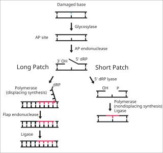
Base excision repair (BER) is a cellular mechanism, studied in the fields of biochemistry and genetics, that repairs damaged DNA throughout the cell cycle. It is responsible primarily for removing small, non-helix-distorting base lesions from the genome. The related nucleotide excision repair pathway repairs bulky helix-distorting lesions. BER is important for removing damaged bases that could otherwise cause mutations by mispairing or lead to breaks in DNA during replication. BER is initiated by DNA glycosylases, which recognize and remove specific damaged or inappropriate bases, forming AP sites. These are then cleaved by an AP endonuclease. The resulting single-strand break can then be processed by either short-patch or long-patch BER.

A nick is a discontinuity in a double stranded DNA molecule where there is no phosphodiester bond between adjacent nucleotides of one strand typically through damage or enzyme action. Nicks allow DNA strands to untwist during replication, and are also thought to play a role in the DNA mismatch repair mechanisms that fix errors on both the leading and lagging daughter strands.

UvrABC endonuclease is a multienzyme complex in bacteria involved in DNA repair by nucleotide excision repair, and it is, therefore, sometimes called an excinuclease. This UvrABC repair process, sometimes called the short-patch process, involves the removal of twelve nucleotides where a genetic mutation has occurred followed by a DNA polymerase, replacing these aberrant nucleotides with the correct nucleotides and completing the DNA repair. The subunits for this enzyme are encoded in the uvrA, uvrB, and uvrC genes. This enzyme complex is able to repair many different types of damage, including cyclobutyl dimer formation.

<i>Hin</i>dIII

HindIII (pronounced "Hin D Three") is a type II site-specific deoxyribonuclease restriction enzyme isolated from Haemophilus influenzae that cleaves the DNA palindromic sequence AAGCTT in the presence of the cofactor Mg2+ via hydrolysis.

Nucleotidyltransferase

Nucleotidyltransferases are transferase enzymes of phosphorus-containing groups, e.g., substituents of nucleotidylic acids or simply nucleoside monophosphates. The general reaction of transferring a nucleoside monophosphate moiety from A to B, can be written as:

Deoxyribonuclease IV (phage-T4-induced) is a kind of Endonuclease that catalyzes the degradation nucleotides in DsDNA by attacking the 5'-terminal end.

APEX1

DNA-(apurinic or apyrimidinic site) lyase is an enzyme that in humans is encoded by the APEX1 gene.

LIG1

DNA ligase 1 is an enzyme that in humans is encoded by the LIG1 gene. DNA ligase I is the only known eukaryotic DNA ligase involved in both DNA replication and repair, making it the most studied of the ligases.

In enzymology, DNA-(apurinic or apyrimidinic site) lyase, also referred to as DNA-(apurinic or apyrimidinic site) 5'-phosphomonoester-lyase or DNA AP lyase is a class of enzyme that catalyzes the chemical reaction of the cleavage of the C3'-O-P bond 3' from the apurinic or apyrimidinic site in DNA via beta-elimination reaction, leaving a 3'-terminal unsaturated sugar and a product with a terminal 5'-phosphate. In the 1970s, this class of enzyme was found to repair at apurinic or apyrimidinic DNA sites in E. coli and in mammalian cells. The major active enzyme of this class in bacteria, and specifically, E. coli is endonuclease type III. This enzyme is part of a family of lyases that cleave carbon-oxygen bonds.

NTHL1

Endonuclease III-like protein 1 is an enzyme that in humans is encoded by the NTHL1 gene.

T7 DNA polymerase

T7 DNA polymerase is an enzyme used during the DNA replication of the T7 bacteriophage. During this process, the DNA polymerase “reads” existing DNA strands and creates two new strands that match the existing ones. The T7 DNA polymerase requires a host factor, E. coli thioredoxin, in order to carry out its function. This helps stabilize the binding of the necessary protein to the primer-template to improve processivity by more than 100-fold, which is a feature unique to this enzyme. It is a member of the Family A DNA polymerases, which include E. coli DNA polymerase I and Taq DNA polymerase.

FPG IleRS zinc finger

The FPG IleRS zinc finger domain represents a zinc finger domain found at the C-terminal in both DNA glycosylase/AP lyase enzymes and in isoleucyl tRNA synthetase. In these two types of enzymes, the C-terminal domain forms a zinc finger.

H2TH domain

In molecular biology, the H2TH domain is a DNA-binding domain found in DNA glycosylase/AP lyase enzymes, which are involved in base excision repair of DNA damaged by oxidation or by mutagenic agents. Most damage to bases in DNA is repaired by the base excision repair pathway. These enzymes are primarily from bacteria, and have both DNA glycosylase activity EC 3.2.2.- and AP lyase activity EC 4.2.99.18. Examples include formamidopyrimidine-DNA glycosylases and endonuclease VIII (Nei).

XPG I protein domain

In molecular biology, the XPG-I is a protein domain found on Xeroderma Pigmentosum Complementation Group G (XPG) protein. The XPG protein is an endonuclease which repairs DNA damage caused by ultraviolet light. The XPG protein repairs DNA by a process called, Nucleotide excision repair. Mutations in the protein commonly cause Xeroderma Pigmentosum which often lead to skin cancer.

References

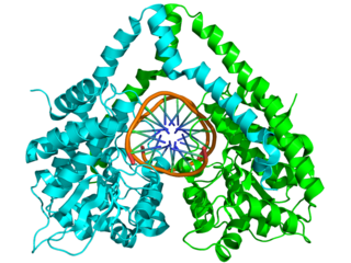
Molecular graphics images were produced using the UCSF Chimera package from the Resource for Biocomputing, Visualization, and Informatics at the University of California, San Francisco (supported by NIH P41 RR-01081). [9]

  1. 1 2 3 4 5 Clifford D. Mol; Tahide Izumi; Sankar Mitra; John A. Tainer (2000). "DNA-bound structures and mutants reveal abasic DNA binding by APE1 DNA repair and coordination". Nature. 403 (6768): 451–456. doi:10.1038/35000249. PMID   10667800. S2CID   4373743.
  2. Levin, Joshua D; Demple, Bruce (1990). "Analysis of class II (hydrolytic) and class I (beta-lyase) apurinic/apyrimidinic endonucleases with a synthetic DNA substrate". Nucleic Acids Research. 18 (17): 5069–75. doi:10.1093/nar/18.17.5069. PMC   332125 . PMID   1698278.
  3. Gary M. Myles; Aziz Sancar (1989). "DNA Repair". Chemical Research in Toxicology . 2 (4): 197–226. doi:10.1021/tx00010a001. PMID   2519777.
  4. 1 2 3 4 5 Burkovics P, Szukacsov V, Unk I, Haracska L (2006). "Human Ape2 protein has a 3'-5' exonuclease activity that acts preferentially on mismatched base pairs". Nucleic Acids Res. 34 (9): 2508–15. doi:10.1093/nar/gkl259. PMC   1459411 . PMID   16687656.
  5. George W. Teebor; Dina R. Marensein; David M. Wilson III (2004). "Human AP endonuclease (APE1) demonstrates endonucleolytic activity against AP sites in single-stranded DNA". DNA Repair. 3 (5): 527–533. doi:10.1016/j.dnarep.2004.01.010. PMID   15084314.
  6. Mark R. Kelley; Melissa L. Fishel (2007). "The DNA base excision repair protein Ape1/Ref-1 as a Therapeutic and chemopreventive target". Molecular Aspects of Medicine. 28 (3–4): 375–395. doi:10.1016/j.mam.2007.04.005. PMID   17560642.
  7. Mark R. Kelley; Meihua Luo; Sarah Delaphlane; Aihua Jiang; April Reed; Ying He; Melissa Fishel; Rodney L. Nyland II; Richard F. Broch; Xizoxi Qiao; Millie M. Georgiadis (2008). "Role of the Multifunctional DNA Repair and Redox Signaling Protein Ape1/Ref-1 in Cancer and Endothelial Cells: Small-Molecule Inhibition of the Redox Function of Ape1". Antioxidants & Redox Signaling. 10 (11): 1–12. doi:10.1089/ars.2008.2120. PMC   2587278 . PMID   18627350.
  8. Stavnezer J, Linehan EK, Thompson MR, Habboub G, Ucher AJ, Kadungure T, Tsuchimoto D, Nakabeppu Y, Schrader CE (2014). "Differential expression of APE1 and APE2 in germinal centers promotes error-prone repair and A:T mutations during somatic hypermutation". Proc. Natl. Acad. Sci. U.S.A. 111 (25): 9217–22. doi: 10.1073/pnas.1405590111 . PMC   4078814 . PMID   24927551.
  9. E.F. Pettersen; T.D. Goddard; C.C. Huang; G.S. Couch; D.M. Greenblat; E.C. Meng; T.E. Ferrin (2004). "UCSF Chimera - A Visualization System for Exploratory Research and Analysis" (PDF). J. Comput. Chem. 25 (13): 1605–1612. doi:10.1002/jcc.20084. PMID   15264254. S2CID   8747218.