
A leiomyoma, also known as a fibroid, is a benign smooth muscle tumor that very rarely becomes cancer (0.1%). They can occur in any organ, but the most common forms occur in the uterus, small bowel, and the esophagus. Polycythemia may occur due to increased erythropoietin production as part of a paraneoplastic syndrome.

Fibromas are benign tumors that are composed of fibrous or connective tissue. They can grow in all organs, arising from mesenchyme tissue. The term "fibroblastic" or "fibromatous" is used to describe tumors of the fibrous connective tissue. When the term fibroma is used without modifier, it is usually considered benign, with the term fibrosarcoma reserved for malignant tumors.

A dermatofibroma, or benign fibrous histiocytomas, is a benign nodule in the skin, typically on the legs, elbows or chest of an adult. It is usually painless.

A neoplasm is a type of abnormal and excessive growth of tissue. The process that occurs to form or produce a neoplasm is called neoplasia. The growth of a neoplasm is uncoordinated with that of the normal surrounding tissue, and persists in growing abnormally, even if the original trigger is removed. This abnormal growth usually forms a mass, when it may be called a tumour or tumor.

A benign tumor is a mass of cells (tumor) that does not invade neighboring tissue or metastasize. Compared to malignant (cancerous) tumors, benign tumors generally have a slower growth rate. Benign tumors have relatively well differentiated cells. They are often surrounded by an outer surface or stay contained within the epithelium. Common examples of benign tumors include moles and uterine fibroids.

A hamartoma is a mostly benign, local malformation of cells that resembles a neoplasm of local tissue but is usually due to an overgrowth of multiple aberrant cells, with a basis in a systemic genetic condition, rather than a growth descended from a single mutated cell (monoclonality), as would typically define a benign neoplasm/tumor. Despite this, many hamartomas are found to have clonal chromosomal aberrations that are acquired through somatic mutations, and on this basis the term hamartoma is sometimes considered synonymous with neoplasm. Hamartomas are by definition benign, slow-growing or self-limiting, though the underlying condition may still predispose the individual towards malignancies.
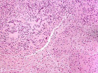
A neuroma is a growth or tumor of nerve tissue. Neuromas tend to be benign ; many nerve tumors, including those that are commonly malignant, are nowadays referred to by other terms.

Hidradenoma refers to a benign adnexal tumor of the apical sweat gland. These are 1–3 cm translucent blue cystic nodules. It usually presents as a single, small skin-colored lesion, and may be considered closely related to or a variant of poromas. Hidradenomas are often sub-classified based on subtle histologic differences, for example:

Myoepithelial cells are cells usually found in glandular epithelium as a thin layer above the basement membrane but generally beneath the luminal cells. These may be positive for alpha smooth muscle actin and can contract and expel the secretions of exocrine glands. They are found in the sweat glands, mammary glands, lacrimal glands, and salivary glands. Myoepithelial cells in these cases constitute the basal cell layer of an epithelium that harbors the epithelial progenitor. In the case of wound healing, myoepithelial cells reactively proliferate. Presence of myoepithelial cells in a hyperplastic tissue proves the benignity of the gland and, when absent, indicates cancer. Only rare cancers like adenoid cystic carcinomas contains myoepithelial cells as one of the malignant components.

Granular cell tumor is a tumor that can develop on any skin or mucosal surface, but occurs on the tongue 40% of the time.

Salivary gland tumours, also known as mucous gland adenomas or neoplasms, are tumours that form in the tissues of salivary glands. The salivary glands are classified as major or minor. The major salivary glands consist of the parotid, submandibular, and sublingual glands. The minor salivary glands consist of 800 to 1000 small mucus-secreting glands located throughout the lining of the oral cavity. Patients with these types of tumours may be asymptomatic.

A mucinous neoplasm is an abnormal and excessive growth of tissue (neoplasia) with associated mucin. It arises from epithelial cells that line certain internal organs and skin, and produce mucin. A malignant mucinous neoplasm is called a mucinous carcinoma. For example, for ovarian mucinous tumors, approximately 75% are benign, 10% are borderline and 15% are malignant.
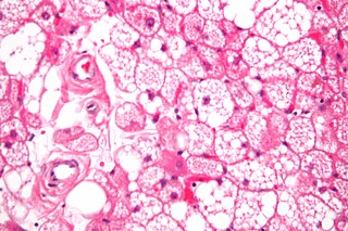
A hibernoma is a benign neoplasm of vestigial brown fat. The term was originally used by the French anatomist Louis Gery in 1914.
Neoplasms of the nailbed may often present with paronychia, ingrown nail, onycholysis, pyogenic granuloma, nail-plate dystrophy, longitudinal erythronychia, bleeding, and discolorations. There are various benign and malignant neoplasms that may occur in or overlying the nail matrix and in the nailbed, and symptoms may include pain, itching, and throbbing.
Clear cell acanthoma is a benign clinical and histological lesion initially described as neoplastic, which some authors now regard as a reactive dermatosis. It usually presents as a moist solitary firm, brown-red, well-circumscribed, 5 mm to 2 cm nodule or plaque on the lower extremities of middle-aged to elderly individuals. The lesion has a crusted, scaly peripheral collarette and vascular puncta on the surface. It is characterized by slow growth, and may persist for years. The clinical differential diagnosis includes: dermatofibroma, inflamed seborrheic keratosis, pyogenic granuloma, basal-cell carcinoma, squamous cell carcinoma, verruca vulgaris, psoriatic plaque, and melanoma.

Mucinous cystadenoma is a benign cystic tumor lined by a mucinous epithelium. It is a type of cystic adenoma (cystadenoma).

Poromas are rare, benign, cutaneous adnexal tumors. Cutaneous adnexal tumors are a group of skin tumors consisting of tissues that have differentiated towards one or more of the four primary adnexal structures found in normal skin: hair follicles, sebaceous sweat glands, apocrine sweat glands, and eccrine sweat glands. Poromas are eccrine or apocrine sweat gland tumors derived from the cells in the terminal portion of these glands' ducts. This part of the sweat gland duct is termed the acrosyringium and had led to grouping poromas in the acrospiroma class of skin tumors. Here, poromas are regarded as distinct sweat gland tumors that differ from other sweat gland tumors by their characteristic clinical presentations, microscopic histopathology, and the genetic mutations that their neoplastic cells have recently been found to carry.

Fibroma of tendon sheath is a benign tumor that presents as a small subcutaneous nodule that slowly increases in size. The tumors often have a multinodular growth pattern, with individual nodules being composed of bland, slender, spindle-shaped cells (myofibroblasts) in a dense, fibrous matrix.” A common microscopic finding is the presence of elongated, slit-like blood vessels. The lesions nearly always arise in the distal portions of the extremities. They often occur on the fingers, hands, toes, or feet. Although they are benign, they may recur in up to 40% of cases.
