Related Research Articles
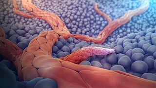
Angiogenesis is the physiological process through which new blood vessels form from pre-existing vessels, formed in the earlier stage of vasculogenesis. Angiogenesis continues the growth of the vasculature mainly by processes of sprouting and splitting, but processes such as coalescent angiogenesis, vessel elongation and vessel cooption also play a role. Vasculogenesis is the embryonic formation of endothelial cells from mesoderm cell precursors, and from neovascularization, although discussions are not always precise. The first vessels in the developing embryo form through vasculogenesis, after which angiogenesis is responsible for most, if not all, blood vessel growth during development and in disease.

Metastasis is a pathogenic agent's spread from an initial or primary site to a different or secondary site within the host's body; the term is typically used when referring to metastasis by a cancerous tumor. The newly pathological sites, then, are metastases (mets). It is generally distinguished from cancer invasion, which is the direct extension and penetration by cancer cells into neighboring tissues.

Angiomotin (AMOT) is a protein that in humans is encoded by the AMOT gene. It belongs to the motin family of angiostatin binding proteins, which includes angiomotin, angiomotin-like 1 (AMOTL1) and angiomotin-like 2 (AMOTL2) characterized by coiled-coil domains at N-terminus and consensus PDZ-binding domain at the C-terminus. Angiomotin is expressed predominantly in endothelial cells of capillaries as well as angiogenic tissues such as placenta and solid tumor.
An angiogenesis inhibitor is a substance that inhibits the growth of new blood vessels (angiogenesis). Some angiogenesis inhibitors are endogenous and a normal part of the body's control and others are obtained exogenously through pharmaceutical drugs or diet.

Endostatin is a naturally occurring, 20-kDa C-terminal fragment derived from type XVIII collagen. It is reported to serve as an anti-angiogenic agent, similar to angiostatin and thrombospondin.
Matrigel is the trade name for the solubilized basement membrane matrix secreted by Engelbreth-Holm-Swarm (EHS) mouse sarcoma cells produced by Corning Life Sciences. Matrigel resembles the laminin/collagen IV-rich basement membrane extracellular environment found in many tissues and is used by cell biologists as a substrate for culturing cells.

Angiogenin (ANG) also known as ribonuclease 5 is a small 123 amino acid protein that in humans is encoded by the ANG gene. Angiogenin is a potent stimulator of new blood vessels through the process of angiogenesis. Ang hydrolyzes cellular RNA, resulting in modulated levels of protein synthesis and interacts with DNA causing a promoter-like increase in the expression of rRNA. Ang is associated with cancer and neurological disease through angiogenesis and through activating gene expression that suppresses apoptosis.

Cytochalasin E, a member of the cytochalasin group, is an inhibitor of actin polymerization in blood platelets. It inhibits angiogenesis and tumor growth. Unlike cytochalasin A and cytochalasin B, it does not inhibit glucose transport. Cytochalasin E, however, was noted to decrease glucose absorption in mice around the intestinal tissues by increasing the Km needed for glucose to reach the Vmax, which meant that a higher concentration of glucose was required in its presence to attain Vmax. Since Vmax remained the same according to another study, it is evident that CE is indeed a competitive inhibitor at the intestinal receptor sites for glucose.

Endoglin (ENG) is a type I membrane glycoprotein located on cell surfaces and is part of the TGF beta receptor complex. It is also commonly referred to as CD105, END, FLJ41744, HHT1, ORW and ORW1. It has a crucial role in angiogenesis, therefore, making it an important protein for tumor growth, survival and metastasis of cancer cells to other locations in the body.

Microvesicles are a type of extracellular vesicle (EV) that are released from the cell membrane. In multicellular organisms, microvesicles and other EVs are found both in tissues and in many types of body fluids. Delimited by a phospholipid bilayer, microvesicles can be as small as the smallest EVs or as large as 1000 nm. They are considered to be larger, on average, than intracellularly-generated EVs known as exosomes. Microvesicles play a role in intercellular communication and can transport molecules such as mRNA, miRNA, and proteins between cells.

72 kDa type IV collagenase also known as matrix metalloproteinase-2 (MMP-2) and gelatinase A is an enzyme that in humans is encoded by the MMP2 gene. The MMP2 gene is located on chromosome 16 at position 12.2.

Pigment epithelium-derived factor (PEDF) also known as serpin F1 (SERPINF1), is a multifunctional secreted protein that has anti-angiogenic, anti-tumorigenic, and neurotrophic functions. Found in vertebrates, this 50 kDa protein is being researched as a therapeutic candidate for treatment of such conditions as choroidal neovascularization, heart disease, and cancer. In humans, pigment epithelium-derived factor is encoded by the SERPINF1 gene.

Sulfatase 1, also known as SULF1, is an enzyme which in humans is encoded by the SULF1 gene.

Vascular endothelial growth factor A (VEGF-A) is a protein that in humans is encoded by the VEGFA gene.
EGF-like domain-containing protein 7 is a protein that in humans is encoded by the EGFL7 gene. Intron 7 of EGFL7 hosts the miR-126 microRNA gene.
Semicarbazide-cadmium therapy was an experimental cancer therapy that was tested in several clinical trials in the Soviet Union during the 1960s. Semicarbazide is an irreversible inhibitor of semicarbazide-sensitive amine oxidase (SSAO), an enzyme possibly involved in exacerbation of inflammation. Cadmium is a heavy metal and can also induce apoptosis.
Angiogenesis is the process of forming new blood vessels from existing blood vessels, formed in vasculogenesis. It is a highly complex process involving extensive interplay between cells, soluble factors, and the extracellular matrix (ECM). Angiogenesis is critical during normal physiological development, but it also occurs in adults during inflammation, wound healing, ischemia, and in pathological conditions such as rheumatoid arthritis, hemangioma, and tumor growth. Proteolysis has been indicated as one of the first and most sustained activities involved in the formation of new blood vessels. Numerous proteases including matrix metalloproteinases (MMPs), a disintegrin and metalloproteinase domain (ADAM), a disintegrin and metalloproteinase domain with throbospondin motifs (ADAMTS), and cysteine and serine proteases are involved in angiogenesis. This article focuses on the important and diverse roles that these proteases play in the regulation of angiogenesis.
Tumstatin is a protein fragment cleaved from collagen that serves as both an antiangiogenic and proapoptotic agent. It has similar function to canstatin, endostatin, restin, and arresten, which also affect angiogenesis. Angiogenesis is the growth of new blood vessels from pre-existing blood vessels, and is important in tumor growth and metastasis. Angiogenesis is stimulated by many growth factors, the most prevalent of which is vascular endothelial growth factor (VEGF).

HU-336 is a strongly antiangiogenic compound, significantly inhibiting angiogenesis at concentrations as low as 300 nM. It inhibits angiogenesis by directly inducing apoptosis of vascular endothelial cells without changing the expression of pro- and antiangiogenic cytokines and their receptors. HU-336 is highly effective against tumor xenografts in nude mice. Although it is technically the oxidized quinone of delta-8 THC, it is entirely non psychoactive.

The tumor microenvironment (TME) is the environment around a tumor, including the surrounding blood vessels, immune cells, fibroblasts, signaling molecules and the extracellular matrix (ECM). The tumor and the surrounding microenvironment are closely related and interact constantly. Tumors can influence the microenvironment by releasing extracellular signals, promoting tumor angiogenesis and inducing peripheral immune tolerance, while the immune cells in the microenvironment can affect the growth and evolution of cancerous cells.
References
- ↑ Roiz, Levava; Smirnoff, Patricia; Bar-Eli, Menashe; Schwartz, Betty; Shoseyov, Oded (15 May 2006). "ACTIBIND, an actin-binding fungal T2-RNase with antiangiogenic and anticarcinogenic characteristics". Cancer. 106 (10): 2295–2308. doi:10.1002/cncr.21878. hdl: 10146/64474 . ISSN 1097-0142. PMID 16586499. S2CID 2693317.
- 1 2 3 4 5 6 7 8 9 10 11 12 Roiz, Levava; Smirnoff, Patricia; Bar-Eli, Menashe; Schwartz, Betty; Shoseyov, Oded (15 May 2006). "ACTIBIND, an actin-binding fungal T2-RNase with antiangiogenic and anticarcinogenic characteristics". Cancer. 106 (10): 2295–2308. doi:10.1002/cncr.21878. ISSN 0008-543X. PMID 16586499. S2CID 2693317.
- ↑ "Pseudopodia – an overview | ScienceDirect Topics". sciencedirect.com. Retrieved 21 April 2023.
- 1 2 "ACTIBIND, a T2 RNase, Competes with Angiogenin and Inhibits Human Melanoma Growth, Angiogenesis, and Metastasis". aacrjournals.org. Retrieved 21 April 2023.
- 1 2 "angiogenesis". cancer.gov. 2 February 2011. Retrieved 21 April 2023.
- ↑ Kerr, J. F.; Wyllie, A. H.; Currie, A. R. (August 1972). "Apoptosis: a basic biological phenomenon with wide-ranging implications in tissue kinetics". British Journal of Cancer. 26 (4): 239–257. doi:10.1038/bjc.1972.33. ISSN 0007-0920. PMC 2008650 . PMID 4561027.
- 1 2 "Xenograft Mouse Models". Melior Discovery. Retrieved 22 April 2023.
- ↑ "Athymic Nude Mouse | Charles River". criver.com. Retrieved 22 April 2023.
- Roiz, L.; Smirnoff, P.; Bar-Eli, M.; Schwartz, B.; Shoseyov, O. (2006). "ACTIBIND, an actin-binding fungal T2-RNase with antiangiogenic and anticarcinogenic characteristics". Cancer. 106 (10): 2295–2308. doi:10.1002/cncr.21878. hdl: 10146/64474 . PMID 16586499. S2CID 2693317.