Alu element

Last updated

An Alu element is a short stretch of DNA originally characterized by the action of the Arthrobacter luteus (Alu) restriction endonuclease. [1] Alu elements are the most abundant transposable elements in the human genome, present in excess of one million copies. [2] Alu elements were thought to be selfish or parasitic DNA, because their sole known function is self reproduction. However, they are likely to play a role in evolution and have been used as genetic markers. [3] [4] They are derived from the small cytoplasmic 7SL RNA, a component of the signal recognition particle. Alu elements are highly conserved within primate genomes and originated in the genome of an ancestor of Supraprimates. [5]

Contents

Alu insertions have been implicated in several inherited human diseases and in various forms of cancer.

The study of Alu elements has also been important in elucidating human population genetics and the evolution of primates, including the evolution of humans.

Karyotype from a female human lymphocyte (46, XX). Chromosomes were hybridized with a probe for Alu elements (green) and counterstained with TOPRO-3 (red). Alu elements were used as a marker for chromosomes and chromosome bands rich in genes. PLoSBiol3.5.Fig7ChromosomesAluFish.jpg
Karyotype from a female human lymphocyte (46, XX). Chromosomes were hybridized with a probe for Alu elements (green) and counterstained with TOPRO-3 (red). Alu elements were used as a marker for chromosomes and chromosome bands rich in genes.

Alu family

The Alu family is a family of repetitive elements in primate genomes, including the human genome. [6] Modern Alu elements are about 300 base pairs long and are therefore classified as short interspersed nuclear elements (SINEs) among the class of repetitive DNA elements. The typical structure is 5' - Part A - A5TACA6 - Part B - PolyA Tail - 3', where Part A and Part B (also known as "left arm" and "right arm") are similar nucleotide sequences. Expressed another way, it is believed modern Alu elements emerged from a head to tail fusion of two distinct FAMs (fossil antique monomers) over 100 million years ago, hence its dimeric structure of two similar, but distinct monomers (left and right arms) joined by an A-rich linker. Both monomers are thought to have evolved from 7SL, also known as SRP RNA. [7] The length of the polyA tail varies between Alu families.

There are over one million Alu elements interspersed throughout the human genome, and it is estimated that about 10.7% of the human genome consists of Alu sequences. However, less than 0.5% are polymorphic (i.e., occurring in more than one form or morph). [8] In 1988, Jerzy Jurka and Temple Smith discovered that Alu elements were split in two major subfamilies known as AluJ (named after Jurka) and AluS (named after Smith), and other Alu subfamilies were also independently discovered by several groups. [9] Later on, a sub-subfamily of AluS which included active Alu elements was given the separate name AluY. Dating back 65 million years, the AluJ lineage is the oldest and least active in the human genome. The younger AluS lineage is about 30 million years old and still contains some active elements. Finally, the AluY elements are the youngest of the three and have the greatest disposition to move along the human genome. [10] The discovery of Alu subfamilies led to the hypothesis of master/source genes, and provided the definitive link between transposable elements (active elements) and interspersed repetitive DNA (mutated copies of active elements). [11]

B1 elements in rats and mice are similar to Alus in that they also evolved from 7SL RNA, but they only have one left monomer arm. 95% percent of human Alus are also found in chimpanzees, and 50% of B elements in mice are also found in rats. These elements are mostly found in introns and upstream regulatory elements of genes. [12]

The ancestral form of Alu and B1 is the fossil Alu monomer (FAM). Free-floating forms of the left and right arms exist, termed free left Alu monomers (FLAMs) and free right Alu monomers (FRAMs) respectively. [13] A notable FLAM in primates is the BC200 lncRNA.

Sequence features

Genetic structure of murine LINE1 and SINEs, including Alu. LINE1s and SINEs.png
Genetic structure of murine LINE1 and SINEs, including Alu.

Two main promoter "boxes" are found in Alu: a 5' A box with the consensus TGGCTCACGCC, and a 3' B box with the consensus GTTCGAGAC (IUPAC nucleic acid notation). tRNAs, which are transcribed by RNA polymerase III, have a similar but stronger promoter structure. [14] Both boxes are located in the left arm. [7]

Alu elements contain four or fewer Retinoic Acid response element hexamer sites in its internal promoter, with the last one overlapping with the "B box". [15] In this 7SL (SRP) RNA example below, functional hexamers are underlined using a solid line, with the non-functional third hexamer denoted using a dotted line:

GCCGGGCGCGGTGGCGCGTGCCTGTAGTCCCAGCTACTCGGGAGGCTGAGGCTGGAGGATCGCTTGAGTCCAGGAGTTCTGGGCTGTAGTGCGCTATGCCGATCGGAATAGCCACTGCACTCCAGCCTGGGCAACATAGCGAGACCCCGTCTC.

The recognition sequence of the Alu I endonuclease is 5' ag/ct 3'; that is, the enzyme cuts the DNA segment between the guanine and cytosine residues (in lowercase above). [16]

Alu elements

Alu elements are responsible for regulation of tissue-specific genes. They are also involved in the transcription of nearby genes and can sometimes change the way a gene is expressed. [17]

Alu elements are retrotransposons and look like DNA copies made from RNA polymerase III-encoded RNAs. Alu elements do not encode for protein products. They are replicated as any other DNA sequence, but depend on LINE retrotransposons for generation of new elements. [18]

Alu element replication and mobilization begins by interactions with signal recognition particles (SRPs), which aid newly translated proteins to reach their final destinations. [19] Alu RNA forms a specific RNA:protein complex with a protein heterodimer consisting of SRP9 and SRP14. [19] SRP9/14 facilitates Alu's attachment to ribosomes that capture nascent L1 proteins. Thus, an Alu element can take control of the L1 protein's reverse transcriptase, ensuring that the Alu's RNA sequence gets copied into the genome rather than the L1's mRNA. [10]

Alu elements in primates form a fossil record that is relatively easy to decipher because Alu element insertion events have a characteristic signature that is both easy to read and faithfully recorded in the genome from generation to generation. The study of Alu Y elements (the more recently evolved) thus reveals details of ancestry because individuals will most likely only share a particular Alu element insertion if they have a common ancestor. This is because insertion of an Alu element occurs only 100 - 200 times per million years, and no known mechanism of deletion of one has been found. Therefore, individuals with an element likely descended from an ancestor with one—and vice versa, for those without. In genetics, the presence or lack thereof of a recently inserted Alu element may be a good property to consider when studying human evolution. [20] Most human Alu element insertions can be found in the corresponding positions in the genomes of other primates, but about 7,000 Alu insertions are unique to humans. [21]

Impact in humans

Alu elements have been proposed to affect gene expression and been found to contain functional promoter regions for steroid hormone receptors. [15] [22] Due to the abundant content of CpG dinucleotides found in Alu elements, these regions serve as a site of methylation, contributing to up to 30% of the methylation sites in the human genome. [23] Alu elements are also a common source of mutations in humans; however, such mutations are often confined to non-coding regions of pre-mRNA (introns), where they have little discernible impact on the bearer. [24] Mutations in the introns (or non-coding regions of RNA) have little or no effect on phenotype of an individual if the coding portion of individual's genome does not contain mutations. The Alu insertions that can be detrimental to the human body are inserted into coding regions (exons) or into mRNA after the process of splicing. [25]

However, the variation generated can be used in studies of the movement and ancestry of human populations, [26] and the mutagenic effect of Alu [27] and retrotransposons in general [28] has played a major role in the evolution of the human genome. There are also a number of cases where Alu insertions or deletions are associated with specific effects in humans:

Associations with human disease

Alu insertions are sometimes disruptive and can result in inherited disorders. However, most Alu variation acts as markers that segregate with the disease so the presence of a particular Alu allele does not mean that the carrier will definitely get the disease. The first report of Alu-mediated recombination causing a prevalent inherited predisposition to cancer was a 1995 report about hereditary nonpolyposis colorectal cancer . [29] In the human genome, the most recently active have been the 22 AluY and 6 AluS Transposon Element subfamilies due to their inherited activity to cause various cancers. Thus due to their major heritable damage it is important to understand the causes that affect their transpositional activity. [30]

The following human diseases have been linked with Alu insertions: [26] [31]

And the following diseases have been associated with single-nucleotide DNA variations in Alu elements affecting transcription levels: [33]

The following disease have been associated with repeat expansion of AAGGG pentamere in Alu element :

Associated human mutations

Related Research Articles

<span class="mw-page-title-main">Genome</span> All genetic material of an organism

In the fields of molecular biology and genetics, a genome is all the genetic information of an organism. It consists of nucleotide sequences of DNA. The nuclear genome includes protein-coding genes and non-coding genes, other functional regions of the genome such as regulatory sequences, and often a substantial fraction of junk DNA with no evident function. Almost all eukaryotes have mitochondria and a small mitochondrial genome. Algae and plants also contain chloroplasts with a chloroplast genome.

<span class="mw-page-title-main">Transposable element</span> Semiparasitic DNA sequence

A transposable element is a nucleic acid sequence in DNA that can change its position within a genome, sometimes creating or reversing mutations and altering the cell's genetic identity and genome size. Transposition often results in duplication of the same genetic material. In the human genome, L1 and Alu elements are two examples. Barbara McClintock's discovery of them earned her a Nobel Prize in 1983. Its importance in personalized medicine is becoming increasingly relevant, as well as gaining more attention in data analytics given the difficulty of analysis in very high dimensional spaces.

<span class="mw-page-title-main">Human genome</span> Complete set of nucleic acid sequences for humans

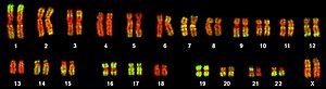
The human genome is a complete set of nucleic acid sequences for humans, encoded as DNA within the 23 chromosome pairs in cell nuclei and in a small DNA molecule found within individual mitochondria. These are usually treated separately as the nuclear genome and the mitochondrial genome. Human genomes include both protein-coding DNA sequences and various types of DNA that does not encode proteins. The latter is a diverse category that includes DNA coding for non-translated RNA, such as that for ribosomal RNA, transfer RNA, ribozymes, small nuclear RNAs, and several types of regulatory RNAs. It also includes promoters and their associated gene-regulatory elements, DNA playing structural and replicatory roles, such as scaffolding regions, telomeres, centromeres, and origins of replication, plus large numbers of transposable elements, inserted viral DNA, non-functional pseudogenes and simple, highly repetitive sequences. Introns make up a large percentage of non-coding DNA. Some of this non-coding DNA is non-functional junk DNA, such as pseudogenes, but there is no firm consensus on the total amount of junk DNA.

Non-coding DNA (ncDNA) sequences are components of an organism's DNA that do not encode protein sequences. Some non-coding DNA is transcribed into functional non-coding RNA molecules. Other functional regions of the non-coding DNA fraction include regulatory sequences that control gene expression; scaffold attachment regions; origins of DNA replication; centromeres; and telomeres. Some non-coding regions appear to be mostly nonfunctional, such as introns, pseudogenes, intergenic DNA, and fragments of transposons and viruses. Regions that are completely nonfunctional are called junk DNA.

<span class="mw-page-title-main">Pseudogene</span> Functionless relative of a gene

Pseudogenes are nonfunctional segments of DNA that resemble functional genes. Most arise as superfluous copies of functional genes, either directly by gene duplication or indirectly by reverse transcription of an mRNA transcript. Pseudogenes are usually identified when genome sequence analysis finds gene-like sequences that lack regulatory sequences needed for transcription or translation, or whose coding sequences are obviously defective due to frameshifts or premature stop codons. Pseudogenes are a type of junk DNA.

Repeated sequences are short or long patterns of nucleic acids that occur in multiple copies throughout the genome. In many organisms, a significant fraction of the genomic DNA is repetitive, with over two-thirds of the sequence consisting of repetitive elements in humans. Some of these repeated sequences are necessary for maintaining important genome structures such as telomeres or centromeres.

<span class="mw-page-title-main">Retrotransposon</span> Type of genetic component

Retrotransposons are a type of genetic component that copy and paste themselves into different genomic locations (transposon) by converting RNA back into DNA through the reverse transcription process using an RNA transposition intermediate.

Interspersed repetitive DNA is found in all eukaryotic genomes. They differ from tandem repeat DNA in that rather than the repeat sequences coming right after one another, they are dispersed throughout the genome and nonadjacent. The sequence that repeats can vary depending on the type of organism, and many other factors. Certain classes of interspersed repeat sequences propagate themselves by RNA mediated transposition; they have been called retrotransposons, and they constitute 25–40% of most mammalian genomes. Some types of interspersed repetitive DNA elements allow new genes to evolve by uncoupling similar DNA sequences from gene conversion during meiosis.

<span class="mw-page-title-main">Endogenous retrovirus</span> Inherited retrovirus encoded in an organisms genome

Endogenous retroviruses (ERVs) are endogenous viral elements in the genome that closely resemble and can be derived from retroviruses. They are abundant in the genomes of jawed vertebrates, and they comprise up to 5–8% of the human genome.

Retrotransposon markers are components of DNA which are used as cladistic markers. They assist in determining the common ancestry, or not, of related taxa. The "presence" of a given retrotransposon in related taxa suggests their orthologous integration, a derived condition acquired via a common ancestry, while the "absence" of particular elements indicates the plesiomorphic condition prior to integration in more distant taxa. The use of presence/absence analyses to reconstruct the systematic biology of mammals depends on the availability of retrotransposons that were actively integrating before the divergence of a particular species.

Exon shuffling is a molecular mechanism for the formation of new genes. It is a process through which two or more exons from different genes can be brought together ectopically, or the same exon can be duplicated, to create a new exon-intron structure. There are different mechanisms through which exon shuffling occurs: transposon mediated exon shuffling, crossover during sexual recombination of parental genomes and illegitimate recombination.

<span class="mw-page-title-main">Mobile genetic elements</span> DNA sequence whose position in the genome is variable

Mobile genetic elements (MGEs), sometimes called selfish genetic elements, are a type of genetic material that can move around within a genome, or that can be transferred from one species or replicon to another. MGEs are found in all organisms. In humans, approximately 50% of the genome is thought to be MGEs. MGEs play a distinct role in evolution. Gene duplication events can also happen through the mechanism of MGEs. MGEs can also cause mutations in protein coding regions, which alters the protein functions. These mechanisms can also rearrange genes in the host genome generating variation. These mechanism can increase fitness by gaining new or additional functions. An example of MGEs in evolutionary context are that virulence factors and antibiotic resistance genes of MGEs can be transported to share genetic code with neighboring bacteria. However, MGEs can also decrease fitness by introducing disease-causing alleles or mutations. The set of MGEs in an organism is called a mobilome, which is composed of a large number of plasmids, transposons and viruses.

In the fields of bioinformatics and computational biology, Genome survey sequences (GSS) are nucleotide sequences similar to expressed sequence tags (ESTs) that the only difference is that most of them are genomic in origin, rather than mRNA.

A conserved non-coding sequence (CNS) is a DNA sequence of noncoding DNA that is evolutionarily conserved. These sequences are of interest for their potential to regulate gene production.

An endogenous viral element (EVE) is a DNA sequence derived from a virus, and present within the germline of a non-viral organism. EVEs may be entire viral genomes (proviruses), or fragments of viral genomes. They arise when a viral DNA sequence becomes integrated into the genome of a germ cell that goes on to produce a viable organism. The newly established EVE can be inherited from one generation to the next as an allele in the host species, and may even reach fixation.

<span class="mw-page-title-main">Long interspersed nuclear element</span>

Long interspersed nuclear elements (LINEs) are a group of non-LTR retrotransposons that are widespread in the genome of many eukaryotes. LINEs contain an internal Pol II promoter to initiate transcription into mRNA, and encode one or two proteins, ORF1 and ORF2. The functional domains present within ORF1 vary greatly among LINEs, but often exhibit RNA/DNA binding activity. ORF2 is essential to successful retrotransposition, and encodes a protein with both reverse transcriptase and endonuclease activity.

<span class="mw-page-title-main">LINE1</span> Group of transposable elements

LINE1 is a family of related class I transposable elements in the DNA of some organisms, classified with the long interspersed elements (LINEs). L1 transposons comprise approximately 17% of the human genome. These active L1s can interrupt the genome through insertions, deletions, rearrangements, and copy number variations. L1 activity has contributed to the instability and evolution of genomes and is tightly regulated in the germline by DNA methylation, histone modifications, and piRNA. L1s can further impact genome variation through mispairing and unequal crossing over during meiosis due to its repetitive DNA sequences.

<span class="mw-page-title-main">Short interspersed nuclear element</span>

Short interspersed nuclear elements (SINEs) are non-autonomous, non-coding transposable elements (TEs) that are about 100 to 700 base pairs in length. They are a class of retrotransposons, DNA elements that amplify themselves throughout eukaryotic genomes, often through RNA intermediates. SINEs compose about 13% of the mammalian genome.

DNA transposons are DNA sequences, sometimes referred to "jumping genes", that can move and integrate to different locations within the genome. They are class II transposable elements (TEs) that move through a DNA intermediate, as opposed to class I TEs, retrotransposons, that move through an RNA intermediate. DNA transposons can move in the DNA of an organism via a single-or double-stranded DNA intermediate. DNA transposons have been found in both prokaryotic and eukaryotic organisms. They can make up a significant portion of an organism's genome, particularly in eukaryotes. In prokaryotes, TE's can facilitate the horizontal transfer of antibiotic resistance or other genes associated with virulence. After replicating and propagating in a host, all transposon copies become inactivated and are lost unless the transposon passes to a genome by starting a new life cycle with horizontal transfer. It is important to note that DNA transposons do not randomly insert themselves into the genome, but rather show preference for specific sites.

Haig Hagop Kazazian Jr. was an American professor in the Department of Genetic Medicine at Johns Hopkins University School of Medicine in Baltimore, Maryland. Kazazian was an elected member of the National Academy of Sciences and the American Academy of Arts and Sciences.

References

  1. Schmid, Carl W; Deininger, Prescott L (1975). "Sequence organization of the human genome". Cell. 6 (3): 345–58. doi:10.1016/0092-8674(75)90184-1. PMID   1052772. S2CID   42804857.
  2. Szmulewicz, Martin N; Novick, Gabriel E; Herrera, Rene J (1998). "Effects of Alu insertions on gene function". Electrophoresis. 19 (8–9): 1260–4. doi:10.1002/elps.1150190806. PMID   9694261. S2CID   45917758.
  3. Kidwell, Margaret G; Lisch, Damon R (2001). "Perspective: Transposable Elements, Parasitic Dna, and Genome Evolution". Evolution. 55 (1): 1–24. doi:10.1554/0014-3820(2001)055[0001:ptepda]2.0.co;2. PMID   11263730. S2CID   25273865.
  4. Pray, Leslie (2008). "Functions and Utility of Alu Jumping Genes". Scitable.com. Nature. Retrieved 26 June 2019.
  5. Kriegs, Jan Ole; Churakov, Gennady; Jurka, Jerzy; Brosius, Jürgen; Schmitz, Jürgen (2007). "Evolutionary history of 7SL RNA-derived SINEs in Supraprimates". Trends in Genetics. 23 (4): 158–61. doi:10.1016/j.tig.2007.02.002. PMID   17307271.
  6. Arcot, Santosh S.; Wang, Zhenyuan; Weber, James L.; Deininger, Prescott L.; Batzer, Mark A. (September 1995). "Alu Repeats: A Source for the Genesis of Primate Microsatellites". Genomics. 29 (1): 136–144. doi:10.1006/geno.1995.1224. ISSN   0888-7543. PMID   8530063.
  7. 1 2 Häsler, Julien; Strub, Katharina (2006). "Alu elements as regulators of gene expression". Nucleic Acids Research. 34 (19): 5491–7. doi:10.1093/nar/gkl706. PMC   1636486 . PMID   17020921.
  8. Roy-Engel, A. M; Carroll, M. L; Vogel, E; Garber, R. K; Nguyen, S. V; Salem, A. H; Batzer, M. A; Deininger, P. L (2001). "Alu insertion polymorphisms for the study of human genomic diversity". Genetics. 159 (1): 279–90. doi:10.1093/genetics/159.1.279. PMC   1461783 . PMID   11560904.
  9. Jurka, J; Smith, T (1988). "A fundamental division in the Alu family of repeated sequences". Proceedings of the National Academy of Sciences. 85 (13): 4775–8. Bibcode:1988PNAS...85.4775J. doi: 10.1073/pnas.85.13.4775 . PMC   280518 . PMID   3387438.
  10. 1 2 Bennett, E. A; Keller, H; Mills, R. E; Schmidt, S; Moran, J. V; Weichenrieder, O; Devine, S. E (2008). "Active Alu retrotransposons in the human genome". Genome Research. 18 (12): 1875–83. doi:10.1101/gr.081737.108. PMC   2593586 . PMID   18836035.
  11. Richard Shen, M; Batzer, Mark A; Deininger, Prescott L (1991). "Evolution of the master Alu gene(s)". Journal of Molecular Evolution. 33 (4): 311–20. Bibcode:1991JMolE..33..311R. doi:10.1007/bf02102862. PMID   1774786. S2CID   13091552.
  12. Tsirigos, Aristotelis; Rigoutsos, Isidore; Stormo, Gary D. (18 December 2009). "Alu and B1 Repeats Have Been Selectively Retained in the Upstream and Intronic Regions of Genes of Specific Functional Classes". PLOS Computational Biology. 5 (12): e1000610. Bibcode:2009PLSCB...5E0610T. doi: 10.1371/journal.pcbi.1000610 . PMC   2784220 . PMID   20019790.
  13. Kojima, K. K. (16 August 2010). "Alu Monomer Revisited: Recent Generation of Alu Monomers". Molecular Biology and Evolution. 28 (1): 13–15. doi: 10.1093/molbev/msq218 . PMID   20713470.
  14. Conti, A; Carnevali, D; Bollati, V; Fustinoni, S; Pellegrini, M; Dieci, G (January 2015). "Identification of RNA polymerase III-transcribed Alu loci by computational screening of RNA-Seq data". Nucleic Acids Research. 43 (2): 817–35. doi:10.1093/nar/gku1361. PMC   4333407 . PMID   25550429.
  15. 1 2 Vansant, G; Reynolds, W. F (1995). "The consensus sequence of a major Alu subfamily contains a functional retinoic acid response element". Proceedings of the National Academy of Sciences. 92 (18): 8229–33. Bibcode:1995PNAS...92.8229V. doi: 10.1073/pnas.92.18.8229 . PMC   41130 . PMID   7667273.
  16. Ullu E, Tschudi C (1984). "Alu sequences are processed 7SL RNA genes". Nature. 312 (5990): 171–2. Bibcode:1984Natur.312..171U. doi:10.1038/312171a0. PMID   6209580. S2CID   4328237.
  17. Britten, R. J (1996). "DNA sequence insertion and evolutionary variation in gene regulation". Proceedings of the National Academy of Sciences of the United States of America. 93 (18): 9374–7. Bibcode:1996PNAS...93.9374B. doi: 10.1073/pnas.93.18.9374 . PMC   38434 . PMID   8790336.
  18. Kramerov, D; Vassetzky, N (2005). "Short Retroposons in Eukaryotic Genomes". International Review of Cytology. 247: 165–221. doi:10.1016/S0074-7696(05)47004-7. PMID   16344113.
  19. 1 2 Weichenrieder, Oliver; Wild, Klemens; Strub, Katharina; Cusack, Stephen (2000). "Structure and assembly of the Alu domain of the mammalian signal recognition particle". Nature. 408 (6809): 167–73. Bibcode:2000Natur.408..167W. doi:10.1038/35041507. PMID   11089964. S2CID   4427070.
  20. Terreros, Maria C.; Alfonso-Sanchez, Miguel A.; Novick; Luis; Lacau; Lowery; Regueiro; Herrera (September 11, 2009). "Insights on human evolution: an analysis of Alu insertion polymorphisms". Journal of Human Genetics. 54 (10): 603–611. doi: 10.1038/jhg.2009.86 . PMID   19745832. S2CID   8153502.
  21. Chimpanzee Sequencing Analysis Consortium (2005). "Initial sequence of the chimpanzee genome and comparison with the human genome". Nature. 437 (7055): 69–87. Bibcode:2005Natur.437...69.. doi: 10.1038/nature04072 . PMID   16136131. S2CID   2638825.
  22. Norris, J; Fan, D; Aleman, C; Marks, J. R; Futreal, P. A; Wiseman, R. W; Iglehart, J. D; Deininger, P. L; McDonnell, D. P (1995). "Identification of a new subclass of Alu DNA repeats that can function as estrogen receptor-dependent transcriptional enhancers". The Journal of Biological Chemistry. 270 (39): 22777–82. doi: 10.1074/jbc.270.39.22777 . PMID   7559405. S2CID   45796017.
  23. Schmid, C. W (1998). "Does SINE evolution preclude Alu function?". Nucleic Acids Research. 26 (20): 4541–50. doi:10.1093/nar/26.20.4541. PMC   147893 . PMID   9753719.
  24. Lander, Eric S; Linton, Lauren M; Birren, Bruce; Nusbaum, Chad; Zody, Michael C; Baldwin, Jennifer; Devon, Keri; Dewar, Ken; Doyle, Michael; Fitzhugh, William; Funke, Roel; Gage, Diane; Harris, Katrina; Heaford, Andrew; Howland, John; Kann, Lisa; Lehoczky, Jessica; Levine, Rosie; McEwan, Paul; McKernan, Kevin; Meldrim, James; Mesirov, Jill P; Miranda, Cher; Morris, William; Naylor, Jerome; Raymond, Christina; Rosetti, Mark; Santos, Ralph; Sheridan, Andrew; et al. (2001). "Initial sequencing and analysis of the human genome" (PDF). Nature. 409 (6822): 860–921. Bibcode:2001Natur.409..860L. doi:10.1038/35057062. PMID   11237011.
  25. Deininger, Prescott L; Batzer, Mark A (1999). "Alu Repeats and Human Disease". Molecular Genetics and Metabolism. 67 (3): 183–93. doi:10.1006/mgme.1999.2864. PMID   10381326. S2CID   15651921.
  26. 1 2 Batzer, Mark A; Deininger, Prescott L (2002). "Alu Repeats and Human Genomic Diversity". Nature Reviews Genetics. 3 (5): 370–9. doi:10.1038/nrg798. PMID   11988762. S2CID   205486422.
  27. Shen, S; Lin, L; Cai, J. J; Jiang, P; Kenkel, E. J; Stroik, M. R; Sato, S; Davidson, B. L; Xing, Y (2011). "Widespread establishment and regulatory impact of Alu exons in human genes". Proceedings of the National Academy of Sciences. 108 (7): 2837–42. Bibcode:2011PNAS..108.2837S. doi: 10.1073/pnas.1012834108 . PMC   3041063 . PMID   21282640.
  28. Cordaux, Richard; Batzer, Mark A (2009). "The impact of retrotransposons on human genome evolution". Nature Reviews Genetics. 10 (10): 691–703. doi:10.1038/nrg2640. PMC   2884099 . PMID   19763152.
  29. Nyström-Lahti, Minna; Kristo, Paula; Nicolaides, Nicholas C; Chang, Sheng-Yung; Aaltonen, Lauri A; Moisio, Anu-Liisa; Järvinen, Heikki J; Mecklin, Jukka-Pekka; Kinzler, Kenneth W; Vogelstein, Bert; de la Chapelle, Albert; Peltomäki, Päivi (1995). "Founding mutations and Alu-mediated recombination in hereditary colon cancer". Nature Medicine. 1 (11): 1203–6. doi:10.1038/nm1195-1203. PMID   7584997. S2CID   39468812.
  30. Jin, Lingling; McQuillan, Ian; Li, Longhai (2017). "Computational identification of harmful mutation regions to the activity of transposable elements". BMC Genomics. 18 (Suppl 9): 862. doi: 10.1186/s12864-017-4227-z . PMC   5773891 . PMID   29219079.
  31. Deininger, Prescott (2011). "Alu elements: Know the SINEs". Genome Biology. 12 (12): 236. doi: 10.1186/gb-2011-12-12-236 . PMC   3334610 . PMID   22204421.
  32. Fukuda, Shinichi; Varshney, Akhil; Fowler, Benjamin J.; Wang, Shao-bin; Narendran, Siddharth; Ambati, Kameshwari; Yasuma, Tetsuhiro; Magagnoli, Joseph; Leung, Hannah; Hirahara, Shuichiro; Nagasaka, Yosuke (2021-02-09). "Cytoplasmic synthesis of endogenous Alu complementary DNA via reverse transcription and implications in age-related macular degeneration". Proceedings of the National Academy of Sciences. 118 (6): e2022751118. Bibcode:2021PNAS..11822751F. doi: 10.1073/pnas.2022751118 . ISSN   0027-8424. PMC   8017980 . PMID   33526699. S2CID   231761522.
  33. "SNP in the promoter region of the myeloperoxidase MPO gene". SNPedia. Archived from the original on 2010-05-21. Retrieved 2010-03-14.[ unreliable medical source? ]
  34. Cortese, A.; Simone, R.; Sullivan, R.; Vandrovcova, J.; Tariq, H.; Yau, W. Y.; Humphrey, J.; Jaunmuktane, Z.; Sivakumar, P.; Polke, J.; Ilyas, M.; Tribollet, E.; Tomaselli, P. J.; Devigili, G.; Callegari, I.; Versino, M.; Salpietro, V.; Efthymiou, S.; Kaski, D.; Wood, N. W.; Andrade, N. S.; Buglo, E.; Rebelo, A.; Rossor, A. M.; Bronstein, A.; Fratta, P.; Marques, W. J.; Züchner, S.; Reilly, M. M.; Houlden, H. (2019). "Biallelic expansion of an intronic repeat in RFC1 is a common cause of late-onset ataxia". Nat Genet. 51 (4): 649–658. doi:10.1038/s41588-019-0372-4. PMC   6709527 . PMID   30926972.
  35. Puthucheary, Zudin; Skipworth, James RA; Rawal, Jai; Loosemore, Mike; Van Someren, Ken; Montgomery, Hugh E (2011). "The ACE Gene and Human Performance". Sports Medicine. 41 (6): 433–48. doi:10.2165/11588720-000000000-00000. PMID   21615186. S2CID   42531424.
  36. Dulai, K. S; von Dornum, M; Mollon, J. D; Hunt, D. M (1999). "The evolution of trichromatic color vision by opsin gene duplication in New World and Old World primates". Genome Research. 9 (7): 629–38. doi: 10.1101/gr.9.7.629 . PMID   10413401. S2CID   10637615.