
The stomach is a muscular, hollow organ in the upper gastrointestinal tract of humans and many other animals, including several invertebrates. The stomach has a dilated structure and functions as a vital organ in the digestive system. The stomach is involved in the gastric phase of digestion, following the cephalic phase in which the sight and smell of food and the act of chewing are stimuli. In the stomach a chemical breakdown of food takes place by means of secreted digestive enzymes and gastric acid.

The pyloruspyloric region or pyloric part connects the stomach to the duodenum. The pylorus is considered as having two parts, the pyloric antrum and the pyloric canal. The pyloric canal ends as the pyloric orifice, which marks the junction between the stomach and the duodenum. The orifice is surrounded by a sphincter, a band of muscle, called the pyloric sphincter. The word pylorus comes from Greek πυλωρός, via Latin. The word pylorus in Greek means "gatekeeper", related to "gate" and is thus linguistically related to the word "pylon".

Pyloric stenosis is a narrowing of the opening from the stomach to the first part of the small intestine. Symptoms include projectile vomiting without the presence of bile. This most often occurs after the baby is fed. The typical age that symptoms become obvious is two to twelve weeks old.

The common hepatic artery is a short blood vessel that supplies oxygenated blood to the liver, pylorus of the stomach, duodenum, pancreas, and gallbladder.

The right gastric artery usually arises from the proper hepatic artery. It descends to the pyloric end of the stomach before passing from right to left along its lesser curvature, supplying it with branches, and finally anastomosing with the left gastric artery.

The foregut in humans is the anterior part of the alimentary canal, from the distal esophagus to the first half of the duodenum, at the entrance of the bile duct. Beyond the stomach, the foregut is attached to the abdominal walls by mesentery. The foregut arises from the endoderm, developing from the folding primitive gut, and is developmentally distinct from the midgut and hindgut. Although the term “foregut” is typically used in reference to the anterior section of the primitive gut, components of the adult gut can also be described with this designation. Pain in the epigastric region, just below the intersection of the ribs, typically refers to structures in the adult foregut.

Gastric glands are glands in the lining of the stomach that play an essential role in the process of digestion. Their secretions make up the digestive gastric juice. The gastric glands open into gastric pits (foveolae) in the mucosa. The gastric mucosa is covered in surface mucous cells that produce the mucus necessary to protect the stomach epithelial lining from gastric acid secreted by parietal cells in the glands, and from pepsin a secreted digestive enzyme. Surface mucous cells follow the indentations and partly line the gastric pits. Other mucus secreting cells are found in the necks of the glands. These are mucous neck cells that produce a different kind of mucus.

The transpyloric plane, also known as Addison's plane, is an imaginary horizontal plane, located halfway between the suprasternal notch of the manubrium and the upper border of the symphysis pubis at the level of the first lumbar vertebrae, L1. It lies roughly a hand's breadth beneath the xiphisternum or midway between the xiphisternum and the umbilicus. The plane in most cases cuts through the pylorus of the stomach, the tips of the ninth costal cartilages and the lower border of the first lumbar vertebra.

The muscular layer is a region of muscle in many organs in the vertebrate body, adjacent to the submucosa. It is responsible for gut movement such as peristalsis. The Latin, tunica muscularis, may also be used.

The anterior vagal trunk is one of the two divisions into which the vagus nerve splits as it passes through the esophageal hiatus to enter the abdominal cavity. The anterior and posterior vagal trunks represent the inferior continuation of the esophageal nervous plexus inferior to the diaphragm. The majority of nerve fibres in the anterior vagal trunk are derived from the left vagus nerve.

The gastric mucosa is the mucous membrane layer of the stomach, which contains the gastric pits, to which the gastric glands empty. In humans, it is about one mm thick, and its surface is smooth, soft, and velvety. It consists of simple secretory columnar epithelium, an underlying supportive layer of loose connective tissue called the lamina propria, and the muscularis mucosae, a thin layer of muscle that separates the mucosa from the underlying submucosa.
Incisura is Latin for "notch", and may refer to:

The right gastric vein drains blood from the lesser curvature of the stomach into the hepatic portal vein. It is part of the portal circulation.
The curvatures of the stomach are the long, convex, lateral surface, and the shorter, concave, medial surface of the stomach, which are referred to as the greater and lesser curvatures, respectively. The greater curvature, which begins at the cardiac notch, and arches backwards, passing inferiorly to the left, is four or five times longer than the lesser curvature, which attaches to the hepatogastric ligament and is supplied by the left gastric artery and right gastric branch of the hepatic artery.
Gastric volvulus or volvulus of stomach is a twisting of all or part of the stomach by more than 180 degrees with obstruction of the flow of material through the stomach, variable loss of blood supply and possible tissue death. The twisting can occur around the long axis of the stomach: this is called organoaxial or around the axis perpendicular to this, called mesenteroaxial. Obstruction is more likely in organoaxial twisting than with mesenteroaxial while the latter is more associated with ischemia. About one third of the cases are associated with a hiatus hernia. Treatment is surgical.
The anterior gastric branches of anterior vagal trunk are branches of the anterior vagal trunk which supply the stomach.
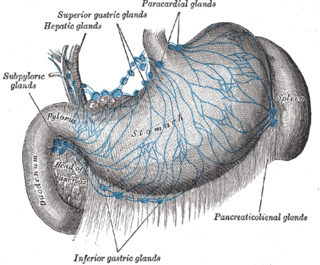
The gastric lymph nodes are lymph nodes which drain the stomach and consist of two sets, superior and inferior:
Pyloromyotomy is a surgical procedure in which a portion of the muscle fibers of the pyloric muscle are cut. This is typically done in cases where the contents from the stomach are inappropriately stopped by the pyloric muscle, causing the stomach contents to build up in the stomach and unable to be appropriately digested. The procedure is typically performed in cases of "hypertrophic pyloric stenosis" in young children. In most cases, the procedure can be performed with either an open approach or a laparoscopic approach and the patients typically have good outcomes with minimal complications.

Pyloroplasty is a surgery performed to widen the opening at the lower part of the stomach, also known as the pylorus. When the pylorus thickens, it becomes difficult for food to pass through. The surgery is performed to widen the band of muscle known as the pyloric sphincter, a ring of smooth, muscular fibers that surrounds the pylorus and helps to regulate digestion and prevent reflux. The widening of the pyloric sphincter enables the contents of the stomach to pass into the first part of the small intestine known as the duodenum.

Antrectomy, also called distal gastrectomy, is a type of gastric resection surgery that involves the removal of the stomach antrum to treat gastric diseases causing the damage, bleeding, or blockage of the stomach. This is performed using either the Billroth I (BI) or Billroth II (BII) reconstruction method. Quite often, antrectomy is used alongside vagotomy to maximise its safety and effectiveness. Modern antrectomies typically have a high success rate and low mortality rate, but the exact numbers depend on the specific conditions being treated.
