Anterior cruciate ligament

Last updated
Anterior cruciate ligament
Knee diagram.svg
Diagram of the right knee. Anterior cruciate ligament labeled at center left.
Details
From Lateral condyle of the femur
To Intercondyloid eminence of the tibia
Identifiers
Latin ligamentum cruciatum anterius
MeSH D016118
TA98 A03.6.08.007
TA2 1890
FMA 44614
Anatomical terminology

The anterior cruciate ligament (ACL) is one of a pair of cruciate ligaments (the other being the posterior cruciate ligament) in the human knee. The two ligaments are also called "cruciform" ligaments, as they are arranged in a crossed formation. In the quadruped stifle joint (analogous to the knee), based on its anatomical position, it is also referred to as the cranial cruciate ligament. [1] The term cruciate translates to cross. This name is fitting because the ACL crosses the posterior cruciate ligament to form an "X". It is composed of strong, fibrous material and assists in controlling excessive motion. This is done by limiting mobility of the joint. The anterior cruciate ligament is one of the four main ligaments of the knee, providing 85% of the restraining force to anterior tibial displacement at 30 and 90° of knee flexion. [2] The ACL is the most injured ligament of the four located in the knee.

Contents

Structure

The ACL originates from deep within the notch of the distal femur. Its proximal fibers fan out along the medial wall of the lateral femoral condyle. [3] The two bundles of the ACL are the anteromedial and the posterolateral, named according to where the bundles insert into the tibial plateau. [4] [5] The tibial plateau is a critical weight-bearing region on the upper extremity of the tibia. The ACL attaches in front of the intercondyloid eminence of the tibia, where it blends with the anterior horn of the medial meniscus.

Purpose

The purpose of the ACL is to resist the motions of anterior tibial translation and internal tibial rotation; this is important to have rotational stability. [6] This function prevents anterior tibial subluxation of the lateral and medial tibiofemoral joints, which is important for the pivot-shift phenomenon. [6] The ACL has mechanoreceptors that detect changes in direction of movement, position of the knee joint, and changes in acceleration, speed, and tension. [7] A key factor in instability after ACL injuries is having altered neuromuscular function secondary to diminished somatosensory information. [7] For athletes who participate in sports involving cutting, jumping, and rapid deceleration, the knee must be stable in terminal extension, which is the screw-home mechanism. [7]

Clinical significance

Injury

MRI of anterior cruciate ligament tear VKB-Riss MRT T1 PDW sag.jpg
MRI of anterior cruciate ligament tear

An ACL tear is one of the most common knee injuries, with over 100,000 tears occurring annually in the US. [8] Most ACL tears are a result of a non-contact mechanism such as a sudden change in a direction causing the knee to rotate inward. As the knee rotates inward, additional strain is placed on the ACL, since the femur and tibia, which are the two bones that articulate together forming the knee joint, move in opposite directions, causing the ACL to tear. Most athletes require reconstructive surgery on the ACL, in which the torn or ruptured ACL is completely removed and replaced with a piece of tendon or ligament tissue from the patient (autograft) or from a donor (allograft). [9] Conservative treatment has poor outcomes in ACL injury, since the ACL is unable to form a fibrous clot, as it receives most of its nutrients from synovial fluid; this washes away the reparative cells, making the formation of fibrous tissue difficult. The two most common sources for tissue are the patellar ligament and the hamstrings tendon. [10] The patellar ligament is often used, since bone plugs on each end of the graft are extracted, which helps integrate the graft into the bone tunnels during reconstruction. [11] The surgery is arthroscopic, meaning that a tiny camera is inserted through a small surgical cut. [9] The camera sends video to a large monitor so the surgeon can see any damage to the ligaments. In the event of an autograft, the surgeon makes a larger cut to get the needed tissue. In the event of an allograft, in which material is donated, this is not necessary, since no tissue is taken directly from the patient's own body. [12] The surgeon drills a hole forming the tibial bone tunnel and femoral bone tunnel, allowing for the patient's new ACL graft to be guided through. [12] Once the graft is pulled through the bone tunnels, two screws are placed into the tibial and femoral bone tunnel. [12] Recovery time usually ranges between one and two years, but is sometimes longer, depending if the patient chose an autograft or allograft. A week or so after the occurrence of the injury, the athlete is usually deceived by the fact that he/she is walking normally and not feeling much pain. [12] This is dangerous, as some athletes start resuming some of their activities such as jogging, which with a wrong move or twist, could damage the bones, as the graft has not completely become integrated into the bone tunnels. Injured athletes must understand the significance of each step of an ACL injury to avoid complications and ensure a proper recovery.

Nonoperative treatment of the ACL

ACL reconstruction is the most common treatment for an ACL tear, but it is not the only treatment available for individuals. Some may find it more beneficial to complete a nonoperative rehabilitation program. Individuals who are going to continue with physical activity that involves cutting and pivoting, and individuals who are no longer participating in those specific activities both are candidates for the nonoperative route. [13] In comparing operative and nonoperative approaches to ACL tears, few differences were noted between surgical and nonsurgical groups, with no significant differences in regard to knee function or muscle strength reported by the patients.[ citation needed ]

The main goals to achieve during rehabilitation (rehab) of an ACL tear is to regain sufficient functional stability, maximize full muscle strength, and decrease risk of reinjury.[ citation needed ] Typically, three phases are involved in nonoperative treatment - the acute phase, the neuromuscular training phase, and the return to sport phase. During the acute phase, the rehab is focusing on the acute symptoms that occur right after the injury and are causing an impairment. The use of therapeutic exercises and appropriate therapeutic modalities is crucial during this phase to assist in repairing the impairments from the injury. The neuromuscular training phase is used to focus on the patient regaining full strength in both the lower extremity and the core muscles. This phase begins when the patient regains full range of motion, no effusion, and adequate lower extremity strength. During this phase, the patient completes advanced balance, proprioception, cardiovascular conditioning, and neuromuscular interventions. [13] In the final, return to sport phase, the patient focuses on sport-specific activities and agility. A functional performance brace is suggested to be used during the phase to assist with stability during pivoting and cutting activities. [13]

Operative treatment of the ACL

Anterior cruciate ligament surgery is a complex operation that requires expertise in the field of orthopedic and sports medicine. Many factors should be considered when discussing surgery, including the athlete's level of competition, age, previous knee injury, other injuries sustained, leg alignment, and graft choice. Typically, four graft types are possible, the bone-patella tendon-bone graft, the semitendinosus and gracilis tendons (quadrupled hamstring tendon), quadriceps tendon, and an allograft. [14] Although extensive research has been conducted on which grafts are the best, the surgeon typically chooses the type of graft with which he or she is most comfortable. If rehabilitated correctly, the reconstruction should last. In fact, 92.9% of patients are happy with graft choice. [14]

Prehabilitation has become an integral part of the ACL reconstruction process. This means that the patient exercises before getting surgery to maintain factors such as range of motion and strength. Based on a single leg hop test and self-reported assessment, prehab improved function; these effects were sustained 12 weeks postoperatively. [15]

Postsurgical rehabilitation is essential in the recovery from the reconstruction. This typically takes a patient 6 to 12 months to return to life as it was prior to the injury. [16] The rehab can be divided into protection of the graft, improving range of motion, decrease swelling, and regaining muscle control. [16] Each phase has different exercises based on the patients' needs. For example, while the ligament is healing, a patient's joint should not be used for full weight-bearing, but the patient should strengthen the quadriceps and hamstrings by doing quad sets and weight shifting drills. Phase two would require full weight-bearing and correcting gait patterns, so exercises such as core strengthening and balance exercises would be appropriate. In phase three, the patient begins running, and can do aquatic workouts to help with reducing joint stresses and cardiorespiratory endurance. Phase four includes multiplanar movements, thus enhancing a running program and beginning agility and plyometric drills. Lastly, phase five focuses on sport- or life-specific motions, depending on the patient. [16]

A 2010 Los Angeles Times review of two medical studies discussed whether ACL reconstruction was advisable. One study found that children under 14 who had ACL reconstruction fared better after early surgery than those who underwent a delayed surgery. For adults 18 to 35, though, patients who underwent early surgery followed by rehabilitation fared no better than those who had rehabilitative therapy and a later surgery. [17]

The first report focused on children and the timing of an ACL reconstruction. ACL injuries in children are a challenge because children have open growth plates in the bottom of the femur or thigh bone and on the top of the tibia or shin. An ACL reconstruction typically crosses the growth plates, posing a theoretical risk of injury to the growth plate, stunting leg growth, or causing the leg to grow at an unusual angle. [18]

The second study noted focused on adults. It found no significant statistical difference in performance and pain outcomes for patients who receive early ACL reconstruction vs. those who receive physical therapy with an option for later surgery. This would suggest that many patients without instability, buckling, or giving way after a course of rehabilitation can be managed nonoperatively, but was limited to outcomes after two years and did not involve patients who were serious athletes. [17] Patients involved in sports requiring significant cutting, pivoting, twisting, or rapid acceleration or deceleration may not be able to participate in these activities without ACL reconstruction. [19]

ACL injuries in women

Risk differences between outcomes in men and women can be attributed to a combination of multiple factors, including anatomical, hormonal, genetic, positional, neuromuscular, and environmental factors. [20] The size of the anterior cruciate ligament is often the most reported difference. Studies look at the length, cross-sectional area, and volume of ACLs. Researchers use cadavers, and in vivo placement to study these factors, and most studies confirm that women have smaller anterior cruciate ligaments. Other factors that could contribute to higher risks of ACL tears in women include patient weight and height, the size and depth of the intercondylar notch, the diameter of the ACL, the magnitude of the tibial slope, the volume of the tibial spines, the convexity of the lateral tibiofemoral articular surfaces, and the concavity of the medial tibial plateau. [21] While anatomical factors are most talked about, extrinsic factors, including dynamic movement patterns, might be the most important risk factor when it comes to ACL injury. [22]

See also

Related Research Articles

<span class="mw-page-title-main">Knee</span> Leg joint in primates

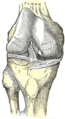
In humans and other primates, the knee joins the thigh with the leg and consists of two joints: one between the femur and tibia, and one between the femur and patella. It is the largest joint in the human body. The knee is a modified hinge joint, which permits flexion and extension as well as slight internal and external rotation. The knee is vulnerable to injury and to the development of osteoarthritis.

<span class="mw-page-title-main">Hamstring</span> Any of the three tendons in the thigh

In human anatomy, a hamstring is any one of the three posterior thigh muscles between the hip and the knee.

<span class="mw-page-title-main">Tibia</span> Leg bone in vertebrates

The tibia, also known as the shinbone or shankbone, is the larger, stronger, and anterior (frontal) of the two bones in the leg below the knee in vertebrates ; it connects the knee with the ankle. The tibia is found on the medial side of the leg next to the fibula and closer to the median plane. The tibia is connected to the fibula by the interosseous membrane of leg, forming a type of fibrous joint called a syndesmosis with very little movement. The tibia is named for the flute tibia. It is the second largest bone in the human body, after the femur. The leg bones are the strongest long bones as they support the rest of the body.

<span class="mw-page-title-main">Posterior cruciate ligament</span> One of four major ligaments of the knee

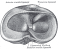
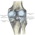
The posterior cruciate ligament (PCL) is a ligament in each knee of humans and various other animals. It works as a counterpart to the anterior cruciate ligament (ACL). It connects the posterior intercondylar area of the tibia to the medial condyle of the femur. This configuration allows the PCL to resist forces pushing the tibia posteriorly relative to the femur.

<span class="mw-page-title-main">Segond fracture</span> Avulsion fracture of the lateral tibial condyle of the knee

The Segond fracture is a type of avulsion fracture from the lateral tibial plateau of the knee, immediately below the articular surface of the tibia.

The drawer test is used in the initial clinical assessment of suspected rupture of the cruciate ligaments in the knee.

<span class="mw-page-title-main">Anterior cruciate ligament reconstruction</span> Surgical process

Anterior cruciate ligament reconstruction is a surgical tissue graft replacement of the anterior cruciate ligament, located in the knee, to restore its function after an injury. The torn ligament can either be removed from the knee, or preserved before reconstruction through an arthroscopic procedure. ACL repair is also a surgical option. This involves repairing the ACL by re-attaching it, instead of performing a reconstruction. Theoretical advantages of repair include faster recovery and a lack of donor site morbidity, but randomised controlled trials and long-term data regarding re-rupture rates using contemporary surgical techniques are lacking.

<span class="mw-page-title-main">Semitendinosus muscle</span> One of the hamstring muscles; posterior part of the thigh

The semitendinosus is a long superficial muscle in the back of the thigh. It is so named because it has a very long tendon of insertion. It lies posteromedially in the thigh, superficial to the semimembranosus.

<span class="mw-page-title-main">Medial meniscus</span> Cartilage formation in the human knee

The medial meniscus is a fibrocartilage semicircular band that spans the knee joint medially, located between the medial condyle of the femur and the medial condyle of the tibia. It is also referred to as the internal semilunar fibrocartilage. The medial meniscus has more of a crescent shape while the lateral meniscus is more circular. The anterior aspects of both menisci are connected by the transverse ligament. It is a common site of injury, especially if the knee is twisted.

<span class="mw-page-title-main">Cruciate ligament</span> Type of ligament shaped like an X

Cruciate ligaments are pairs of ligaments arranged like a letter X. They occur in several joints of the body, such as the knee joint, wrist joint and the atlanto-axial joint. In a fashion similar to the cords in a toy Jacob's ladder, the crossed ligaments stabilize the joint while allowing a very large range of motion.

<span class="mw-page-title-main">Anterior cruciate ligament injury</span> Ligament injury near the knee

An anterior cruciate ligament injury occurs when the anterior cruciate ligament (ACL) is either stretched, partially torn, or completely torn. The most common injury is a complete tear. Symptoms include pain, an audible cracking sound during injury, instability of the knee, and joint swelling. Swelling generally appears within a couple of hours. In approximately 50% of cases, other structures of the knee such as surrounding ligaments, cartilage, or meniscus are damaged.

<span class="mw-page-title-main">Patellar tendon</span> Tendon in the human knee

The patellar tendon is the distal portion of the common tendon of the quadriceps femoris, which is continued from the patella to the tibial tuberosity. It is also sometimes called the patellar ligament as it forms a bone to bone connection when the patella is fully ossified.

<span class="mw-page-title-main">Unhappy triad</span> Medical condition of the knee

The unhappy triad, also known as a blown knee among other names, is an injury to the anterior cruciate ligament, medial collateral ligament, and meniscus. Analysis during the 1990s indicated that this 'classic' O'Donoghue triad is actually an unusual clinical entity among athletes with knee injuries. Some authors mistakenly believe that in this type of injury, "combined anterior cruciate and medial collateral ligament disruptions that were incurred during athletic endeavors" always present with concomitant medial meniscus injury. However, the 1990 analysis showed that lateral meniscus tears are more common than medial meniscus tears in conjunction with sprains of the ACL.

<span class="mw-page-title-main">Meniscus tear</span> Rupturing of the fibrocartilage strips in the knee called menisci

A tear of a meniscus is a rupturing of one or more of the fibrocartilage strips in the knee called menisci. When doctors and patients refer to "torn cartilage" in the knee, they actually may be referring to an injury to a meniscus at the top of one of the tibiae. Menisci can be torn during innocuous activities such as walking or squatting. They can also be torn by traumatic force encountered in sports or other forms of physical exertion. The traumatic action is most often a twisting movement at the knee while the leg is bent. In older adults, the meniscus can be damaged following prolonged 'wear and tear'. Especially acute injuries can lead to displaced tears which can cause mechanical symptoms such as clicking, catching, or locking during motion of the joint. The joint will be in pain when in use, but when there is no load, the pain goes away.

The medial patellofemoral ligament (MPFL) is one of several ligaments on the medial aspect of the knee. It originates in the superomedial aspect of the patella and inserts in the space between the adductor tubercle and the medial femoral epicondyle. The ligament itself extends from the femur to the superomedial patella, and its shape is similar to a trapezoid. It keeps the patella in place, but its main function is to prevent lateral displacement of the patella.

Posterolateral corner injuries of the knee are injuries to a complex area formed by the interaction of multiple structures. Injuries to the posterolateral corner can be debilitating to the person and require recognition and treatment to avoid long term consequences. Injuries to the PLC often occur in combination with other ligamentous injuries to the knee; most commonly the anterior cruciate ligament (ACL) and posterior cruciate ligament (PCL). As with any injury, an understanding of the anatomy and functional interactions of the posterolateral corner is important to diagnosing and treating the injury.

<span class="mw-page-title-main">Medial knee injuries</span> Medical condition

Medial knee injuries are the most common type of knee injury. The medial ligament complex of the knee consists of:

High tibial osteotomy is an orthopaedic surgical procedure which aims to correct a varus deformation with compartmental osteoarthritis. Since the inception of the procedure, advancements to technique, fixation devices, and a better understanding of patient selection has allowed HTO to become more popular in younger, more active patients hoping to combat arthritis. The idea behind the procedure is to realign the weight-bearing line of the knee. By realigning the knee, the force produced from weight-bearing is shifted from the arthritic, medial compartment to the healthy, lateral compartment. This decrease in force or load in the diseased part of the knee joint decreases knee pain and can delay the development or progression of osteoarthritis in the medial compartment.

<span class="mw-page-title-main">Posterior cruciate ligament injury</span> Medical condition

The function of the posterior cruciate ligament (PCL) is to prevent the femur from sliding off the anterior edge of the tibia and to prevent the tibia from displacing posterior to the femur. Common causes of PCL injuries are direct blows to the flexed knee, such as the knee hitting the dashboard in a car accident or falling hard on the knee, both instances displacing the tibia posterior to the femur.

Artificial ligaments are devices used to replace damaged ligaments. Today, the most common use of artificial ligaments is in anterior cruciate ligament reconstruction. Although autotransplantation remains the most common method of ligament reconstruction, numerous materials and structures were developed to optimize the artificial ligament since its creation in the World War I era. Many modern artificial ligaments are made of synthetic polymers, such as polyethylene terephthalate. Various coatings have been added to improve the biocompatibility of the synthetic polymers. Early artificial ligaments developed in the 1980s were ineffective due to material deterioration. Currently, the Ligament Advanced Reinforcement System (LARS) artificial ligament has been utilized extensively in clinical applications. Tissue engineering is a growing area of research which aims to regenerate and restore ligament function.

References

  1. "Canine Cranial Cruciate Ligament Disease" (PDF). Melbourne Veterinary Referral Centre. pp. 1–2. Archived from the original (PDF) on 19 July 2008. Retrieved September 8, 2009.
  2. Ellison, A. E.; Berg, E. E. (1985). "Embryology, anatomy, and function of the anterior cruciate ligament". The Orthopedic Clinics of North America. 16 (1): 3–14. doi:10.1016/S0030-5898(20)30463-6. PMID   3969275.
  3. Petersen, W.; Tillmann, B. (August 2002). "[Anatomy and function of the anterior cruciate ligament]". Der Orthopade. 31 (8): 710–718. doi:10.1007/s00132-002-0330-0. ISSN   0085-4530. PMID   12426749. S2CID   45919449.
  4. Duthon, V. B.; Barea, C.; Abrassart, S.; Fasel, J. H.; Fritschy, D.; Ménétrey, J. (March 2006). "Anatomy of the anterior cruciate ligament". Knee Surgery, Sports Traumatology, Arthroscopy. 14 (3): 204–213. doi:10.1007/s00167-005-0679-9. ISSN   0942-2056. PMID   16235056. S2CID   25658911.
  5. Petersen, Wolf; Zantop, Thore (January 2007). "Anatomy of the anterior cruciate ligament with regard to its two bundles". Clinical Orthopaedics and Related Research. 454: 35–47. doi:10.1097/BLO.0b013e31802b4a59. ISSN   0009-921X. PMID   17075382.
  6. 1 2 Noyes, Frank R. (January 2009). "The Function of the Human Anterior Cruciate Ligament and Analysis of Single- and Double-Bundle Graft Reconstructions". Sports Health. 1 (1): 66–75. doi:10.1177/1941738108326980. ISSN   1941-7381. PMC   3445115 . PMID   23015856.
  7. 1 2 3 Liu-Ambrose, T. (December 2003). "The anterior cruciate ligament and functional stability of the knee joint". British Columbia Medical Journal. 45 (10): 495–499. Retrieved 2018-11-15.
  8. Cimino, Francesca; Volk, Bradford Scott; Setter, Don (2010-10-15). "Anterior Cruciate Ligament Injury: Diagnosis, Management, and Prevention". American Family Physician. 82 (8): 917–922. ISSN   0002-838X. PMID   20949884.
  9. 1 2 "ACL reconstruction - Mayo Clinic". www.mayoclinic.org. Retrieved 2018-11-15.
  10. Samuelsen, Brian T.; Webster, Kate E.; Johnson, Nick R.; Hewett, Timothy E.; Krych, Aaron J. (October 2017). "Hamstring Autograft versus Patellar Tendon Autograft for ACL Reconstruction: Is There a Difference in Graft Failure Rate? A Meta-analysis of 47,613 Patients". Clinical Orthopaedics and Related Research. 475 (10): 2459–2468. doi:10.1007/s11999-017-5278-9. ISSN   1528-1132. PMC   5599382 . PMID   28205075.
  11. "When Would You Use Patellar Tendon Autograft as Your Main Graft Selection?". www.healio.com. Retrieved 2018-11-15.
  12. 1 2 3 4 "ACL Injury: Does It Require Surgery? - OrthoInfo - AAOS" . Retrieved 2018-11-15.
  13. 1 2 3 Paterno, Mark V. (2017-07-29). "Non-operative Care of the Patient with an ACL-Deficient Knee". Current Reviews in Musculoskeletal Medicine. 10 (3): 322–327. doi:10.1007/s12178-017-9431-6. ISSN   1935-973X. PMC   5577432 . PMID   28756525.
  14. 1 2 Macaulay, Alec A.; Perfetti, Dean C.; Levine, William N. (January 2012). "Anterior Cruciate Ligament Graft Choices". Sports Health. 4 (1): 63–68. doi:10.1177/1941738111409890. ISSN   1941-7381. PMC   3435898 . PMID   23016071.
  15. Shaarani, Shahril R.; O'Hare, Christopher; Quinn, Alison; Moyna, Niall; Moran, Raymond; O'Byrne, John M. (September 2013). "Effect of prehabilitation on the outcome of anterior cruciate ligament reconstruction". The American Journal of Sports Medicine. 41 (9): 2117–2127. doi:10.1177/0363546513493594. ISSN   1552-3365. PMID   23845398. S2CID   38240767.
  16. 1 2 3 "Rehabilitation Guidelines for ACL Reconstruction in the Adult Athlete (Skeletally Mature)" (PDF). UW Health. Archived from the original (PDF) on 2020-11-12. Retrieved 2018-12-06.
  17. 1 2 Stein, Jeannine (2010-07-22). "Studies on ACL surgery". The Los Angeles Times. Archived from the original on July 28, 2010. Retrieved 2010-07-23.
  18. "ACL Tears: To reconstruct or not, and if so, when?". howardluksmd.com. Archived from the original on January 25, 2013. Retrieved 2010-07-23.
  19. Frobell, Richard B.; Roos, Ewa M.; Roos, Harald P.; Ranstam, Jonas; Lohmander, L. Stefan (2010). "A Randomized Trial of Treatment for Acute Anterior Crut Tears". New England Journal of Medicine. 363 (4): 331–342. doi: 10.1056/NEJMoa0907797 . PMID   20660401.
  20. Chandrashekara, Naveen; Mansourib, Hossein; Slauterbeckc, James; Hashemia, Javad (2006). "Sex-based differences in the tensile properties of the human anterior cruciate ligament". Journal of Biomechanics. 39 (16): 2943–2950. doi:10.1016/j.jbiomech.2005.10.031. PMID   16387307.
  21. Schneider, Antione; Si-Mohamed, Salim; Magnussen, Robert; Lustig, Sebastien; Neyret, Philippe; Servien, Elvire (October 2017). "Tibiofemoral joint congruence is lower in females with ACL injuries than males with ACL injuries". Knee Surgery, Sports Traumatology, Arthroscopy. 25 (5): 1375–1383. doi:10.1007/s00167-017-4756-7. PMID   29052744. S2CID   4968334.
  22. Lloyd Ireland, Mary (2002). "The female ACL: why is it more prone to injury?". Orthopedic Clinics of North America. 33 (2): 637–651. doi:10.1016/S0030-5898(02)00028-7. PMC   4805849 . PMID   12528906.