
The endometrium is the inner epithelial layer, along with its mucous membrane, of the mammalian uterus. It has a basal layer and a functional layer; the functional layer thickens and then is shed during menstruation in humans and some other mammals, including apes, Old World monkeys, some species of bat, and the elephant shrew. In most other mammals, the endometrium is reabsorbed in the estrous cycle. During pregnancy, the glands and blood vessels in the endometrium further increase in size and number. Vascular spaces fuse and become interconnected, forming the placenta, which supplies oxygen and nutrition to the embryo and fetus. The speculated presence of an endometrial microbiota has been argued against.

Endometrial cancer is a cancer that arises from the endometrium. It is the result of the abnormal growth of cells that have the ability to invade or spread to other parts of the body. The first sign is most often vaginal bleeding not associated with a menstrual period. Other symptoms include pain with urination, pain during sexual intercourse, or pelvic pain. Endometrial cancer occurs most commonly after menopause.

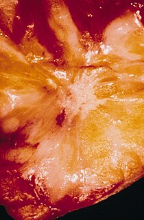
Molar pregnancy is an abnormal form of pregnancy in which a non-viable fertilized egg implants in the uterus and will fail to come to term. A molar pregnancy is a gestational trophoblastic disease which grows into a mass in the uterus that has swollen chorionic villi. These villi grow in clusters that resemble grapes. A molar pregnancy can develop when a fertilized egg does not contain an original maternal nucleus. The products of conception may or may not contain fetal tissue. It is characterized by the presence of a hydatidiform mole. Molar pregnancies are categorized as partial moles or complete moles, with the word mole being used to denote simply a clump of growing tissue, or a growth.

Surface epithelial-stromal tumors are a class of ovarian neoplasms that may be benign or malignant. Neoplasms in this group are thought to be derived from the ovarian surface epithelium or from ectopic endometrial or Fallopian tube (tubal) tissue. Tumors of this type are also called ovarian adenocarcinoma. This group of tumors accounts for 90% to 95% of all cases of ovarian cancer. Serum CA-125 is often elevated but is only 50% accurate so it is not a useful tumor marker to assess the progress of treatment.

The decidua is the modified mucosal lining of the uterus that forms in preparation for pregnancy. It is formed in a process called decidualization under the influence of progesterone. Endometrial cells become highly characteristic. The decidua forms the maternal part of the placenta and remains for the duration of the pregnancy. It is shed off during childbirth — hence why the term is used, "decidua" having the meaning of falling away, as in the word deciduous.

A koilocyte is a squamous epithelial cell that has undergone a number of structural changes, which occur as a result of infection of the cell by human papillomavirus.

In humans, implantation is the stage of pregnancy at which the embryo adheres to the wall of the uterus. At this stage of prenatal development, the conceptus is called a blastocyst. It is by this adhesion that the embryo receives oxygen and nutrients from the mother to be able to grow.
Endometrial intraepithelial neoplasia (EIN) is a premalignant lesion of the uterine lining that predisposes to endometrioid endometrial adenocarcinoma. It is composed of a collection of abnormal endometrial cells, arising from the glands that line the uterus, which have a tendency over time to progress to the most common form of uterine cancer—endometrial adenocarcinoma, endometrioid type.
Decidualization is a process that results in significant changes to cells of the endometrium in preparation for, and during, pregnancy. This includes morphological and functional changes to endometrial stromal cells (ESCs), the presence of decidual white blood cells (leukocytes), and vascular changes to maternal arteries. The sum of these changes results in the endometrium changing into a structure called the decidua. In humans, the decidua is shed during the third phase of birth.

Uterine glands or endometrial glands are tubular glands, lined by ciliated columnar epithelium, found in the functional layer of the endometrium that lines the uterus. Their appearance varies during the menstrual cycle. During the proliferative phase, uterine glands appear long due to estrogen secretion by the ovaries. During the secretory phase, the uterine glands become very coiled with wide lumens and produce a glycogen-rich secretion. This change corresponds with an increase in blood flow to spiral arteries due to increased progesterone secretion from the corpus luteum. During the pre-menstrual phase, progesterone secretion decreases as the corpus luteum degenerates, which results in decreased blood flow to the spiral arteries. The functional layer of the uterus containing the glands becomes necrotic, and eventually sloughs off during the menstrual phase of the cycle.

Endometrial hyperplasia is a condition of excessive proliferation of the cells of the endometrium, or inner lining of the uterus.

Juxtaposed with another zinc finger protein 1 (JAZF1) also known as TAK1-interacting protein 27 (TIP27) or zinc finger protein 802 (ZNF802) is a protein that in humans is encoded by the JAZF1 gene. Variants are associated with an increased risk of prostate cancer, an increased risk of type 2 diabetes, and an increased height.

High-grade prostatic intraepithelial neoplasia (HGPIN) is an abnormality of prostatic glands and believed to precede the development of prostate adenocarcinoma.

Uterine serous carcinoma (USC), is an uncommon form of endometrial cancer that typically arises in postmenopausal women. It is typically diagnosed on endometrial biopsy, prompted by post-menopausal bleeding.

Javier Arias Stella was a Peruvian pathologist and politician. After earning his doctorate from the San Marcos National University, Arias Stella began his career as a pathology lecturer at the same institution, eventually rising to the senior lecturer position. He left to become a co-founder of the new Cayetano Heredia University in 1961, becoming head of pathology there in 1975. His research included the eponymous Arias-Stella reaction, in which he discovered that a reaction during pregnancy that was often mistaken for cancer, was in fact a benign reaction of hormones from placental tissue. He also performed research into how altitude changes in the Andes affected anatomy and histology in human beings.
A radial scar of the breast, formally radial scar of the breast, is a benign breast lesion that can radiologically mimic malignancy, i.e. cancer.

Nuclear atypia refers to abnormal appearance of cell nuclei. It is a term used in cytopathology and histopathology. Atypical nuclei are often pleomorphic.

Pleomorphism is a term used in histology and cytopathology to describe variability in the size, shape and staining of cells and/or their nuclei. Several key determinants of cell and nuclear size, like ploidy and the regulation of cellular metabolism, are commonly disrupted in tumors. Therefore, cellular and nuclear pleomorphism is one of the earliest hallmarks of cancer progression and a feature characteristic of malignant neoplasms and dysplasia. Certain benign cell types may also exhibit pleomorphism, e.g. neuroendocrine cells, Arias-Stella reaction.
Microglandular hyperplasia (MGH) of the cervix is an epithelial benign abnormality (lesion) associated with gland proliferation. It can terminate in mature squamous metaplasia, and it is suspected reserve cells are involved in this process, perhaps in the form of reserve cell hyperplasia with glandular differentiation.
Endometrial cups form during pregnancy in mares and are the source of equine chorionic gonadotropin (eCG) and a placenta-associated structure, which is derived from the fetus. Their purpose is to increase the immunological tolerance of the mare in order to protect the developing foal.
