
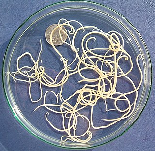
Ascaris lumbricoides is a large parasitic worm that causes ascariasis in humans. A roundworm of genus Ascaris, it is the most common parasitic worm in humans. An estimated 807 million–1.2 billion people are infected with A. lumbricoides worldwide. People living in tropical and subtropical countries are at greater risk of infection.

Trichuris trichiura, Trichocephalus trichiuris or whipworm, is a parasitic roundworm that causes trichuriasis when it infects a human large intestine. It is commonly known as the whipworm which refers to the shape of the worm; it looks like a whip with wider "handles" at the posterior end.

Trichuriasis, also known as whipworm infection, is an infection by the parasitic worm Trichuris trichiura (whipworm). If infection is only with a few worms, there are often no symptoms. In those who are infected with many worms, there may be abdominal pain, fatigue and diarrhea. The diarrhea sometimes contains blood. Infections in children may cause poor intellectual and physical development. Low red blood cell levels may occur due to loss of blood.

Ascariasis is a disease caused by the parasitic roundworm Ascaris lumbricoides. Infections have no symptoms in more than 85% of cases, especially if the number of worms is small. Symptoms increase with the number of worms present and may include shortness of breath and fever in the beginning of the disease. These may be followed by symptoms of abdominal swelling, abdominal pain, and diarrhea. Children are most commonly affected, and in this age group the infection may also cause poor weight gain, malnutrition, and learning problems.
Toxocariasis is an illness of humans caused by the dog roundworm and, less frequently, the cat roundworm. These are the most common intestinal roundworms of dogs, coyotes, wolves and foxes and domestic cats, respectively. Humans are among the many "accidental" or paratenic hosts of these roundworms.

Baylisascaris is a genus of roundworms that infect more than fifty animal species.

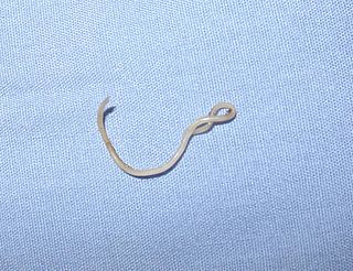
Necator americanus is a species of hookworm commonly known as the New World hookworm. Like other hookworms, it is a member of the phylum Nematoda. It is an obligatory parasitic nematode that lives in the small intestine of human hosts. Necatoriasis—a type of helminthiasis—is the term for the condition of being host to an infestation of a species of Necator. Since N. americanus and Ancylostoma duodenale are the two species of hookworms that most commonly infest humans, they are usually dealt with under the collective heading of "hookworm infection". They differ most obviously in geographical distribution, structure of mouthparts, and relative size.
Ascaris is a nematode genus of parasitic worms known as the "small intestinal roundworms", which is a type of parasitic worm. One species, Ascaris lumbricoides, affects humans and causes the disease ascariasis. Another species, Ascaris suum, typically infects pigs. Other ascarid genera infect other animals, such as Parascaris equorum, the equine roundworm, and Toxocara and Toxascaris, which infect dogs and cats.

Parasitic worms, also known as helminths, are large macroparasites; adults can generally be seen with the naked eye. Many are intestinal worms that are soil-transmitted and infect the gastrointestinal tract. Other parasitic worms such as schistosomes reside in blood vessels.

Eucestoda, commonly referred to as tapeworms, is the larger of the two subclasses of flatworms in the class Cestoda. Larvae have six posterior hooks on the scolex (head), in contrast to the ten-hooked Cestodaria. All tapeworms are endoparasites of vertebrates, living in the digestive tract or related ducts. Examples are the pork tapeworm with a human definitive host, and pigs as the secondary host, and Moniezia expansa, the definitive hosts of which are ruminants.
Spirometra erinaceieuropaei is a parasitic tapeworm that infects domestic animals and humans. The medical term for this infection in humans and other animals is sparganosis. Morphologically, these worms are similar to other worms in the genus Spirometra. They have a long body consisting of three sections: the scolex, the neck, and the strobilia. They have a complex life cycle that consists of three hosts, and can live in varying environments and bodily tissues. Humans can contract this parasite in three main ways. Historically, humans are considered a paratenic host; however, the first case of an adult S. erinaceieuropaei infection in humans was reported in 2017. Spirometra tapeworms exist worldwide and infection is common in animals, but S. erinaceieuropaei infections are rare in humans. Treatment for infection typically includes surgical removal and anti-worm medication.
Gnathostoma spinigerum is a parasitic nematode that causes gnathostomiasis in humans, also known as its clinical manifestations are creeping eruption, larva migrans, Yangtze edema, Choko-Fuschu Tua chid and wandering swelling. Gnathostomiasis in animals can be serious, and even fatal. The first described case of gnathostomiasis was in a young tiger that died in the London Zoo in 1835. The larval nematode is acquired by eating raw or undercooked fish and meat.
Toxocara canis is a worldwide-distributed helminth parasite that primarily infects dogs and other canids, but can also infect other animals including humans. The name is derived from the Greek word "toxon," meaning bow or quiver, and the Latin word "caro," meaning flesh. T. canis live in the small intestine of the definitive host. This parasite is very common in puppies and somewhat less common in adult dogs. In adult dogs, infection is usually asymptomatic but may be characterized by diarrhea. By contrast, untreated infection with Toxocara canis can be fatal in puppies, causing diarrhea, vomiting, pneumonia, enlarged abdomen, flatulence, poor growth rate, and other complications.
The 1970 ascariasis poisoning incident was a poisoning incident that took place in Quebec in February, 1970. At least seven people claimed to have been infected with parasitic worm eggs by Eric Kranz, a former postgraduate student from Hempstead, New York. The victims were Canadians Richard Davis, William Butler, David Fisk, and Keith Fern, with three other friends and acquaintances reported to be mildly infested. Doctors said that one of the men may have been affected by as many as 400,000 larvae.
Baylisascaris procyonis, also known by the common name raccoon roundworm, is a roundworm nematode, found ubiquitously in raccoons, the definitive hosts. It is named after H. A. Baylis, who studied them in the 1920s–30s, and Greek askaris. Baylisascaris larvae in paratenic hosts can migrate, causing larva migrans. Baylisascariasis as the zoonotic infection of humans is rare, though extremely dangerous due to the ability of the parasite's larvae to migrate into brain tissue and cause damage. Concern for human infection has been increasing over the years due to the urbanization of rural areas, resulting in the increase in proximity and potential human interaction with raccoons.
Gnathostoma hispidum is a nematode (roundworm) that infects many vertebrate animals including humans. Infection of Gnathostoma hispidum, like many species of Gnathostoma causes the disease gnathostomiasis due to the migration of immature worms in the tissues.

Soil-transmitted helminthiasis is a type of worm infection (helminthiasis) caused by different species of roundworms. It is caused specifically by those worms which are transmitted through soil contaminated with faecal matter and are therefore called soil-transmitted helminths. Three types of soil-transmitted helminthiasis can be distinguished: ascariasis, hookworm infection and whipworm infection. These three types of infection are therefore caused by the large roundworm A. lumbricoides, the hookworms Necator americanus or Ancylostoma duodenale and by the whipworm Trichuris trichiura.
Paragordius varius is a parasite species in the horsehair worm group (Nematomorpha). They cycle between terrestrial and aquatic habitats and are most commonly known for their ability to manipulate their definitive host to jump into a pool of water, which allows them to complete their life cycle. Adults are over 10 cm long and 400 μm in diameter. P. varius is usually found in water or wet areas. The definitive hosts are mainly terrestrial arthropods, most often carabid beetles, crickets and praying mantids.

Anisakis simplex, known as the herring worm, is a species of nematode in the genus Anisakis. Like other nematodes, it infects and settles in the organs of marine animals, such as salmon, mackerels and squids. It is commonly found in cold marine waters, such as the Pacific Ocean and Atlantic Ocean.
Nematode infection in dogs - the infection of dogs with parasitic nemamotodes - are, along with tapeworm infections and infections with protozoa, frequent parasitoses in veterinary practice. Nematodes, as so-called endoparasites, colonize various internal organs - most of them the digestive tract - and the skin. To date, about 30 different species of nematode have been identified in domestic dogs; they are essentially also found in wild dog species. However, the majority of them often cause no or only minor symptoms of disease in adult animals. The infection therefore does not necessarily have to manifest itself in a worm disease (helminthosis). For most nematodes, an infection can be detected by examining the feces for eggs or larvae. Roundworm infection in dogs and the hookworm in dogs is of particular health significance in Central Europe, as they can also be transmitted to humans (zoonosis). Regular deworming can significantly reduce the frequency of infection and thus the risk of infection for humans and dogs.


