Bone marrow examination

Last updated
Bone marrow examination
Acute leukemia-ALL.jpg
A Wright's stained bone marrow aspirate smear from a patient with leukemia.
MeSH D001856

Bone marrow examination refers to the pathologic analysis of samples of bone marrow obtained by bone marrow biopsy (often called trephine biopsy) and bone marrow aspiration. Bone marrow examination is used in the diagnosis of a number of conditions, including leukemia, multiple myeloma, lymphoma, anemia, and pancytopenia. The bone marrow produces the cellular elements of the blood, including platelets, red blood cells and white blood cells. While much information can be gleaned by testing the blood itself (drawn from a vein by phlebotomy), it is sometimes necessary to examine the source of the blood cells in the bone marrow to obtain more information on hematopoiesis; this is the role of bone marrow aspiration and biopsy.

Contents

Components of the procedure

Section of bone marrow core biopsy as seen under the microscope (stained with H&E). Bone marrow core biopsy microscopy (trephine) H&E panorama by gabriel caponetti.jpg
Section of bone marrow core biopsy as seen under the microscope (stained with H&E).
The preferred sites for the procedure Site for procedure of Bone Marrow.jpg
The preferred sites for the procedure

Bone marrow samples can be obtained by aspiration and trephine biopsy. Sometimes, a bone marrow examination will include both an aspirate and a biopsy. The aspirate yields semi-liquid bone marrow, which can be examined by a pathologist under a light microscope and analyzed by flow cytometry, chromosome analysis, or polymerase chain reaction (PCR). Frequently, a trephine biopsy is also obtained, which yields a narrow, cylindrically shaped solid piece of bone marrow, 2 mm wide and 2 cm long (80 μL), which is examined microscopically (sometimes with the aid of immunohistochemistry) for cellularity and infiltrative processes. An aspiration, using a 20 mL syringe, yields approximately 300 μL of bone marrow. [1] A volume greater than 300 μL is not recommended, since it may dilute the sample with peripheral blood. [1]

Comparison
AspirationBiopsy
Advantages
  • Gives cell and stroma constitution
  • Represents all cells
  • Explains cause of "dry tap" (aspiration gives no blood cells)
DrawbacksDoes not represent all cellsSlow processing

Aspiration does not always represent all cells since some such as lymphoma stick to the trabecula, and would thus be missed by a simple aspiration.[ citation needed ]

Site of procedure

Bone marrow aspiration and trephine biopsy are usually performed on the back of the hipbone, or posterior iliac crest. An aspirate can also be obtained from the sternum (breastbone). For the sternal aspirate, the patient lies on their back, with a pillow under the shoulder to raise the chest. A trephine biopsy should never be performed on the sternum, due to the risk of injury to blood vessels, lungs or the heart. Bone marrow aspiration may also be performed on the tibial (shinbone) site in children up to 2 years of age while spinous process aspiration is frequently done in a lumbar puncture position and on the L3-L4 vertebrae.[ citation needed ]

Anesthesia is used to reduce surface pain at the spot where the needle is inserted. Pain may result from the procedure's insult to the marrow, which cannot be anesthetized, as well as short periods of pain from the anesthetic process itself. The experience is not uniform; different patients report different levels of pain, and some do not report any pain at certain expected points. [2]

Test performance

A needle used for bone marrow aspiration, with removable stylet. Bone marrow biopsy needle.jpg
A needle used for bone marrow aspiration, with removable stylet.
Bone marrow aspirate. Bone marrow biopsy.jpg
Bone marrow aspirate.

A bone marrow biopsy may be done in a health care provider's office or in a hospital. Informed consent for the procedure is typically required. The patient is asked to lie on their abdomen (prone position) or on their side (lateral decubitus position). The skin is cleansed, and a local anesthetic such as lidocaine or procaine is injected to numb the area. Patients may also be pretreated with analgesics and/or anti-anxiety medications, although this is not a routine practice.

Typically, the aspirate is performed first. An aspirate needle is inserted through the skin using manual pressure and force until it hits the bone. Then, with a twisting motion of clinician's hand and wrist, the needle is advanced through the bony cortex (the hard outer layer of the bone) and into the marrow cavity. Once the needle is in the marrow cavity, a syringe is attached and used to aspirate ("suck out") liquid bone marrow. A twisting motion is performed during the aspiration to avoid excess content of blood in the sample, which might be the case if an excessively large sample from one single point is taken. Subsequently, the biopsy is performed if indicated. A different, larger trephine needle is inserted and anchored in the bony cortex. The needle is then advanced with a twisting motion and rotated to obtain a solid piece of bone marrow. This piece is then removed along with the needle. The entire procedure, once preparation is complete, typically takes 10–15 minutes.

If several samples are taken, the needle is removed between the samples to avoid blood coagulation.

After the procedure is complete, the patient is typically asked to lie flat for 5–10 minutes to provide pressure over the procedure site. After that, assuming no bleeding is observed, the patient can get up and go about their normal activities. Paracetamol (aka acetaminophen) or other simple analgesics can be used to ease soreness, which is common for 2–3 days after the procedure. Any worsening pain, redness, fever, bleeding or swelling may suggest a complication. Patients are also advised to avoid washing the procedure site for at least 24 hours after the procedure is completed.

Contraindications

A volunteer donating bone marrow for scientific research. Bone marrow aspiration.jpg
A volunteer donating bone marrow for scientific research.

There are few contraindications to bone marrow examination. It is important to note that thrombocytopenia or bleeding disorders are not contraindications as long as the procedure is performed by a skilled clinician. [3] Bone marrow aspiration and biopsy can be safely performed even in the setting of extreme thrombocytopenia (low platelet count). If there is a skin or soft tissue infection over the hip, a different site should be chosen for bone marrow examination.[ citation needed ]

Complications

While mild soreness lasting 12–24 hours is common after a bone marrow examination, serious complications are extremely rare. In a large review, an estimated 55,000 bone marrow examinations were performed, with 26 serious adverse events (0.05%), including one fatality. [4] The same author collected data on over 19,000 bone marrow examinations performed in the United Kingdom in 2003, and found 16 adverse events (0.08% of total procedures), the most common of which was bleeding. In this report, complications, while rare, were serious in individual cases. [5]

Reference ranges for white blood cells in bone marrow

Range (%) [6] Reference range (%) [6] Median (%) [6] Mean (%) [6]
Erythroblasts (total)8.5–56.515.8–46.23231.7
Male11.0–54.516.2–46.63332.4
Female8.5–56.515.6–45.029.530.1
Proerythroblast0–5.00–3.00.50.6
Basophilic0–21.00.5–13.53.54.5
Polychromatophilic3.0–47.07.8–34.52020.2
Orthochromatic0–21.00.5–16.55.56.3
Granulocytes (total)24.5–72.534.8–66.349.549.9
Male26.0–70.535.5–64.549.549.7
Female24.5–72.534.1–67.45050.5
Neutrophils (total)
Myeloblast0–8.50–5.01.51.6
Promyelocyte0–8.50–5.511.3
Myelocyte1.0–31.75.8–24.013.513.7
Metamyelocyte0.5–18.01.0–12.04.55
Band2.0–34.06.5–26.214.515.2
Segmented1.0–27.52.5–19.79.59.8
Eosinophils (total)0–18.00.5–7.02.53
Basophils (and mast cells)0–3.00–1.500.3
GE-ratio0.5–5.90.8–4.11.61.7
Monocytes 0–8.50–6.022.2
ME ratio0.6–6.20.8–4.11.61.8
Male0.6–6.10.8–4.01.61.8
Female0.6–6.20.8–4.21.72
Lymphocytes 1.0–38.55.5–23.213.213.6
Plasma cells 0–17.50–7.02.32.6

See also

Related Research Articles

<span class="mw-page-title-main">Amniocentesis</span> Sampling of amniotic fluid done mainly to detect fetal chromosomal abnormalities

Amniocentesis is a medical procedure used primarily in the prenatal diagnosis of genetic conditions. It has other uses such as in the assessment of infection and fetal lung maturity. Prenatal diagnostic testing, which includes amniocentesis, is necessary to conclusively diagnose the majority of genetic disorders, with amniocentesis being the gold-standard procedure after 15 weeks' gestation.

<span class="mw-page-title-main">Bone marrow</span> Semi-solid tissue in the spongy portions of bones

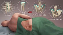
Bone marrow is a semi-solid tissue found within the spongy portions of bones. In birds and mammals, bone marrow is the primary site of new blood cell production. It is composed of hematopoietic cells, marrow adipose tissue, and supportive stromal cells. In adult humans, bone marrow is primarily located in the ribs, vertebrae, sternum, and bones of the pelvis. Bone marrow comprises approximately 5% of total body mass in healthy adult humans, such that a man weighing 73 kg (161 lbs) will have around 3.7 kg (8 lbs) of bone marrow.

<span class="mw-page-title-main">Thrombocytopenia</span> Abnormally low levels of platelets in the blood

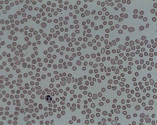
In hematology, thrombocytopenia is a condition characterized by abnormally low levels of platelets in the blood. Low levels of platelets in turn may lead to prolonged or excessive bleeding. It is the most common coagulation disorder among intensive care patients and is seen in a fifth of medical patients and a third of surgical patients.

<span class="mw-page-title-main">Esophagogastroduodenoscopy</span> Diagnostic endoscopic procedure

Esophagogastroduodenoscopy (EGD) or oesophagogastroduodenoscopy (OGD), also called by various other names, is a diagnostic endoscopic procedure that visualizes the upper part of the gastrointestinal tract down to the duodenum. It is considered a minimally invasive procedure since it does not require an incision into one of the major body cavities and does not require any significant recovery after the procedure. However, a sore throat is common.

<span class="mw-page-title-main">Plateletpheresis</span> Method of collecting platelets from blood

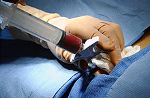
Plateletpheresis is the process of collecting thrombocytes, more commonly called platelets, a component of blood involved in blood clotting. The term specifically refers to the method of collecting the platelets, which is performed by a device used in blood donation that separates the platelets and returns other portions of the blood to the donor. Platelet transfusion can be a life-saving procedure in preventing or treating serious complications from bleeding and hemorrhage in patients who have disorders manifesting as thrombocytopenia or platelet dysfunction. This process may also be used therapeutically to treat disorders resulting in extraordinarily high platelet counts such as essential thrombocytosis.

Lymph node biopsy is a test in which a lymph node or a piece of a lymph node is removed for examination under a microscope.

<span class="mw-page-title-main">Fine-needle aspiration</span> Diagnostic medical procedure

Fine-needle aspiration (FNA) is a diagnostic procedure used to investigate lumps or masses. In this technique, a thin, hollow needle is inserted into the mass for sampling of cells that, after being stained, are examined under a microscope (biopsy). The sampling and biopsy considered together are called fine-needle aspiration biopsy (FNAB) or fine-needle aspiration cytology (FNAC). Fine-needle aspiration biopsies are very safe minor surgical procedures. Often, a major surgical biopsy can be avoided by performing a needle aspiration biopsy instead, eliminating the need for hospitalization. In 1981, the first fine-needle aspiration biopsy in the United States was done at Maimonides Medical Center. Today, this procedure is widely used in the diagnosis of cancer and inflammatory conditions. Fine needle aspiration is generally considered a safe procedure. Complications are infrequent.

<span class="mw-page-title-main">Endometrial biopsy</span> Diagnostic medical procedure

The endometrial biopsy is a medical procedure that involves taking a tissue sample of the lining of the uterus. The tissue subsequently undergoes a histologic evaluation which aids the physician in forming a diagnosis.

Arthrocentesis, or joint aspiration, is the clinical procedure performed to diagnose and, in some cases, treat musculoskeletal conditions. The procedure entails using a syringe to collect synovial fluid from or inject medication into the joint capsule. Laboratory analysis of synovial fluid can further help characterize the diseased joint and distinguish between gout, arthritis, and synovial infections such as septic arthritis.

<span class="mw-page-title-main">Skin biopsy</span> Removal of skin cells for medical examination

Skin biopsy is a biopsy technique in which a skin lesion is removed to be sent to a pathologist to render a microscopic diagnosis. It is usually done under local anesthetic in a physician's office, and results are often available in 4 to 10 days. It is commonly performed by dermatologists. Skin biopsies are also done by family physicians, internists, surgeons, and other specialties. However, performed incorrectly, and without appropriate clinical information, a pathologist's interpretation of a skin biopsy can be severely limited, and therefore doctors and patients may forgo traditional biopsy techniques and instead choose Mohs surgery.

<span class="mw-page-title-main">Basophilia</span> Medical condition

Basophilia is the condition of having greater than 200 basophils/μL in the venous blood. Basophils are the least numerous of the myelogenous cells, and it is rare for their numbers to be abnormally high without changes to other blood components. Rather, basophilia is most often coupled with other white blood cell conditions such as eosinophilia, high levels of eosinophils in the blood. Basophils are easily identifiable by a blue coloration of the granules within each cell, marking them as granulocytes, in addition to segmented nuclei.

Acute myelomonocytic leukemia (AMML) is a form of acute myeloid leukemia that involves a proliferation of CFU-GM myeloblasts and monoblasts. AMML occurs with a rapid increase amount in white blood cell count and is defined by more than 20% of myeloblast in the bone marrow. It is classified under "M4" in the French-American-British classification (FAB). It is classified under "AML, not otherwise classified" in the WHO classification.

<span class="mw-page-title-main">Congenital amegakaryocytic thrombocytopenia</span> Medical condition

Congenital amegakaryocytic thrombocytopenia (CAMT) is a rare autosomal recessive bone marrow failure syndrome characterized by severe thrombocytopenia, which can progress to aplastic anemia and leukemia. CAMT usually manifests as thrombocytopenia in the initial month of life or in the fetal phase. Typically CAMPT presents with petechiae, cerebral bleeds, recurrent rectal bleeding, or pulmonary hemorrhage.

<span class="mw-page-title-main">Renal biopsy</span> Removal of kidney tissue for medical examination

Renal biopsy is a medical procedure in which a small piece of kidney is removed from the body for examination, usually under a microscope. Microscopic examination of the tissue can provide information needed to diagnose, monitor or treat problems of the kidney.

Gestational (incidental) thrombocytopenia is a condition that commonly affects pregnant women. Thrombocytopenia is defined as the drop in platelet count from the normal range of 150,000–400,000/μL to a count lower than 150,000/μL. There is still ongoing research to determine the reason for the lowering of platelet count in women with a normal pregnancy. Some researchers speculate the cause to be dependent on dilution, decreased production of platelets, or an increased turnover event. Although women with normal pregnancy experience a low platelet count, women experiencing a continuous drop in platelet will be diagnosed with thrombocytopenia and women with levels greater than 70,000/μL will be diagnosed with gestational thrombocytopenia.

<span class="mw-page-title-main">Jamshidi needle</span>

The Jamshidi needle is a trephine needle for performing bone marrow biopsy, whereby a cylindrical sample of tissue, a core biopsy specimen, is obtained. It is a cylindrical needle with a tapered cutting tip. The tapered end reduces the potential of crush artifact. It is the most commonly used needle for performing bone marrow biopsies. The device is named for its inventor Khosrow Jamshidi who is an Iranian physician.

<span class="mw-page-title-main">Breast biopsy</span> Surgical diagnostic procedure for breast tumours

A breast biopsy is usually done after a suspicious lesion is discovered on either mammography or ultrasound to get tissue for pathological diagnosis. Several methods for a breast biopsy now exist. The most appropriate method of biopsy for a patient depends upon a variety of factors, including the size, location, appearance and characteristics of the abnormality. The different types of breast biopsies include fine-needle aspiration (FNA), vacuum-assisted biopsy, core needle biopsy, and surgical excision biopsy. Breast biopsies can be done utilizing ultrasound, MRI or a stereotactic biopsy imaging guidance. Vacuum assisted biopsies are typically done using stereotactic techniques when the suspicious lesion can only be seen on mammography. On average, 5–10 biopsies of a suspicious breast lesion will lead to the diagnosis of one case of breast cancer. Needle biopsies have largely replaced open surgical biopsies in the initial assessment of imaging as well as palpable abnormalities in the breast.

<span class="mw-page-title-main">Sampling (medicine)</span> Collection of bodily substances for medical assessment

In medicine, sampling is gathering of matter from the body to aid in the process of a medical diagnosis and/or evaluation of an indication for treatment, further medical tests or other procedures. In this sense, the sample is the gathered matter, and the sampling tool or sampler is the person or material to collect the sample.

Suprapubic aspiration is a procedure to take a urine sample. It involves putting a needle through the skin just above the pubic bone into the bladder. It is typically used as a method to collect urine in child less than 2 years of age who is not yet toilet trained in an effort to diagnose a urinary tract infection.

A bone biopsy is a procedure in which a small bone sample is removed from the outer layers of bone for examination, unlike a bone marrow biopsy, which involves the innermost part of the bone. The bone biopsy sample retains the architecture of bone when seen using histopathological examination slide.

References

  1. 1 2 eMedicine "Specialties > Hematology > Diagnostic Procedures > Bone Marrow Aspiration and Biopsy". Article last updated April 7, 2008.
  2. What to Expect During a Bone Marrow Aspirate and Biopsy Archived 2016-03-04 at the Wayback Machine , Roswell Park Comprehensive Cancer Center, n.d. Accessed 2014-08-08.
  3. Malempati, Suman; Joshi, Sarita; Lai, Susanna; Braner, Dana A.V.; Tegtmeyer, Ken (2009). "Bone Marrow Aspiration and Biopsy". New England Journal of Medicine. 361 (15): e28. doi:10.1056/NEJMvcm0804634. PMID   19812396.
  4. Bain BJ (2003). "Bone marrow biopsy morbidity and mortality". Br. J. Haematol. 121 (6): 949–51. doi: 10.1046/j.1365-2141.2003.04329.x . PMID   12786808.
  5. Bain BJ (2005). "Bone marrow biopsy morbidity: review of 2003". J. Clin. Pathol. 58 (4): 406–8. doi:10.1136/jcp.2004.022178. PMC   1770618 . PMID   15790706.
  6. 1 2 3 4 Parmentier S, Kramer M, Weller S, Schuler U, Ordemann R, Rall G; et al. (2020). "Reevaluation of reference values for bone marrow differential counts in 236 healthy bone marrow donors". Ann Hematol. 99 (12): 2723–2729. doi:10.1007/s00277-020-04255-4. PMC   7683448 . PMID   32935189.{{cite journal}}: CS1 maint: multiple names: authors list (link) Attribution 4.0 International (CC BY 4.0) license