Related Research Articles

Meiosis is a special type of cell division of germ cells in sexually-reproducing organisms that produces the gametes, such as sperm or egg cells. It involves two rounds of division that ultimately result in four cells with only one copy of each chromosome (haploid). Additionally, prior to the division, genetic material from the paternal and maternal copies of each chromosome is crossed over, creating new combinations of code on each chromosome. Later on, during fertilisation, the haploid cells produced by meiosis from a male and female will fuse to create a cell with two copies of each chromosome again, the zygote.

In cell biology, mitosis is a part of the cell cycle in which replicated chromosomes are separated into two new nuclei. Cell division by mitosis gives rise to genetically identical cells in which the total number of chromosomes is maintained. Therefore, mitosis is also known as equational division. In general, mitosis is preceded by S phase of interphase and is often followed by telophase and cytokinesis; which divides the cytoplasm, organelles and cell membrane of one cell into two new cells containing roughly equal shares of these cellular components. The different stages of mitosis altogether define the mitotic (M) phase of an animal cell cycle—the division of the mother cell into two daughter cells genetically identical to each other.

Microtubules are polymers of tubulin that form part of the cytoskeleton and provide structure and shape to eukaryotic cells. Microtubules can be as long as 50 micrometres, as wide as 23 to 27 nm and have an inner diameter between 11 and 15 nm. They are formed by the polymerization of a dimer of two globular proteins, alpha and beta tubulin into protofilaments that can then associate laterally to form a hollow tube, the microtubule. The most common form of a microtubule consists of 13 protofilaments in the tubular arrangement.
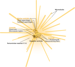
In cell biology, the centrosome is an organelle that serves as the main microtubule organizing center (MTOC) of the animal cell, as well as a regulator of cell-cycle progression. The centrosome provides structure for the cell. The centrosome is thought to have evolved only in the metazoan lineage of eukaryotic cells. Fungi and plants lack centrosomes and therefore use other structures to organize their microtubules. Although the centrosome has a key role in efficient mitosis in animal cells, it is not essential in certain fly and flatworm species.
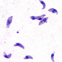
Toxoplasma gondii is an obligate intracellular parasitic protozoan that causes toxoplasmosis. Found worldwide, T. gondii is capable of infecting virtually all warm-blooded animals, but felids are the only known definitive hosts in which the parasite may undergo sexual reproduction.

In cell biology, the spindle apparatus refers to the cytoskeletal structure of eukaryotic cells that forms during cell division to separate sister chromatids between daughter cells. It is referred to as the mitotic spindle during mitosis, a process that produces genetically identical daughter cells, or the meiotic spindle during meiosis, a process that produces gametes with half the number of chromosomes of the parent cell.

Telophase is the final stage in both meiosis and mitosis in a eukaryotic cell. During telophase, the effects of prophase and prometaphase are reversed. As chromosomes reach the cell poles, a nuclear envelope is re-assembled around each set of chromatids, the nucleoli reappear, and chromosomes begin to decondense back into the expanded chromatin that is present during interphase. The mitotic spindle is disassembled and remaining spindle microtubules are depolymerized. Telophase accounts for approximately 2% of the cell cycle's duration.
A kinetochore is a disc-shaped protein structure associated with duplicated chromatids in eukaryotic cells where the spindle fibers attach during cell division to pull sister chromatids apart. The kinetochore assembles on the centromere and links the chromosome to microtubule polymers from the mitotic spindle during mitosis and meiosis. The term kinetochore was first used in a footnote in a 1934 Cytology book by Lester W. Sharp and commonly accepted in 1936. Sharp's footnote reads: "The convenient term kinetochore has been suggested to the author by J. A. Moore", likely referring to John Alexander Moore who had joined Columbia University as a freshman in 1932.

Aurora kinase A also known as serine/threonine-protein kinase 6 is an enzyme that in humans is encoded by the AURKA gene.
An apicoplast is a derived non-photosynthetic plastid found in most Apicomplexa, including Toxoplasma gondii, and Plasmodium falciparum and other Plasmodium spp., but not in others such as Cryptosporidium. It originated from algae through secondary endosymbiosis; there is debate as to whether this was a green or red alga. The apicoplast is surrounded by four membranes within the outermost part of the endomembrane system. The apicoplast hosts important metabolic pathways like fatty acid synthesis, isoprenoid precursor synthesis and parts of the heme biosynthetic pathway.

Serine/threonine-protein kinase Nek2 is an enzyme that in humans is encoded by the NEK2 gene.

Centromere protein F is a protein that in humans is encoded by the CENPF gene. It is involved in chromosome segregation during cell division. It also has a role in the orientation of microtubules to form cellular cilia.

Pericentrin (kendrin), also known as PCNT and pericentrin-B (PCNTB), is a protein which in humans is encoded by the PCNT gene on chromosome 21. This protein localizes to the centrosome and recruits proteins to the pericentriolar matrix (PCM) to ensure proper centrosome and mitotic spindle formation, and thus, uninterrupted cell cycle progression. This gene is implicated in many diseases and disorders, including congenital disorders such as microcephalic osteodysplastic primordial dwarfism type II (MOPDII) and Seckel syndrome.

Centrosome-associated protein CEP250 is a protein that in humans is encoded by the CEP250 gene. This gene encodes a core centrosomal protein required for centriole-centriole cohesion during interphase of the cell cycle. The encoded protein dissociates from the centrosomes when parental centrioles separate at the beginning of mitosis. The protein associates with and is phosphorylated by NIMA-related kinase 2, which is also associated with the centrosome. Furthermore, CEP135 is also required for the centriolar localization of CEP250.

Mitotic Catastrophe has been defined as either a cellular mechanism to prevent potentially cancerous cells from proliferating or as a mode of cellular death that occurs following improper cell cycle progression or entrance. Mitotic catastrophe can be induced by prolonged activation of the spindle assembly checkpoint, errors in mitosis, or DNA damage and functioned to prevent genomic instability. It is a mechanism that is being researched as a potential therapeutic target in cancers, and numerous approved therapeutics induce mitotic catastrophe.

Toll-like receptor 11 (TLR11) is a protein that in mice and rats is encoded by the gene TLR11, whereas in humans it is represented by a pseudogene. TLR11 belongs to the toll-like receptor (TLR) family and the interleukin-1 receptor/toll-like receptor superfamily. In mice, TLR11 has been shown to recognise (bacterial) flagellin and (eukaryotic) profilin present on certain microbes, it helps propagate a host immune response. TLR11 plays a fundamental role in both the innate and adaptive immune responses, through the activation of Tumor necrosis factor-alpha, the Interleukin 12 (IL-12) response, and Interferon-gamma (IFN-gamma) secretion. TLR11 mounts an immune response to multiple microbes, including Toxoplasma gondii, Salmonella species, and uropathogenic E. coli, and likely many other species due to the highly conserved nature of flagellin and profilin.
A series of biochemical switches control transitions between and within the various phases of the cell cycle. The cell cycle is a series of complex, ordered, sequential events that control how a single cell divides into two cells, and involves several different phases. The phases include the G1 and G2 phases, DNA replication or S phase, and the actual process of cell division, mitosis or M phase. During the M phase, the chromosomes separate and cytokinesis occurs.
MORN1 containing repeat 1, also known as Morn1, is a protein that in humans is encoded by the MORN1 gene.

Centrosomes are the major microtubule organizing centers (MTOC) in mammalian cells. Failure of centrosome regulation can cause mistakes in chromosome segregation and is associated with aneuploidy. A centrosome is composed of two orthogonal cylindrical protein assemblies, called centrioles, which are surrounded by a protein dense amorphous cloud of pericentriolar material (PCM). The PCM is essential for nucleation and organization of microtubules. The centrosome cycle is important to ensure that daughter cells receive a centrosome after cell division. As the cell cycle progresses, the centrosome undergoes a series of morphological and functional changes. Initiation of the centrosome cycle occurs early in the cell cycle in order to have two centrosomes by the time mitosis occurs.

The parasitophorous vacuole (PV) is a structure produced by apicomplexan parasites in the cells of its host. The PV allows the parasite to develop while protected from the phagolysosomes of the host cell.
References
- 1 2 3 Brooks CF, Francia ME, Gissot M, Croken MM, Kim K, Striepen B (March 2011). "Toxoplasma gondii sequesters centromeres to a specific nuclear region throughout the cell cycle". Proceedings of the National Academy of Sciences of the United States of America. 108 (9): 3767–3772. doi: 10.1073/pnas.1006741108 . PMC 3048097 . PMID 21321216.
- ↑ Naumov A, Kratzer S, Ting LM, Kim K, Suvorova ES, White MW (August 2017). "The Toxoplasma Centrocone Houses Cell Cycle Regulatory Factors". mBio. 8 (4): e00579–17. doi:10.1128/mBio.00579-17. PMC 5565962 . PMID 28830940.
- ↑ Gubbels MJ, Vaishnava S, Boot N, Dubremetz JF, Striepen B (June 2006). "A MORN-repeat protein is a dynamic component of the Toxoplasma gondii cell division apparatus". Journal of Cell Science. 119 (Pt 11): 2236–2245. doi:10.1242/jcs.02949. PMID 16684814.
- ↑ Farrell M, Gubbels MJ (January 2014). "The Toxoplasma gondii kinetochore is required for centrosome association with the centrocone (spindle pole)". Cellular Microbiology. 16 (1): 78–94. doi:10.1111/cmi.12185. PMC 3933516 . PMID 24015880.
| | This cell biology article is a stub. You can help Wikipedia by expanding it. |