Chameleon vision

Last updated

The chameleon is among the most highly visually-oriented lizards, using this sense in prey capture, mating behavior, and predator avoidance. [1] Unique features of chameleon vision include a negative lens, a positive cornea, and monocular focusing. The development of the chameleon visual system could have evolved to aid in prey capture and/or in predator avoidance.

Contents

Chameleon eye Chamaleon2.jpg
Chameleon eye

The angle, or amplitude, of eye movement in chameleons is very large for a vertebrate [1] and the eyes move independently of each other. [2] This allows a chameleon to watch an approaching object while simultaneously scanning the rest of its environment. [1] Chameleon eyes protrude laterally from the head, giving the lizard panoramic sight. [2] An eyelid fused to the pupil protects the eyes, leaving only a small part exposed. [2] With a negative (nearsighted or concave) lens and a positive (farsighted or convex) cornea, chameleons use a method of monocular focusing to judge distance called corneal accommodation. Each eye focuses independently, which is achieved by the chameleon eye’s unique anatomy of separated nodal and center points of the eye. [3] Finally, “striated rather than smooth ciliary muscle in sauropsids” allows for rapid focusing. [3]

Negative lens

Chameleon eyes feature a negative lens, meaning that the lens is concave. This increases retinal image size, allowing more precise focusing. [3] [4] In fact, image magnification in chameleons is higher in a scaled comparison to all other vertebrates eyes. [4]

Positive cornea

While the lens are negative, the cornea of chameleon eyes are positive, meaning that it is convex. The increased power of the cornea also contributes to more precise focusing than in other vertebrates. [3] The cornea improves sight resolution in a narrower field of vision. [3]

Monocular focusing and corneal accommodation

The combination of a negative lens and a positive cornea in the chameleon eye allow for accurate focusing by corneal accommodation. [4] Using corneal accommodation for depth perception [5] makes the chameleon the only vertebrate to focus monocularly. [1] While sight is primarily independent in the two chameleon eyes, the eye that first detects prey will guide accommodation in the other eye. [3] Contrary to the previous belief that chameleons used stereopsis (both eyes) for depth perception, research has shown monocular focusing to be more likely. [6] Depending on the chameleon's step in the predation sequence, corneal accommodation can be coupled, meaning the eyes independently focus on the same object. [3] When scanning the environment and in judging distance to prey, vision and accommodation are uncoupled: the eyes are focusing on different objects, such as the environment and the newly-sighted prey. Immediately before the chameleon's characteristic tongue is extended, accommodation in both eyes is coupled: both eyes focus independently on the prey. [3] Imprecise alignment of the images from each eye, as demonstrated by measuring various angles from eye to target, shows that stereopsis is unlikely for depth perception the chameleon. [3]

Nodal point separation

The nodal point in the eye is the point at which "lines connecting points in the scene and corresponding points in the image intersect." [5] In chameleons, the nodal point is located a significant distance before the center of rotation, the point around which the eye rotates in the eye socket. As a result of this nodal point separation, images of objects move more or less on the retina based on their distance from the chameleon. The position of an image on the retina is the "primary means by which chameleons judge distance." [3] Therefore, the rotation of one eye informs the chameleon of the "relative distances of different objects." [5] An important effect of the ability to judge distance with one eye is that the head does not have to be turned to allow stereoptic viewing of the object. [7]

Evolution

Chameleons as an evolutionary transition to stereopsis

A suggested theory for the evolution of squamate vision is that corneal accommodation and monocular depth perception are "primitive" mechanisms in comparison to binocular vision and stereopsis. [3] Chameleons use an alternative strategy to stereopsis in functional coupling of the eyes immediately before the tongue shot. This differs from stereopsis in that the images from both eyes are not reconciled into one. However, it is possible that this was first used for neural static reduction. [3] This suggests that chameleons could be seen as a transition between independent and coupled eye use. [8] However, it is also possible that the chameleon vision system is an alternative, equally successful mode of prey capture and predator avoidance, and perhaps more appropriate for the chameleon's niche as a camouflaged, arboreal hunter than other vision systems.

Prey/predator causes of chameleon eye development

The chameleon, a camouflaged, slow-moving lizard, is an arboreal hunter that hides and ambushes prey. [2] Prey and predators alike can be sighted and monitored using monocular depth perception. Also, nodal point separation allows distance to be judged with one eye, so minimal head movement is needed by the chameleon in watching its surroundings, reinforcing the chameleon strategy of inconspicuousness. [5]

Prey capture

The specialized strategy by which chameleons capture prey is reflected in their sensory anatomy and use, in particular that of vision. [2] First, prey is sighted and distance assessed using one eye. [6] To avoid detection by prey, a chameleon uses minimal head movement, made possible by nodal point separation. [5] The chameleon then slowly turns its head toward the prey. Both eyes focus independently on the prey before the tongue shot. [3]

Predator avoidance

The chameleon predator avoidance response is vision-mediated. [1] In predator avoidance, chameleons use minimal head movement and a unique method to monitor potential threats. Due to nodal point separation, a chameleon can judge distance to a potential threat with minimal head movement needed. When confronted with a potential threat, chameleons rotate their slender bodies to the opposite side of their perch to avoid detection. [1] They will keep moving around the branch to keep the branch between themselves and the threat and to keep the threat in their line of sight. [1] If the branch is narrow, a chameleon can observe a threat binocularly around the branch. While a wide branch might present a difficulty in depth perception to another lizard as it is forced to view the threat monocularly, a chameleon due to corneal accommodation and nodal point separation can judge distance between itself and a potential threat with only one eye viewing the threat. [1]

Comparison to the sandlance fish

While the chameleon eye is unique in lizards, parallels exist in other animals. In particular, the barred sandburrower fish shares key vision features with the chameleon. This is because the environmental circumstances such as the need for camouflaged quick prey capture that led to the development of the chameleon eye seem to have acted on the sandburrower fish as well. [7] Rapid predatory attacks are made possible through the chameleon and the sandlances' striated corneal muscles allowing for corneal accommodation, a reduced power lens, and increased corneal power. [3] A nearly complete eyelid covering and lack of head movement due to nodal separation reduce conspicuousness to prey and predators. [7]

Related Research Articles

<span class="mw-page-title-main">Eye</span> Organ that detects light and converts it into electro-chemical impulses in neurons

An eye is a sensory organ that allows an organism to perceive visual information. It detects light and converts it into electro-chemical impulses in neurons (neurones). It is part of an organism's visual system.

<span class="mw-page-title-main">Binocular vision</span> Type of vision with two eyes facing the same direction

In biology, binocular vision is a type of vision in which an animal has two eyes capable of facing the same direction to perceive a single three-dimensional image of its surroundings. Binocular vision does not typically refer to vision where an animal has eyes on opposite sides of its head and shares no field of view between them, like in some animals.

<span class="mw-page-title-main">Stereoscopy</span> Technique for creating or enhancing the illusion of depth in an image

Stereoscopy is a technique for creating or enhancing the illusion of depth in an image by means of stereopsis for binocular vision. The word stereoscopy derives from Greek στερεός (stereos) 'firm, solid', and σκοπέω (skopeō) 'to look, to see'. Any stereoscopic image is called a stereogram. Originally, stereogram referred to a pair of stereo images which could be viewed using a stereoscope.

<span class="mw-page-title-main">Depth perception</span> Visual ability to perceive the world in 3D

Depth perception is the ability to perceive distance to objects in the world using the visual system and visual perception. It is a major factor in perceiving the world in three dimensions. Depth perception happens primarily due to stereopsis and accommodation of the eye.

<span class="mw-page-title-main">Visual system</span> Body parts responsible for vision

The visual system is the physiological basis of visual perception. The system detects, transduces and interprets information concerning light within the visible range to construct an image and build a mental model of the surrounding environment. The visual system is associated with the eye and functionally divided into the optical system and the neural system.

<span class="mw-page-title-main">Farsightedness</span> Eye condition in which light is focused behind instead of on the retina

Far-sightedness, also known as long-sightedness, hypermetropia, and hyperopia, is a condition of the eye where distant objects are seen clearly but near objects appear blurred. This blur is due to incoming light being focused behind, instead of on, the retina due to insufficient accommodation by the lens. Minor hypermetropia in young patients is usually corrected by their accommodation, without any defects in vision. But, due to this accommodative effort for distant vision, people may complain of eye strain during prolonged reading. If the hypermetropia is high, there will be defective vision for both distance and near. People may also experience accommodative dysfunction, binocular dysfunction, amblyopia, and strabismus. Newborns are almost invariably hypermetropic, but it gradually decreases as the newborn gets older.

<span class="mw-page-title-main">Autostereogram</span> Visual illusion of 3D scene achieved by unfocusing eyes when viewing specific 2D images

An autostereogram is a two-dimensional (2D) image that can create the optical illusion of a three-dimensional (3D) scene. Autostereograms use only one image to accomplish the effect while normal stereograms require two. The 3D scene in an autostereogram is often unrecognizable until it is viewed properly, unlike typical stereograms. Viewing any kind of stereogram properly may cause the viewer to experience vergence-accommodation conflict.

<span class="mw-page-title-main">Intraocular lens</span> Lens implanted in the eye to treat cataracts or myopia

An Intraocular lens (IOL) is a lens implanted in the eye usually as part of a treatment for cataracts or for correcting other vision problems such as short sightedness and long sightedness; a form of refractive surgery. If the natural lens is left in the eye, the IOL is known as phakic, otherwise it is a pseudophakic lens. Both kinds of IOLs are designed to provide the same light-focusing function as the natural crystalline lens. This can be an alternative to LASIK, but LASIK is not an alternative to an IOL for treatment of cataracts.

<span class="mw-page-title-main">Eye examination</span> Series of tests assessing vision and pertaining to the eyes

An eye examination is a series of tests performed to assess vision and ability to focus on and discern objects. It also includes other tests and examinations pertaining to the eyes. Eye examinations are primarily performed by an optometrist, ophthalmologist, or an orthoptist. Health care professionals often recommend that all people should have periodic and thorough eye examinations as part of routine primary care, especially since many eye diseases are asymptomatic.

Stereopsis is the component of depth perception retrieved by means of binocular disparity through binocular vision. It is not the only contributor to depth perception, but it is a major one. Binocular vision occurs because each eye receives a different image due to their slightly different positions in one's head. These positional differences are referred to as "horizontal disparities" or, more generally, "binocular disparities". Disparities are processed in the visual cortex of the brain to yield depth perception. While binocular disparities are naturally present when viewing a real three-dimensional scene with two eyes, they can also be simulated by artificially presenting two different images separately to each eye using a method called stereoscopy. The perception of depth in such cases is also referred to as "stereoscopic depth".

Monocular vision is vision using only one eye. It is seen in two distinct categories: either a species moves its eyes independently, or a species typically uses two eyes for vision, but is unable to use one due to circumstances such as injury.

<span class="mw-page-title-main">Equine vision</span>

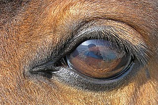
The equine eye is one of the largest of any land mammal. Its visual abilities are directly related to the animal's behavior; for example, it is active during both day and night, and it is a prey animal. Both the strengths and weaknesses of the horse's visual abilities should be taken into consideration when training the animal, as an understanding of the horse's eye can help to discover why the animal behaves the way it does in various situations.

<span class="mw-page-title-main">Vision in toads</span>

The neural basis of prey detection, recognition, and orientation was studied in depth by Jörg-Peter Ewert in a series of experiments that made the toad visual system a model system in neuroethology. He began by observing the natural prey catching behavior of the common European toad.

<span class="mw-page-title-main">Chromostereopsis</span> Visual illusion whereby the impression of depth is conveyed in two-dimensional color images

Chromostereopsis is a visual illusion whereby the impression of depth is conveyed in two-dimensional color images, usually of red–blue or red–green colors, but can also be perceived with red–grey or blue–grey images. Such illusions have been reported for over a century and have generally been attributed to some form of chromatic aberration.

Vision of humans and other organisms depends on several organs such as the lens of the eye, and any vision correcting devices, which use optics to focus the image.

<span class="mw-page-title-main">Vision in fish</span> Sense found in most species of fish

Vision is an important sensory system for most species of fish. Fish eyes are similar to the eyes of terrestrial vertebrates like birds and mammals, but have a more spherical lens. Birds and mammals normally adjust focus by changing the shape of their lens, but fish normally adjust focus by moving the lens closer to or further from the retina. Fish retinas generally have both rod cells and cone cells, and most species have colour vision. Some fish can see ultraviolet and some are sensitive to polarised light.

The eye, like any other optical system, suffers from a number of specific optical aberrations. The optical quality of the eye is limited by optical aberrations, diffraction and scatter. Correction of spherocylindrical refractive errors has been possible for nearly two centuries following Airy's development of methods to measure and correct ocular astigmatism. It has only recently become possible to measure the aberrations of the eye and with the advent of refractive surgery it might be possible to correct certain types of irregular astigmatism.

<span class="mw-page-title-main">Eagle eye</span>

The eagle eye is among the sharpest in the animal kingdom, with an eyesight estimated at 4 to 8 times stronger than that of the average human. Although an eagle may only weigh 10 pounds (4.5 kg), its eyes are roughly the same size as those of a human. Eagle weight varies: a small eagle could weigh 700 grams (1.5 lb), while a larger one could weigh 6.5 kilograms (14 lb); an eagle of about 10 kilograms (22 lb) weight could have eyes as big as that of a human who weighs 200 pounds (91 kg). Although the size of the eagle eye is about the same as that of a human being, the back side shape of the eagle eye is flatter. Their eyes are stated to be larger in size than their brain, by weight. Color vision with resolution and clarity are the most prominent features of eagles' eyes, hence sharp-sighted people are sometimes referred to as "eagle-eyed". Eagles can identify five distinctly colored squirrels and locate their prey even if hidden.

<i>Limnichthys fasciatus</i> Species of fish

Limnichthys fasciatus, the barred sand burrower, is a species of sandburrower. It is noted for its highly developed eyes, with a structure similar to the eyes of a chameleon, which has led it to be described as marine chameleon. Its fully grown length measures between 20 millimetres (0.79 in) and 40 millimetres (1.6 in). The species is native to reefs in the Indo-Pacific. The fish preys on plankton prey by surprise attacking it from a hiding in loose sand, with only the eyes protruding from the sand.

<span class="mw-page-title-main">Vergence-accommodation conflict</span> Visual and perceptual phenomenon

Vergence-accommodation conflict (VAC), also known as accommodation-vergence conflict, is a visual phenomenon that occurs when the brain receives mismatching cues between vergence and accommodation of the eye. This commonly occurs in virtual reality devices, augmented reality devices, 3D movies, and other types of stereoscopic displays and autostereoscopic displays. The effect can be unpleasant and cause eye strain.

References

  1. 1 2 3 4 5 6 7 8 Lustig, Avichai; Hadas Keter-Katz; Gadi Katzir (2012). "Threat perception in the chameleon (Chamaeleo chameleon): evidence for lateralized eye use". Animal Cognition. 15 (4): 609–621. doi:10.1007/s10071-012-0489-7. PMID   22460630.
  2. 1 2 3 4 5 Sandor, Peter S.; Maarten A. Frens; Volker Henn (2001). "Chameleon eye position obeys Listing's law". Vision Research. 41 (17): 2245–2251. doi: 10.1016/s0042-6989(01)00111-0 . PMID   11448716.
  3. 1 2 3 4 5 6 7 8 9 10 11 12 13 14 Ott, M.; F. Schaeffel; W. Kirmse (1998). "Binocular vision and accommodation in prey-catching chameleons". Journal of Comparative Physiology A. 182 (3): 319–330. doi:10.1007/s003590050182.
  4. 1 2 3 Ott, Matthias; Frank Schaeffel (1995). "A negatively powered lens in the chameleon". Nature. 373 (6516): 692–694. Bibcode:1995Natur.373..692O. doi:10.1038/373692a0. PMID   7854450.
  5. 1 2 3 4 5 Srinivasan, Mandyam V. (1999). "Ecology: When one eye is better than two". Nature. 399 (6734): 305–307. Bibcode:1999Natur.399..305S. doi:10.1038/20550.
  6. 1 2 Harkness, Lindesay (1977). "Chameleons use accommodation clues to judge distance". Nature. 267 (5609): 346–349. Bibcode:1977Natur.267..346H. doi:10.1038/267346a0.
  7. 1 2 3 Pettigrew, John D.; Shaun P. Collin; Matthias Ott (1999). "Convergence of specialised behaviour, eye movements and visual optics in the sandlance (Teleostei) and the chameleon (Reptilia)". Current Biology. 9 (8): 421–424. Bibcode:1999CBio....9..421P. doi: 10.1016/s0960-9822(99)80189-4 .
  8. Ott, Matthias (2001). "Chameleons have independent eye movements but synchronise both eyes during saccadic prey tracking". Experimental Brain Research. 139 (2): 173–179. doi:10.1007/s002210100774.