Related Research Articles

Microscopy is the technical field of using microscopes to view objects and areas of objects that cannot be seen with the naked eye. There are three well-known branches of microscopy: optical, electron, and scanning probe microscopy, along with the emerging field of X-ray microscopy.

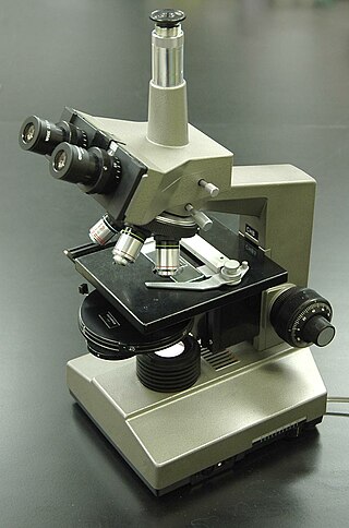
The optical microscope, also referred to as a light microscope, is a type of microscope that commonly uses visible light and a system of lenses to generate magnified images of small objects. Optical microscopes are the oldest design of microscope and were possibly invented in their present compound form in the 17th century. Basic optical microscopes can be very simple, although many complex designs aim to improve resolution and sample contrast.
Interferometry is a technique which uses the interference of superimposed waves to extract information. Interferometry typically uses electromagnetic waves and is an important investigative technique in the fields of astronomy, fiber optics, engineering metrology, optical metrology, oceanography, seismology, spectroscopy, quantum mechanics, nuclear and particle physics, plasma physics, biomolecular interactions, surface profiling, microfluidics, mechanical stress/strain measurement, velocimetry, optometry, and making holograms.

Atomic force microscopy (AFM) or scanning force microscopy (SFM) is a very-high-resolution type of scanning probe microscopy (SPM), with demonstrated resolution on the order of fractions of a nanometer, more than 1000 times better than the optical diffraction limit.
In physics, coherence expresses the potential for two waves to interfere. Two monochromatic beams from a single source always interfere. Physical sources are not strictly monochromatic: they may be partly coherent. Beams from different sources are mutually incoherent.

The Mach–Zehnder interferometer is a device used to determine the relative phase shift variations between two collimated beams derived by splitting light from a single source. The interferometer has been used, among other things, to measure phase shifts between the two beams caused by a sample or a change in length of one of the paths. The apparatus is named after the physicists Ludwig Mach and Ludwig Zehnder; Zehnder's proposal in an 1891 article was refined by Mach in an 1892 article. Demonstrations of Mach–Zehnder interferometry with particles other than photons had been demonstrated as well in multiple experiments.

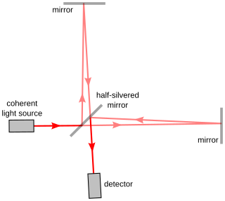
The Michelson interferometer is a common configuration for optical interferometry and was invented by the 19/20th-century American physicist Albert Abraham Michelson. Using a beam splitter, a light source is split into two arms. Each of those light beams is reflected back toward the beamsplitter which then combines their amplitudes using the superposition principle. The resulting interference pattern that is not directed back toward the source is typically directed to some type of photoelectric detector or camera. For different applications of the interferometer, the two light paths can be with different lengths or incorporate optical elements or even materials under test.
Cooke, Troughton & Simms was a British instrument-making firm formed in York in 1922 by the merger of T. Cooke & Sons and Troughton & Simms.

Confocal microscopy, most frequently confocal laser scanning microscopy (CLSM) or laser scanning confocal microscopy (LSCM), is an optical imaging technique for increasing optical resolution and contrast of a micrograph by means of using a spatial pinhole to block out-of-focus light in image formation. Capturing multiple two-dimensional images at different depths in a sample enables the reconstruction of three-dimensional structures within an object. This technique is used extensively in the scientific and industrial communities and typical applications are in life sciences, semiconductor inspection and materials science.
Differential interference contrast (DIC) microscopy, also known as Nomarski interference contrast (NIC) or Nomarski microscopy, is an optical microscopy technique used to enhance the contrast in unstained, transparent samples. DIC works on the principle of interferometry to gain information about the optical path length of the sample, to see otherwise invisible features. A relatively complex optical system produces an image with the object appearing black to white on a grey background. This image is similar to that obtained by phase contrast microscopy but without the bright diffraction halo. The technique was invented by Francis Hughes Smith. The "Smith DIK" was produced by Ernst Leitz Wetzlar in Germany and was difficult to manufacture. DIC was then developed further by Polish physicist Georges Nomarski in 1952.
Phase-contrast microscopy (PCM) is an optical microscopy technique that converts phase shifts in light passing through a transparent specimen to brightness changes in the image. Phase shifts themselves are invisible, but become visible when shown as brightness variations.

The Jamin interferometer is a type of interferometer, related to the Mach–Zehnder interferometer. It was developed in 1856 by the French physicist Jules Jamin.
Acoustic microscopy is microscopy that employs very high or ultra high frequency ultrasound. Acoustic microscopes operate non-destructively and penetrate most solid materials to make visible images of internal features, including defects such as cracks, delaminations and voids.
The N-slit interferometer is an extension of the double-slit interferometer also known as Young's double-slit interferometer. One of the first known uses of N-slit arrays in optics was illustrated by Newton. In the first part of the twentieth century, Michelson described various cases of N-slit diffraction.

Interference reflection microscopy (IRM), also called Reflection Interference Contrast Microscopy (RICM) or Reflection Contrast Microscopy (RCM) depending on the context, is an optical microscopy technique that leverages interference effects to form an image of an object on a glass surface. The intensity of the signal is a measure of proximity of the object to the glass surface. This technique can be used to study events at the cell membrane without the use of a (fluorescent) label as is the case for TIRF microscopy.
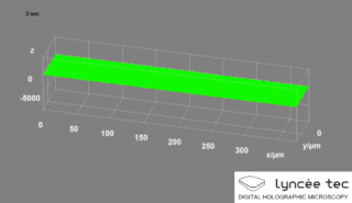
Digital holographic microscopy (DHM) is digital holography applied to microscopy. Digital holographic microscopy distinguishes itself from other microscopy methods by not recording the projected image of the object. Instead, the light wave front information originating from the object is digitally recorded as a hologram, from which a computer calculates the object image by using a numerical reconstruction algorithm. The image forming lens in traditional microscopy is thus replaced by a computer algorithm. Other closely related microscopy methods to digital holographic microscopy are interferometric microscopy, optical coherence tomography and diffraction phase microscopy. Common to all methods is the use of a reference wave front to obtain amplitude (intensity) and phase information. The information is recorded on a digital image sensor or by a photodetector from which an image of the object is created (reconstructed) by a computer. In traditional microscopy, which do not use a reference wave front, only intensity information is recorded and essential information about the object is lost.
Self-mixing or back-injection laser interferometry is an interferometric technique in which a part of the light reflected by a vibrating target is reflected into the laser cavity, causing a modulation both in amplitude and in frequency of the emitted optical beam. In this way, the laser becomes sensitive to the distance traveled by the reflected beam thus becoming a distance, speed or vibration sensor. The advantage compared to a traditional measurement system is a lower cost thanks to the absence of collimation optics and external photodiodes.
A common-path interferometer is a class of interferometers in which the reference beam and sample beams travel along the same path. Examples include the Sagnac interferometer, Zernike phase-contrast interferometer, and the point diffraction interferometer. A common-path interferometer is generally more robust to environmental vibrations than a "double-path interferometer" such as the Michelson interferometer or the Mach–Zehnder interferometer. Although travelling along the same path, the reference and sample beams may travel along opposite directions, or they may travel along the same direction but with the same or different polarization.
Quantum microscopy allows microscopic properties of matter and quantum particles to be measured and imaged. Various types of microscopy use quantum principles. The first microscope to do so was the scanning tunneling microscope, which paved the way for development of the photoionization microscope and the quantum entanglement microscope.
References
- ↑ Dyson J. (1950). "An Interferometer Microscope". Proceedings of the Royal Society A . 204 (1077): 170–187. Bibcode:1950RSPSA.204..170D. doi:10.1098/rspa.1950.0167. S2CID 121877024.
- ↑ Smith F. H. (1954). "Two Half-Shade Devices for Optical Polarizing Instruments". Nature. 173 (4399): 362–363. Bibcode:1954Natur.173..362S. doi:10.1038/173362b0. S2CID 4176399.
- ↑ Smith F. H. (1955). "Microscopic interferometry". Research. 8: 385–395.
- ↑ Huxley, A. F.; Niedergerke, R. (1954). "Structural changes in muscle during contraction; interference microscopy of living muscle fibres". Nature. 173 (4412): 971–973. Bibcode:1954Natur.173..971H. doi:10.1038/173971a0. PMID 13165697. S2CID 4275495.
- ↑ Zicha, D.; Genot, E.; Dunn, G. A.; Kramer, I. M. (1999). "TGFbeta1 induces a cell-cycle-dependent increase in motility of epithelial cells". Journal of Cell Science. 112 (4): 447–454. doi:10.1242/jcs.112.4.447. PMID 9914157.
- ↑ Mahlmann, Daniel M.; Jahnke, Joachim; Loosen, Peter (2008). "Rapid determination of the dry weight of single, living cyanobacterial cells using the Mach–Zehnder double-beam interference microscope". Eur. J. Phycol. 43 (4): 355–364. doi: 10.1080/09670260802168625 . S2CID 84728819.
- ↑ Kaul, R.A.; Mahlmann, D.M.; Loosen, P. (2010). "Mach–Zehnder interference microscopy optically records electrically stimulated cellular activity in unstained nerve cells". Journal of Microscopy. 240 (1): 60–74. doi:10.1111/j.1365-2818.2010.03385.x. PMID 21050214. S2CID 40054949.
- ↑ de Groot, P (2015). "Principles of interference microscopy for the measurement of surface topography". Advances in Optics and Photonics. 7 (1): 1–65. Bibcode:2015AdOP....7....1D. doi:10.1364/AOP.7.000001.