Cnidocyte

Last updated
Nomarski micrograph of a ruthenium red-stained nematocyst from Aiptasia pallida, the pale anemone. The red dye stains the polyanionic venom proteins found inside the capsule of this partially-discharged nematocyst. Nematocyst1.jpg
Nomarski micrograph of a ruthenium red-stained nematocyst from Aiptasia pallida, the pale anemone. The red dye stains the polyanionic venom proteins found inside the capsule of this partially-discharged nematocyst.

A cnidocyte (also known as a cnidoblast) is an explosive cell containing one large secretory organelle called a cnidocyst (also known as a cnida (pl.: cnidae)) that can deliver a sting to other organisms. The presence of this cell defines the phylum Cnidaria (corals, sea anemones, hydrae, jellyfish, etc.). Cnidae are used to capture prey and as a defense against predators. A cnidocyte fires a structure that contains a toxin within the cnidocyst; this is responsible for the stings delivered by a cnidarian. Cnidocytes are single-use cells that need to be continuously replaced.

Contents

Structure and function

Each cnidocyte contains an organelle called a cnida, cnidocyst, nematocyst, ptychocyst or spirocyst. This organelle consists of a bulb-shaped capsule containing a coiled hollow tubule structure attached to it. An immature cnidocyte is referred to as a cnidoblast or nematoblast. The externally oriented side of the cell has a hair-like trigger called a cnidocil, which is a mechano- and chemo-receptor. When the trigger is activated, the tubule shaft of the cnidocyst is ejected and, in the case of the penetrant nematocyst, the forcefully ejected tubule penetrates the target organism. This discharge takes a few microseconds, and is able to reach accelerations of about 40,000  g. [1] [2] Research from 2006 suggests the process occurs in as little as 700 nanoseconds, thus reaching an acceleration of up to 5,410,000 g. [3] After penetration, the toxic content of the nematocyst is injected into the target organism, allowing the sessile cnidarian to capture the immobilized prey. Recently, in two sea anemone species ( Nematostella vectensis and Anthopleura elegantissima), the type I neurotoxin protein Nv1 was shown to be localized in ectodermal gland cells in the tentacles, next to but not in nematocytes. Upon encounter with a crustacean prey, nematocytes discharge and pierce the prey, and Nv1 is massively secreted into the extracellular medium by the nearby gland cells, thus suggesting another mode of entry for toxins. [4]

Cnidocyte capsule composition

The cnidocyte capsule is made of novel Cnidaria-specific genes which combine known protein domains. Minicollagen genes are one of the major structural components of the capsule. They are very short genes containing the characteristic collagen-triple helix sequence, as well as polyproline domains and cystein-rich domains. [5] Trimers of minicollagen proteins assemble through their terminal cystein-rich domain, forming highly organized and rigid supra-structures. Minicollagen 1 Ncol-1 polymers assemble on the inner shell while the outer capsule is composed of polymerized NOWA (Nematocyst Outer Wall Antigen) proteins. Nematogalectin, minicollagen Ncol-15 and chondroitin are novel proteins used to build the tubule shaft. In piercing cnidocytes, the novel protein spinalin is used to make the spines present at the base of the shaft. [6] [7] [8]

Discharge mechanism

Discharge mechanism of a nematocyst Fluids-05-00020-g002.png
Discharge mechanism of a nematocyst

The cnidocyst capsule stores a large concentration of calcium ions, which are released from the capsule into the cytoplasm of the cnidocyte when the trigger is activated. This causes a large concentration gradient of calcium across the cnidocyte plasma membrane. The resulting osmotic pressure causes a rapid influx of water into the cell. This increase in water volume in the cytoplasm forces the coiled cnidae tubule to eject rapidly. Prior to discharge the coiled cnidae tubule exists inside the cell in an "inside out" condition. The back pressure resulting from the influx of water into the cnidocyte together with the opening of the capsule tip structure or operculum, triggers the forceful eversion of the cnidae tubule causing it to right itself as it comes rushing out of the cell with enough force to impale a prey organism.

That force is to be calculated as the mass of the mechanism's stylet multiplied by its acceleration. The pressure that is generated by this impact into its prey is to be calculated by the stylet's force divided by its area. Researchers have calculated an ejected mass of 1 nanogram, an acceleration of 5,410,000 g and a stylet tip radius of 15 ± 8 nm. [3] Therefore, a pressure of more than 7  GPa was estimated at the stylet tip which they write is in the range of technical bullets. [3]

Fluid dynamics in nematocyst discharge

Computational fluid dynamics model parameters of nematocyst discharge Model Overview.png
Computational fluid dynamics model parameters of nematocyst discharge

Few papers have modeled the discharge aside from direct observation. Observational studies typically used a tentacle solution assay with a chemical stimulant to create discharge and cameras to record them. One in 1984 [1] and another in 2006 [3] as imaging technology improved. One study involved computational fluid dynamics where variables such as barb plate size, prey cylindrical diameter and fluid medium Reynolds number were manipulated. [9]

Observational studies indicate that velocities of the barb/stylet decrease throughout the discharge. As such, the incredible maximum acceleration is achieved at the beginning. Dynamic traits such as maximum discharge velocities and trajectory patterns may not correspond to static traits such as tubule lengths and capsule volumes. [10] Therefore, caution is appropriate when using medusan nematocyst assemblages as indicators of prey selection and trophic role. [10] This is possibly the case for other jelly species and hence one cannot generally infer nematocyst static traits to prey size.

Prey detection

Cnidae are "single use" cells, and thus represent a large expenditure of energy to produce. In Hydrozoans, in order to regulate discharge, cnidocytes are connected as "batteries", containing several types of cnidocytes connected to supporting cells and neurons. The supporting cells contain chemosensors, which, together with the mechanoreceptor on the cnidocyte (cnidocil), allow only the right combination of stimuli to cause discharge, such as prey swimming, and chemicals found in prey cuticle or cutaneous tissue. This prevents the cnidarian from stinging itself although sloughed off cnidae can be induced to fire independently.

Types of cnidae

Over 30 types of cnidae are found in different cnidarians. They can be divided into the following groups:

  1. Nematocyst (Penetrant or Piercing [11] ): The penetrant or stenotele is the largest and most complex nematocyst. When discharged, it pierces the skin or chitinous exoskeleton of the prey and injects the venomous fluid, hypnotoxin, that either paralyzes the victim or kills it.
  2. Ptychocysts (Glutinant or Adhesive [11] ): a sticky surface used to stick to prey, referred to as ptychocysts and found on burrowing (tube) anemones, which help create the tube in which the animal lives
  3. Spirocyte (Volvent or Ensnaring [11] ): The volvent or desmoneme is a small and pear-shaped cnidocyte. It contains a short, thick, spineless, smooth and elastic thread tube forming a single loop and closed at the far end. When discharged, it tightly coils around the prey. They are the smallest cnidocytes. A lasso-like string is fired at prey and wraps around a cellular projection on the prey, which are referred to as spirocysts.

Cnidocyte subtypes can be differentially localized in the animal. In the sea anemone Nematostella vectensis , the majority of its non-penetrant sticky cnidocytes, the spirocytes, are found in the tentacles, and are thought to help with prey capture by sticking to the prey. By contrast, the two penetrant types of cnidocytes present in this species display a much broader localization, on the outer epithelial layer of the tentacles and body column, as well as on the pharynx epithelium and within mesenteries. [12]

The diversity of cnidocytes types correlates with the expansion and diversification of structural cnidocyst genes like minicollagen genes. [13] Minicollagen genes form compact gene clusters in Cnidarian genomes, suggesting a diversification through gene duplication and subfunctionalization. Anthozoans display less capsule diversity and a reduced number of minicollagen genes, and medusozoans have more capsule diversity (about 25 types) and a vastly expanded minicollagen genes repertoire. [13] In the sea anemone Nematostella vectensis , some minicollagens display a differential expression pattern in different cnidocytes subtypes. [12] [14]

Cnidocyte development

Cnidocytes are single-use cells that need to be continuously replaced throughout the life of the animal with different mode of renewal across species.

Modes of renewal

Overview of the development of the 4 different capsule types of Hydra polyps Cnidocyte development in hydra.png
Overview of the development of the 4 different capsule types of Hydra polyps

In Hydra polyps, cnidocytes differentiate from a specific population of stem cells, the interstitial cells (I-cells) located within the body column. Developing nematocytes first undergo multiple rounds of mitosis without cytokinesis, giving rise to nematoblast nests with 8, 16, 32 or 64 cells. After this expansion phase, nematoblasts develop their capsules. Nests separate into single nematocytes when the formation of the capsule is complete. [5] Most of them migrate to the tentacles where they are incorporated into battery cells, which hold several nematocytes, and neurons. Battery cells coordinate firing of nematocytes.

In the hydrozoan jellyfish Clytia hemisphaerica , nematogenesis takes place at the base of the tentacles, as well as in the manubrium. At the base of the tentacles, nematoblasts proliferate then differentiate along a proximal-distal gradient, giving rise to mature nematocytes in the tentacles through a conveyor belt system. [15]

In the Anthozoan sea anemone Nematostella vectensis , nematocytes are thought to develop throughout the animal from epithelial progenitors. [16] Furthermore, a single regulatory gene that codes for the transcription factor ZNF845 also called CnZNF1 promotes the development of a cnidocyte and inhibits the development of a RFamide producing neuron cell. [17] This gene evolved in the stem cnidarian through domain shuffling. [17]

Cnidocyst maturation

The nematocyst forms through a multi-step assembly process from a giant post-Golgi vacuole. Vesicles from the Golgi apparatus first fuse onto a primary vesicle: the capsule primordium. Subsequent vesicle fusion enables the formation of a tubule outside of the capsule, which then invaginates into the capsule. Then, an early maturation phase enables the formation of long arrays of barbed spines onto the invaginated tubule through the condensation of spinalin proteins. Finally, a late maturation stage gives rise to undischarged capsules under high osmotic pressure through the synthesis of poly-γ-glutamate into the matrix of the capsule. This trapped osmotic pressure enables rapid thread discharge upon triggering through a massive osmotic shock. [8]

Nematocyst toxicity

Nematocysts from Chironex fleckeri (400x magnification) Chironex fleckeri nematocysts 01.jpg
Nematocysts from Chironex fleckeri (400x magnification)

Nematocysts are very efficient weapons. A single nematocyst has been shown to suffice in paralyzing a small arthropod ( Drosophila larva). The most deadly cnidocytes (to humans, at least) are found on the body of a box jellyfish. [18] [19] [20] One member of this family, the sea wasp, Chironex fleckeri , is "claimed to be the most venomous marine animal known," according to the Australian Institute of Marine Science. It can cause excruciating pain to humans, sometimes followed by death. Other cnidarians, such as the jellyfish Cyanea capillata (the "Lion's Mane" made famous by Sherlock Holmes) or the siphonophore Physalia physalis (Portuguese man o' war, "Bluebottle") can cause extremely painful and sometimes fatal stings. On the other hand, aggregating sea anemones may have the lowest sting intensity, perhaps due to the inability of the nematocysts to penetrate the skin, creating a feeling similar to touching sticky candies. Besides feeding and defense, sea anemone and coral colonies use cnidocytes to sting one another in order to defend or win space. [21] Despite their effectiveness in prey-predator interactions, there is an evolutionary tradeoff as cnidarian venom systems are known to reduce the cnidarian's reproductive fitness and overall growth. [22]

Venom from animals such as cnidarians, scorpions and spiders may be species-specific. A substance that is weakly toxic for humans or other mammals may be strongly toxic to the natural prey or predators of the venomous animal. Such specificity has been used to create new medicines and bioinsecticides, and biopesticides.

Animals in the phylum Ctenophora ("sea-gooseberries" or "comb jellies") are transparent and jelly-like but have no nematocysts, and are harmless to humans.

Certain types of sea slugs, such as the nudibranch aeolids, are known to undergo kleptocnidy (in addition to kleptoplasty), whereby the organisms store nematocysts of digested prey at the tips of their cerata.

See also

Related Research Articles

<span class="mw-page-title-main">Cnidaria</span> Aquatic animal phylum having cnydocytes

Cnidaria is a phylum under kingdom Animalia containing over 11,000 species of aquatic animals found both in fresh water and marine environments, including jellyfish, hydroids, sea anemones, corals and some of the smallest marine parasites. Their distinguishing features are a decentralized nervous system distributed throughout a gelatinous body and the presence of cnidocytes or cnidoblasts, specialized cells with ejectable flagella used mainly for envenomation and capturing prey. Their bodies consist of mesoglea, a non-living, jelly-like substance, sandwiched between two layers of epithelium that are mostly one cell thick. Cnidarians are also some of the only animals that can reproduce both sexually and asexually.

<i>Hydra</i> (genus) Genus of cnidarians

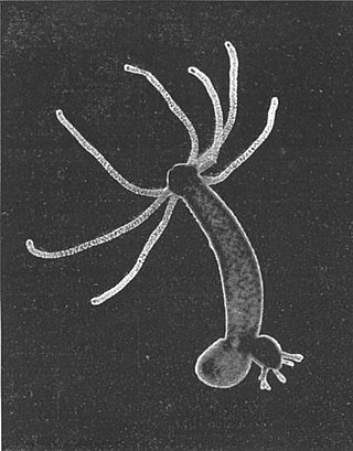
Hydra is a genus of small freshwater hydrozoans of the phylum Cnidaria. They are native to the temperate and tropical regions. The genus was named by Linnaeus in 1758 after the Hydra, which was the many-headed beast of myth defeated by Heracles, as when the animal has a part severed, it will regenerate much like the mythical hydra’s heads. Biologists are especially interested in Hydra because of their regenerative ability; they do not appear to die of old age, or to age at all.

<span class="mw-page-title-main">Venom</span> Toxin secreted by an animal

Venom or zootoxin is a type of toxin produced by an animal that is actively delivered through a wound by means of a bite, sting, or similar action. The toxin is delivered through a specially evolved venom apparatus, such as fangs or a stinger, in a process called envenomation. Venom is often distinguished from poison, which is a toxin that is passively delivered by being ingested, inhaled, or absorbed through the skin, and toxungen, which is actively transferred to the external surface of another animal via a physical delivery mechanism.

<span class="mw-page-title-main">Jellyfish</span> Soft-bodied, aquatic invertebrates

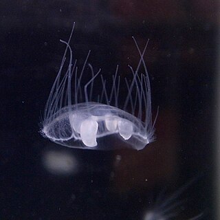
Jellyfish, also known as sea jellies, are the medusa-phase of certain gelatinous members of the subphylum Medusozoa, which is a major part of the phylum Cnidaria.

<span class="mw-page-title-main">Box jellyfish</span> Class of cnidarians distinguished by their cube-shaped medusae

Box jellyfish are cnidarian invertebrates distinguished by their box-like body. Some species of box jellyfish produce potent venom delivered by contact with their tentacles. Stings from some species, including Chironex fleckeri, Carukia barnesi, Malo kingi, and a few others, are extremely painful and often fatal to humans.

<span class="mw-page-title-main">Tentacle</span> Varied organ found in many animals and used for palpation and manipulation

In zoology, a tentacle is a flexible, mobile, and elongated organ present in some species of animals, most of them invertebrates. In animal anatomy, tentacles usually occur in one or more pairs. Anatomically, the tentacles of animals work mainly like muscular hydrostats. Most forms of tentacles are used for grasping and feeding. Many are sensory organs, variously receptive to touch, vision, or to the smell or taste of particular foods or threats. Examples of such tentacles are the eyestalks of various kinds of snails. Some kinds of tentacles have both sensory and manipulatory functions.

<i>Chironex fleckeri</i> Species of jellyfish

Chironex fleckeri, commonly known as the Australian box jelly, and nicknamed the sea wasp, is a species of extremely venomous box jellyfish found in coastal waters from northern Australia and New Guinea to Indonesia, Cambodia, Malaysia and Singapore, the Philippines and Vietnam. It has been described as "the most lethal jellyfish in the world", with at least 64 known deaths in Australia from 1884 to 2021.

<span class="mw-page-title-main">Medusozoa</span> Clade of marine invertebrates

Medusozoa is a clade in the phylum Cnidaria, and is often considered a subphylum. It includes the classes Hydrozoa, Scyphozoa, Staurozoa and Cubozoa, and possibly the parasitic Polypodiozoa. Medusozoans are distinguished by having a medusa stage in their often complex life cycle, a medusa typically being an umbrella-shaped body with stinging tentacles around the edge. With the exception of some Hydrozoa, all are called jellyfish in their free-swimming medusa phase.

<i>Craspedacusta sowerbii</i> Species of jellyfish

Craspedacusta sowerbii or peach blossom jellyfish is a species of freshwater hydrozoan jellyfish, or hydromedusa cnidarian. Hydromedusan jellyfish differ from scyphozoan jellyfish because they have a muscular, shelf-like structure called a velum on the ventral surface, attached to the bell margin. Originally from the Yangtze basin in China, C. sowerbii is an introduced species now found throughout the world in bodies of fresh water.

<span class="mw-page-title-main">Starlet sea anemone</span> Species of sea anemone

The starlet sea anemone is a species of small sea anemone in the family Edwardsiidae native to the east coast of the United States, with introduced populations along the coast of southeast England and the west coast of the United States. Populations have also been located in Nova Scotia, Canada. This sea anemone is found in the shallow brackish water of coastal lagoons and salt marshes where its slender column is usually buried in the mud and its tentacles exposed. Its genome has been sequenced and it is cultivated in the laboratory as a model organism, but the IUCN has listed it as being a "Vulnerable species" in the wild.

<i>Pelagia noctiluca</i> Species of cnidarian

Pelagia noctiluca is a jellyfish in the family Pelagiidae and the only currently recognized species in the genus Pelagia. It is typically known in English as the mauve stinger, but other common names are purple-striped jelly, purple stinger, purple people eater, purple jellyfish, luminous jellyfish and night-light jellyfish. In Greek, pelagia means "(she) of the sea", from pelagos "sea, open sea"; in Latin noctiluca is the combining form of nox, "night"", and lux, "light"; thus, Pelagia noctiluca can be described as a marine organism with the ability to glow in the dark (bioluminescence). It is found worldwide in tropical and warm temperate seas, although it is suspected that records outside the North Atlantic region, which includes the Mediterranean and Gulf of Mexico, represent closely related but currently unrecognized species.

<span class="mw-page-title-main">Sea anemone</span> Marine animals of the order Actiniaria

Sea anemones are a group of predatory marine invertebrates constituting the order Actiniaria. Because of their colourful appearance, they are named after the Anemone, a terrestrial flowering plant. Sea anemones are classified in the phylum Cnidaria, class Anthozoa, subclass Hexacorallia. As cnidarians, sea anemones are related to corals, jellyfish, tube-dwelling anemones, and Hydra. Unlike jellyfish, sea anemones do not have a medusa stage in their life cycle.

<i>Condylactis gigantea</i> Species of sea anemone

Condylactis gigantea is a tropical species of ball anemone that is found in shallow reefs and other shallow inshore areas in the Caribbean Sea – more specifically the West Indies – and the western Atlantic Ocean including southern Florida through the Florida Keys. It is also commonly known as: giant Caribbean sea anemone, giant golden anemone, condylactis anemone, Haitian anemone, pink-tipped anemone, purple-tipped anemone, and Florida condy. This species can easily be seen growing in lagoons or in inner reefs as either individuals or loose groups, but never as colonies. They are often used as a model organism along with others in their genus for facultative symbiosis with monocellular algae.

<span class="mw-page-title-main">Cnidosac</span>

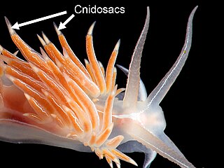
A cnidosac is an anatomical feature that is found in the group of sea slugs known as aeolid nudibranchs, a clade of marine opisthobranch gastropod molluscs. A cnidosac contains cnidocytes, stinging cells that are also known as cnidoblasts or nematocysts. These stinging cells are not made by the nudibranch, but by the species that it feeds upon. However, once the nudibranch is armed with these stinging cells, they are used in its own defense.

<span class="mw-page-title-main">Enthemonae</span> Suborder of sea anemone

The Enthemonae is a suborder of sea anemones in the order Actiniaria. It comprises those sea anemones with typical arrangement of mesenteries for actiniarians.

<span class="mw-page-title-main">Nematocyst (dinoflagellate)</span> Subcellular structure in unicellular algae

A nematocyst is a subcellular structure or organelle containing extrusive filaments found in two families of athecate dinoflagellates, the Warnowiaceae and Polykrikaceae. It is distinct from the similar subcellular structures found in the cnidocyte cells of cnidarians, a group of multicellular organisms including jellyfish and corals; such structures are also often called nematocysts, and cnidocytes are sometimes referred to as nematocytes. It is unclear whether the relationship between dinoflagellate and cnidarian nematocysts is a case of convergent evolution or common descent, although molecular evidence has been interpreted as supporting an endosymbiotic origin for cnidarian nematocysts.

Nematosomes are multicellular motile bodies found in the gastrovascular cavity of the model sea anemone Nematostella vectensis starlet sea anemone. First described by Stephenson in 1935, nematosomes are the defining apomorphy (synapomorphy) of the genus Nematostella but have received relatively little study. Nematosomes can be observed circulating through the body cavity and tentacle lumen of adult anemones, occasionally coming to rest on the gastrodermis. Nematosomes that are dislodged from rest return to circulation. The lifespan of a single nematosome has not been studied.

<span class="mw-page-title-main">Thimble jellyfish</span> Species of cnidarian

The thimble jellyfish is a species of cnidarian found in the warm West Atlantic Ocean, including the Caribbean. It is a tiny jellyfish with a straight-sided, flat-topped bell. This jellyfish is the most common cause of seabather's eruption, a reaction caused by the injection of juvenile jellyfish nematocysts into human skin.

<i>Aiptasia mutabilis</i> Species of sea anemone

Aiptasia mutabilis, also known as the Trumpet anemone, Rock anemone, and Glass anemone, is a species of anemone typically found attached to substrates in cold waters of the Atlantic Ocean. Its unique trumpet shape gives it its common name and it can grow to be 12 cm, having a column between 3 and 6 cm in size. Like many cnidarians, they rely on nematocysts for protection and to capture prey. They are not difficult to care for, and can be kept in a home aquarium, although due to their speed of reproduction, can quickly become overpopulated.

<i>Clytia hemisphaerica</i> Species of hydrozoan

Clytia hemisphaerica is a small hydrozoan-group cnidarian, about 1 cm in diameter, that is found in the Mediterranean Sea and the North-East Atlantic Ocean. Clytia has the free-swimming jellyfish form typical of the Hydrozoa, as well as vegetatively propagating polyps.

References

  1. 1 2 Holstein T.; Tardent P. (1984). "An ultrahigh-speed analysis of exocytosis: nematocyst discharge". Science. 223 (4638): 830–833. Bibcode:1984Sci...223..830H. doi:10.1126/science.6695186. PMID   6695186.
  2. Kass-Simon G.; Scappaticci A. A. Jr. (2002). "The behavioral and developmental physiology of nematocysts" (PDF). Canadian Journal of Zoology. 80 (10): 1772–1794. doi:10.1139/Z02-135 . Retrieved 2012-10-25.
  3. 1 2 3 4 Nüchter Timm; Benoit Martin; Engel Ulrike; Özbek Suat; Holstein Thomas W (2006). "Nanosecond-scale kinetics of nematocyst discharge". Current Biology. 16 (9): R316–R318. doi: 10.1016/j.cub.2006.03.089 . PMID   16682335.
  4. Moran, Yehu; Genikhovich, Grigory; Gordon, Dalia; Wienkoop, Stefanie; Zenkert, Claudia; Ozbek, Suat; Technau, Ulrich; Gurevitz, Michael (2012-04-07). "Neurotoxin localization to ectodermal gland cells uncovers an alternative mechanism of venom delivery in sea anemones". Proceedings. Biological Sciences. 279 (1732): 1351–1358. doi:10.1098/rspb.2011.1731. ISSN   1471-2954. PMC   3282367 . PMID   22048953.
  5. 1 2 Beckmann, Anna; Özbek, Suat (2012-06-05). "The Nematocyst: a molecular map of the Cnidarian stinging organelle". International Journal of Developmental Biology. 56 (6–7–8): 577–582. doi: 10.1387/ijdb.113472ab . ISSN   0214-6282. PMID   22689365.
  6. Shpirer, Erez; Chang, E Sally; Diamant, Arik; Rubinstein, Nimrod; Cartwright, Paulyn; Huchon, Dorothée (2014-09-29). "Diversity and evolution of myxozoan minicollagens and nematogalectins". BMC Evolutionary Biology. 14 (1): 205. Bibcode:2014BMCEE..14..205S. doi: 10.1186/s12862-014-0205-0 . ISSN   1471-2148. PMC   4195985 . PMID   25262812.
  7. Balasubramanian, Prakash G.; Beckmann, Anna; Warnken, Uwe; Schnölzer, Martina; Schüler, Andreas; Bornberg-Bauer, Erich; Holstein, Thomas W.; Özbek, Suat (2012-03-23). "Proteome of Hydra Nematocyst". The Journal of Biological Chemistry. 287 (13): 9672–9681. doi: 10.1074/jbc.M111.328203 . ISSN   0021-9258. PMC   3323026 . PMID   22291027.
  8. 1 2 David, Charles N.; Özbek, Suat; Adamczyk, Patrizia; Meier, Sebastian; Pauly, Barbara; Chapman, Jarrod; Hwang, Jung Shan; Gojobori, Takashi; Holstein, Thomas W. (2008-09-01). "Evolution of complex structures: minicollagens shape the cnidarian nematocyst". Trends in Genetics. 24 (9): 431–438. doi:10.1016/j.tig.2008.07.001. ISSN   0168-9525. PMID   18676050.
  9. Hamlet, Christina; Strychalski, Wanda; Miller, Laura (March 2020). "Fluid Dynamics of Ballistic Strategies in Nematocyst Firing". Fluids. 5 (1): 20. Bibcode:2020Fluid...5...20H. doi: 10.3390/fluids5010020 . ISSN   2311-5521.
  10. 1 2 Colin, Sean P.; Costello, John H. (2007-11-23). "Functional characteristics of nematocysts found on the scyphomedusa Cyanea capillata". Journal of Experimental Marine Biology and Ecology. 351 (1): 114–120. doi:10.1016/j.jembe.2007.06.033. ISSN   0022-0981. S2CID   51791589.
  11. 1 2 3 Babonis, Leslie S.; Enjolras, Camille; Reft, Abigail J.; Foster, Brent M.; Hugosson, Fredrik; Ryan, Joseph F.; Daly, Marymegan; Martindale, Mark Q. (2023-02-16). "Single-cell atavism reveals an ancient mechanism of cell type diversification in a sea anemone". Nature Communications. 14 (1): 885. Bibcode:2023NatCo..14..885B. doi:10.1038/s41467-023-36615-9. ISSN   2041-1723. PMC   9935875 . PMID   36797294.
  12. 1 2 Zenkert, Claudia; Takahashi, Toshio; Diesner, Mark-Oliver; Özbek, Suat (2011-07-28). "Morphological and Molecular Analysis of the Nematostella vectensis Cnidom". PLOS ONE. 6 (7): e22725. Bibcode:2011PLoSO...622725Z. doi: 10.1371/journal.pone.0022725 . ISSN   1932-6203. PMC   3145756 . PMID   21829492.
  13. 1 2 Khalturin, Konstantin; Shinzato, Chuya; Khalturina, Maria; Hamada, Mayuko; Fujie, Manabu; Koyanagi, Ryo; Kanda, Miyuki; Goto, Hiroki; Anton-Erxleben, Friederike; Toyokawa, Masaya; Toshino, Sho (May 2019). "Medusozoan genomes inform the evolution of the jellyfish body plan". Nature Ecology & Evolution. 3 (5): 811–822. Bibcode:2019NatEE...3..811K. doi: 10.1038/s41559-019-0853-y . ISSN   2397-334X. PMID   30988488.
  14. Sebé-Pedrós, Arnau; Saudemont, Baptiste; Chomsky, Elad; Plessier, Flora; Mailhé, Marie-Pierre; Renno, Justine; Loe-Mie, Yann; Lifshitz, Aviezer; Mukamel, Zohar; Schmutz, Sandrine; Novault, Sophie (31 May 2018). "Cnidarian Cell Type Diversity and Regulation Revealed by Whole-Organism Single-Cell RNA-Seq". Cell. 173 (6): 1520–1534.e20. doi: 10.1016/j.cell.2018.05.019 . ISSN   1097-4172. PMID   29856957.
  15. Denker, Elsa; Manuel, Michaël; Leclère, Lucas; Le Guyader, Hervé; Rabet, Nicolas (2008-03-01). "Ordered progression of nematogenesis from stem cells through differentiation stages in the tentacle bulb of Clytia hemisphaerica (Hydrozoa, Cnidaria)". Developmental Biology. 315 (1): 99–113. doi:10.1016/j.ydbio.2007.12.023. ISSN   1095-564X. PMID   18234172.
  16. Babonis, Leslie S.; Martindale, Mark Q. (2017-09-04). "PaxA, but not PaxC, is required for cnidocyte development in the sea anemone Nematostella vectensis". EvoDevo. 8: 14. doi: 10.1186/s13227-017-0077-7 . ISSN   2041-9139. PMC   5584322 . PMID   28878874.
  17. 1 2 Babonis, Leslie S.; Enjolras, Camille; Ryan, Joseph F.; Martindale, Mark Q. (2022-05-10). "A novel regulatory gene promotes novel cell fate by suppressing ancestral fate in the sea anemone Nematostella vectensis". Proceedings of the National Academy of Sciences. 119 (19). Bibcode:2022PNAS..11913701B. doi: 10.1073/pnas.2113701119 . ISSN   0027-8424. PMC   9172639 . PMID   35500123.
  18. Tibballs J (December 2006). "Australian venomous jellyfish, envenomation syndromes, toxins and therapy". Toxicon. 48 (7): 830–59. doi:10.1016/j.toxicon.2006.07.020. PMID   16928389.
  19. Brinkman D, Burnell J (November 2007). "Identification, cloning and sequencing of two major venom proteins from the box jellyfish, Chironex fleckeri". Toxicon. 50 (6): 850–60. doi:10.1016/j.toxicon.2007.06.016. PMID   17688901.
  20. Brinkman D, Burnell J (April 2008). "Partial purification of cytolytic venom proteins from the box jellyfish, Chironex fleckeri". Toxicon. 51 (5): 853–63. doi:10.1016/j.toxicon.2007.12.017. PMID   18243272.
  21. "YouTube". www.youtube.com. Archived from the original on 2014-06-09. Retrieved 6 April 2018.
  22. Surm, Joachim M.; Birch, Sydney; Macrander, Jason; Jaimes-Becerra, Adrian; Fridrich, Arie; Aharoni, Reuven; Rozenblat, Rotem; Sharabany, Julia; Appelbaum, Lior (2023-07-26). Venom tradeoff shapes interspecific interactions, physiology and reproduction (Report). Evolutionary Biology. doi:10.1101/2023.07.24.550294.