Cochlea

Last updated
Cochlea
Cochlea-crosssection.svg
Cross section of the cochlea
Parts of the inner ear, showing the cochlea
Details
Pronunciation /ˈkɒkliə,ˈkkliə/ [1]
Part of Inner ear
System Auditory system
Identifiers
Latin cochlea
MeSH D003051
NeuroLex ID birnlex_1190
TA98 A15.3.03.025
TA2 6964
FMA 60201
Anatomical terminology
3D model of cochlea and semicircular canals 3DPX-002432 Cochlea and semicircular canals Nevit Dilmen.stl
3D model of cochlea and semicircular canals

The cochlea is the part of the inner ear involved in hearing. It is a spiral-shaped cavity in the bony labyrinth, in humans making 2.75 turns around its axis, the modiolus. [2] [3] A core component of the cochlea is the organ of Corti, the sensory organ of hearing, which is distributed along the partition separating the fluid chambers in the coiled tapered tube of the cochlea.

Contents

The name cochlea derives from Ancient Greek κοχλίας (kokhlias) 'spiral, snail shell'.

Structure

Structural diagram of the cochlea showing how fluid pushed in at the oval window moves, deflects the cochlear partition, and bulges back out at the round window. Cochlea.svg
Structural diagram of the cochlea showing how fluid pushed in at the oval window moves, deflects the cochlear partition, and bulges back out at the round window.

The cochlea (pl.: cochleae) is a spiraled, hollow, conical chamber of bone, in which waves propagate from the base (near the middle ear and the oval window) to the apex (the top or center of the spiral). The spiral canal of the cochlea is a section of the bony labyrinth of the inner ear that is approximately 30 mm long and makes 234 turns about the modiolus. The cochlear structures include:

The cochlea is a portion of the inner ear that looks like a snail shell (cochlea is Greek for snail). [4] The cochlea receives sound in the form of vibrations, which cause the stereocilia to move. The stereocilia then convert these vibrations into nerve impulses which are taken up to the brain to be interpreted. Two of the three fluid sections are canals and the third is the 'Organ of Corti' which detects pressure impulses that travel along the auditory nerve to the brain. The two canals are called the vestibular canal and the tympanic canal.

Microanatomy

The walls of the hollow cochlea are made of bone, with a thin, delicate lining of epithelial tissue. This coiled tube is divided through most of its length by an inner membranous partition. Two fluid-filled outer spaces (ducts or scalae) are formed by this dividing membrane. At the top of the snailshell-like coiling tubes, there is a reversal of the direction of the fluid, thus changing the vestibular duct to the tympanic duct. This area is called the helicotrema. This continuation at the helicotrema allows fluid being pushed into the vestibular duct by the oval window to move back out via movement in the tympanic duct and deflection of the round window; since the fluid is nearly incompressible and the bony walls are rigid, it is essential for the conserved fluid volume to exit somewhere.

The lengthwise partition that divides most of the cochlea is itself a fluid-filled tube, the third 'duct'. This central column is called the cochlear duct. Its fluid, endolymph, also contains electrolytes and proteins, but is chemically quite different from perilymph. Whereas the perilymph is rich in sodium ions, the endolymph is rich in potassium ions, which produces an ionic, electrical potential.

The hair cells are arranged in four rows in the Organ of Corti along the entire length of the cochlear coil. Three rows consist of outer hair cells (OHCs) and one row consists of inner hair cells (IHCs). The inner hair cells provide the main neural output of the cochlea. The outer hair cells, instead, mainly 'receive' neural input from the brain, which influences their motility as part of the cochlea's mechanical "pre-amplifier". The input to the OHC is from the olivary body via the medial olivocochlear bundle.

The cochlear duct is almost as complex on its own as the ear itself. The cochlear duct is bounded on three sides by the basilar membrane, the stria vascularis , and Reissner's membrane. The stria vascularis is a rich bed of capillaries and secretory cells; Reissner's membrane is a thin membrane that separates endolymph from perilymph; and the basilar membrane is a mechanically somewhat stiff membrane, supporting the receptor organ for hearing, the Organ of Corti, and determines the mechanical wave propagation properties of the cochlear system.

Sexual dimorphism

Between males and females, there are differences in the shape of the human cochlea. The variation is in the twist at the end of the spiral. Because of this difference, and because the cochlea is one of the more durable bones in the skull, it is used in ascertaining the sexes of human remains found at archaeological sites. [5] [6]

Function

How sounds make their way from the source to the brain

The cochlea is filled with a watery liquid, the endolymph, which moves in response to the vibrations coming from the middle ear via the oval window. As the fluid moves, the cochlear partition (basilar membrane and organ of Corti) moves; thousands of hair cells sense the motion via their stereocilia, and convert that motion to electrical signals that are communicated via neurotransmitters to many thousands of nerve cells. These primary auditory neurons transform the signals into electrochemical impulses known as action potentials, which travel along the auditory nerve to structures in the brainstem for further processing.

Hearing

The stapes (stirrup) ossicle bone of the middle ear transmits vibrations to the fenestra ovalis (oval window) on the outside of the cochlea, which vibrates the perilymph in the vestibular duct (upper chamber of the cochlea). The ossicles are essential for efficient coupling of sound waves into the cochlea, since the cochlea environment is a fluid–membrane system, and it takes more pressure to move sound through fluid–membrane waves than it does through air. A pressure increase is achieved by reducing the area ratio from the tympanic membrane (drum) to the oval window (stapes bone) by 20. As pressure = force/area, results in a pressure gain of about 20 times from the original sound wave pressure in air. This gain is a form of impedance matching – to match the soundwave travelling through air to that travelling in the fluid–membrane system.

At the base of the cochlea, each 'duct' ends in a membranous portal that faces the middle ear cavity: The vestibular duct ends at the oval window, where the footplate of the stapes sits. The footplate vibrates when the pressure is transmitted via the ossicular chain. The wave in the perilymph moves away from the footplate and towards the helicotrema. Since those fluid waves move the cochlear partition that separates the ducts up and down, the waves have a corresponding symmetric part in perilymph of the tympanic duct, which ends at the round window, bulging out when the oval window bulges in.

The perilymph in the vestibular duct and the endolymph in the cochlear duct act mechanically as a single duct, being kept apart only by the very thin Reissner's membrane. The vibrations of the endolymph in the cochlear duct displace the basilar membrane in a pattern that peaks a distance from the oval window depending upon the soundwave frequency. The Organ of Corti vibrates due to outer hair cells further amplifying these vibrations. Inner hair cells are then displaced by the vibrations in the fluid, and depolarise by an influx of K+ via their tip-link-connected channels, and send their signals via neurotransmitter to the primary auditory neurons of the spiral ganglion. [7]

The hair cells in the Organ of Corti are tuned to certain sound frequencies by way of their location in the cochlea, due to the degree of stiffness in the basilar membrane. [8] This stiffness is due to, among other things, the thickness and width of the basilar membrane, [9] which along the length of the cochlea is stiffest nearest its beginning at the oval window, where the stapes introduces the vibrations coming from the eardrum. Since its stiffness is high there, it allows only high-frequency vibrations to move the basilar membrane, and thus the hair cells. The farther a wave travels towards the cochlea's apex (the helicotrema), the less stiff the basilar membrane is; thus lower frequencies travel down the tube, and the less-stiff membrane is moved most easily by them where the reduced stiffness allows: that is, as the basilar membrane gets less and less stiff, waves slow down and it responds better to lower frequencies. In addition, in mammals, the cochlea is coiled, which has been shown to enhance low-frequency vibrations as they travel through the fluid-filled coil. [10] This spatial arrangement of sound reception is referred to as tonotopy.

For very low frequencies (below 20 Hz), the waves propagate along the complete route of the cochlea – differentially up vestibular duct and tympanic duct all the way to the helicotrema. Frequencies this low still activate the Organ of Corti to some extent but are too low to elicit the perception of a pitch. Higher frequencies do not propagate to the helicotrema, due to the stiffness-mediated tonotopy.

A very strong movement of the basilar membrane due to very loud noise may cause hair cells to die. This is a common cause of partial hearing loss and is the reason why users of firearms or heavy machinery often wear earmuffs or earplugs.

Pathway to the brain

To transmit the sensation of sound to the brain, where it can be processed into the perception of hearing, hair cells of the cochlea must convert their mechanical stimulation into the electrical signaling patterns of the nervous system. Hair cells are modified neurons, able to generate action potentials which can be transmitted to other nerve cells. These action potential signals travel through the vestibulocochlear nerve to eventually reach the anterior medulla, where they synapse and are initially processed in the cochlear nuclei. [11]

Some processing occurs in the cochlear nuclei themselves, but the signals must also travel to the superior olivary complex of the pons as well as the inferior colliculi for further processing. [11]

Hair cell amplification

Not only does the cochlea "receive" sound, a healthy cochlea generates and amplifies sound when necessary. Where the organism needs a mechanism to hear very faint sounds, the cochlea amplifies by the reverse transduction of the OHCs, converting electrical signals back to mechanical in a positive-feedback configuration. The OHCs have a protein motor called prestin on their outer membranes; it generates additional movement that couples back to the fluid–membrane wave. This "active amplifier" is essential in the ear's ability to amplify weak sounds. [12] [13]

The active amplifier also leads to the phenomenon of soundwave vibrations being emitted from the cochlea back into the ear canal through the middle ear (otoacoustic emissions).

Otoacoustic emissions

Otoacoustic emissions are due to a wave exiting the cochlea via the oval window, and propagating back through the middle ear to the eardrum, and out the ear canal, where it can be picked up by a microphone. Otoacoustic emissions are important in some types of tests for hearing impairment, since they are present when the cochlea is working well, and less so when it is suffering from loss of OHC activity. Otoacoustic emissions also exhibit sex dimorphisms, since females tend to display higher magnitudes of otoacoustic emissions. Males tend to experience a reduction in otoacoustic emission magnitudes as they age. Women, on the other hand, do not experience a change in otoacoustic emission magnitudes with age. [14]

Role of gap junctions

Gap-junction proteins, called connexins, expressed in the cochlea play an important role in auditory functioning. [15] Mutations in gap-junction genes have been found to cause syndromic and nonsyndromic deafness. [16] Certain connexins, including connexin 30 and connexin 26, are prevalent in the two distinct gap-junction systems found in the cochlea. The epithelial-cell gap-junction network couples non-sensory epithelial cells, while the connective-tissue gap-junction network couples connective-tissue cells. [17] Gap-junction channels recycle potassium ions back to the endolymph after mechanotransduction in hair cells. [18] Importantly, gap junction channels are found between cochlear supporting cells, but not auditory hair cells. [19]

Clinical significance

Physical Damage

Damage to the cochlea can result from different incidents or conditions like a severe head injury, a cholesteatoma, an infection, and/or exposure to loud noise which could kill hair cells in the cochlea.

Hearing loss

Hearing loss associated with the cochlea is often a result of outer hair cells and inner hair cells damage or death. Outer hair cells are more susceptible to damage, which can result in less sensitivity to weak sounds. Frequency sensitivity is also affected by cochlear damage which can impair the patient's ability to distinguish between spectral differences of vowels. The effects of cochlear damage on different aspects of hearing loss like temporal integration, pitch perception, and frequency determination are still being studied, given that multiple factors must be taken into account in regard to cochlear research. [20]

Bionics

In 2009, engineers at the Massachusetts Institute of Technology created an electronic chip that can quickly analyze a very large range of radio frequencies while using only a fraction of the power needed for existing technologies; its design specifically mimics a cochlea. [21] [22]

Other animals

The coiled form of cochlea is unique to mammals. In birds and in other non-mammalian vertebrates, the compartment containing the sensory cells for hearing is occasionally also called "cochlea," despite not being coiled up. Instead, it forms a blind-ended tube, also called the cochlear duct. This difference apparently evolved in parallel with the differences in frequency range of hearing between mammals and non-mammalian vertebrates. The superior frequency range in mammals is partly due to their unique mechanism of pre-amplification of sound by active cell-body vibrations of outer hair cells. Frequency resolution is, however, not better in mammals than in most lizards and birds, but the upper frequency limit is – sometimes much – higher. Most bird species do not hear above 45 kHz, the currently known maximum being ~ 11 kHz in the barn owl. Some marine mammals hear up to 200 kHz. A long coiled compartment, rather than a short and straight one, provides more space for additional octaves of hearing range, and has made possible some of the highly derived behaviors involving mammalian hearing. [23]

As the study of the cochlea should fundamentally be focused at the level of hair cells, it is important to note the anatomical and physiological differences between the hair cells of various species. In birds, for instance, instead of outer and inner hair cells, there are tall and short hair cells. There are several similarities of note in regard to this comparative data. For one, the tall hair cell is very similar in function to that of the inner hair cell, and the short hair cell, lacking afferent auditory-nerve fiber innervation, resembles the outer hair cell. One unavoidable difference, however, is that while all hair cells are attached to a tectorial membrane in birds, only the outer hair cells are attached to the tectorial membrane in mammals.

History

The name cochlea is derived from the Latin word for snail shell, which in turn is from the Greek κοχλίας kokhlias ("snail, screw"), from κόχλος kokhlos ("spiral shell") [24] in reference to its coiled shape; the cochlea is coiled in mammals with the exception of monotremes.

Additional images

See also

Related Research Articles

<span class="mw-page-title-main">Inner ear</span> Innermost part of the vertebrate ear

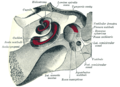
The inner ear is the innermost part of the vertebrate ear. In vertebrates, the inner ear is mainly responsible for sound detection and balance. In mammals, it consists of the bony labyrinth, a hollow cavity in the temporal bone of the skull with a system of passages comprising two main functional parts:

<span class="mw-page-title-main">Vestibulocochlear nerve</span> Cranial nerve VIII, for hearing and balance

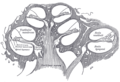
The vestibulocochlear nerve or auditory vestibular nerve, also known as the eighth cranial nerve, cranial nerve VIII, or simply CN VIII, is a cranial nerve that transmits sound and equilibrium (balance) information from the inner ear to the brain. Through olivocochlear fibers, it also transmits motor and modulatory information from the superior olivary complex in the brainstem to the cochlea.

<span class="mw-page-title-main">Basilar membrane</span> Stiff structural element within the cochlea of the inner ear which separates two liquid-filled tubes

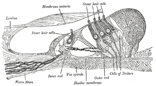
The basilar membrane is a stiff structural element within the cochlea of the inner ear which separates two liquid-filled tubes that run along the coil of the cochlea, the scala media and the scala tympani. The basilar membrane moves up and down in response to incoming sound waves, which are converted to traveling waves on the basilar membrane.

<span class="mw-page-title-main">Organ of Corti</span> Receptor organ for hearing

The organ of Corti, or spiral organ, is the receptor organ for hearing and is located in the mammalian cochlea. This highly varied strip of epithelial cells allows for transduction of auditory signals into nerve impulses' action potential. Transduction occurs through vibrations of structures in the inner ear causing displacement of cochlear fluid and movement of hair cells at the organ of Corti to produce electrochemical signals.

<span class="mw-page-title-main">Auditory system</span> Sensory system used for hearing

The auditory system is the sensory system for the sense of hearing. It includes both the sensory organs and the auditory parts of the sensory system.

<span class="mw-page-title-main">Ear</span> Organ of hearing and balance

An ear is the organ that enables hearing and body balance using the vestibular system. In mammals, the ear is usually described as having three parts: the outer ear, the middle ear and the inner ear. The outer ear consists of the pinna and the ear canal. Since the outer ear is the only visible portion of the ear in most animals, the word "ear" often refers to the external part alone. The middle ear includes the tympanic cavity and the three ossicles. The inner ear sits in the bony labyrinth, and contains structures which are key to several senses: the semicircular canals, which enable balance and eye tracking when moving; the utricle and saccule, which enable balance when stationary; and the cochlea, which enables hearing. The ear is a self cleaning organ through its relationship with earwax and the ear canals. The ears of vertebrates are placed somewhat symmetrically on either side of the head, an arrangement that aids sound localization.

<span class="mw-page-title-main">Hair cell</span> Auditory sensory receptor nerve cells

Hair cells are the sensory receptors of both the auditory system and the vestibular system in the ears of all vertebrates, and in the lateral line organ of fishes. Through mechanotransduction, hair cells detect movement in their environment.

<span class="mw-page-title-main">Endolymph</span> Inner ear fluid

Endolymph is the fluid contained in the membranous labyrinth of the inner ear. The major cation in endolymph is potassium, with the values of sodium and potassium concentration in the endolymph being 0.91 mM and 154 mM, respectively. It is also called Scarpa's fluid, after Antonio Scarpa.

In audiology and psychoacoustics the concept of critical bands, introduced by Harvey Fletcher in 1933 and refined in 1940, describes the frequency bandwidth of the "auditory filter" created by the cochlea, the sense organ of hearing within the inner ear. Roughly, the critical band is the band of audio frequencies within which a second tone will interfere with the perception of the first tone by auditory masking.

<span class="mw-page-title-main">Stereocilia (inner ear)</span> Mechanosensing organelles of hair cells

In the inner ear, stereocilia are the mechanosensing organelles of hair cells, which respond to fluid motion in numerous types of animals for various functions, including hearing and balance. They are about 10–50 micrometers in length and share some similar features of microvilli. The hair cells turn the fluid pressure and other mechanical stimuli into electric stimuli via the many microvilli that make up stereocilia rods. Stereocilia exist in the auditory and vestibular systems.

<span class="mw-page-title-main">Perilymph</span> Extracellular fluid located within the inner ear

Perilymph is an extracellular fluid located within the inner ear. It is found within the scala tympani and scala vestibuli of the cochlea. The ionic composition of perilymph is comparable to that of plasma and cerebrospinal fluid. The major cation in perilymph is sodium, with the values of sodium and potassium concentration in the perilymph being 138 mM and 6.9 mM, respectively. It is also named Cotunnius' liquid and liquor cotunnii for Domenico Cotugno.

<span class="mw-page-title-main">Tympanic duct</span>

The tympanic duct or scala tympani is one of the perilymph-filled cavities in the inner ear of humans. It is separated from the cochlear duct by the basilar membrane, and it extends from the round window to the helicotrema, where it continues as vestibular duct.

<span class="mw-page-title-main">Cochlear duct</span> Cavity in the cochlea of the inner ear

The cochlear duct is an endolymph filled cavity inside the cochlea, located between the tympanic duct and the vestibular duct, separated by the basilar membrane and the vestibular membrane respectively. The cochlear duct houses the organ of Corti.

<span class="mw-page-title-main">Vestibular membrane</span> Membrane in the cochlea in the inner ear

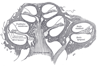
The vestibular membrane, vestibular wall or Reissner's membrane is a membrane inside the cochlea of the inner ear. It separates the cochlear duct from the vestibular duct. It helps to transmit vibrations from fluid in the vestibular duct to the cochlear duct. Together with the basilar membrane, it creates a compartment in the cochlea filled with endolymph, which is important for the function of the spiral organ of Corti. It allows nutrients to travel from the perilymph to the endolymph of the membranous labyrinth. It may be damaged in Ménière's disease. It is named after the German anatomist Ernst Reissner.

<span class="mw-page-title-main">Helicotrema</span> Connection between the scala tympani and the scala vestibuli in the cochlea

The helicotrema is the part of the cochlear labyrinth where the scala tympani and the scala vestibuli meet. It is the main component of the cochlear apex. The hair cells near this area best detect low frequency sounds.

<span class="mw-page-title-main">Hearing</span> Sensory perception of sound by living organisms

Hearing, or auditory perception, is the ability to perceive sounds through an organ, such as an ear, by detecting vibrations as periodic changes in the pressure of a surrounding medium. The academic field concerned with hearing is auditory science.

The cochlear amplifier is a positive feedback mechanism within the cochlea that provides acute sensitivity in the mammalian auditory system. The main component of the cochlear amplifier is the outer hair cell (OHC) which increases the amplitude and frequency selectivity of sound vibrations using electromechanical feedback.

The neural encoding of sound is the representation of auditory sensation and perception in the nervous system. The complexities of contemporary neuroscience are continually redefined. Thus what is known of the auditory system has been continually changing. The encoding of sounds includes the transduction of sound waves into electrical impulses along auditory nerve fibers, and further processing in the brain.

Electrocochleography is a technique of recording electrical potentials generated in the inner ear and auditory nerve in response to sound stimulation, using an electrode placed in the ear canal or tympanic membrane. The test is performed by an otologist or audiologist with specialized training, and is used for detection of elevated inner ear pressure or for the testing and monitoring of inner ear and auditory nerve function during surgery.

Cochlea is Latin for “snail, shell or screw” and originates from the Greek word κοχλίας kokhlias. The modern definition, the auditory portion of the inner ear, originated in the late 17th century. Within the mammalian cochlea exists the organ of Corti, which contains hair cells that are responsible for translating the vibrations it receives from surrounding fluid-filled ducts into electrical impulses that are sent to the brain to process sound.

References

  1. "cochlea". Dictionary.com Unabridged (Online). n.d.
  2. Anne M. Gilroy; Brian R. MacPherson; Lawrence M. Ross (2008). Atlas of anatomy. Thieme. p. 536. ISBN   978-1-60406-151-2.
  3. Moore & Dalley (1999). Clinically Oriented Anatomy (4th ed.). Lippincott Williams & Wilkins. p. 974. ISBN   0-683-06141-0.
  4. The Kingfisher children's encyclopedia. Kingfisher Publications. (3rd ed., fully rev. and updated ed.). New York: Kingfisher. 2012 [2011]. ISBN   9780753468142. OCLC   796083112.{{cite book}}: CS1 maint: others (link)
  5. Cochlear shape reveals that the human organ of hearing is sex-typed from birth. J. Braga, C. Samir, L. Risser, J. Dumoncel, D. Descouens, J. F. Thackeray, P. Balaresque, A. Oettlé, J.-M. Loubes & A. Fradi, Scientific Reports, 26 July 2019. https://doi.org/10.1038/s41598-019-47433-9
  6. Braga, J., Samir, C., Risser, L. et al. Cochlear shape reveals that the human organ of hearing is sex-typed from birth. Sci Rep 9, 10889 (2019). https://doi.org/10.1038/s41598-019-47433-9
  7. Nin, Fumiaki; Hibino, Hiroshi; Doi, Katsumi; Suzuki, Toshihiro; Hisa, Yasuo; Kurachi, Yoshihisa (5 February 2008). "The endocochlear potential depends on two K + diffusion potentials and an electrical barrier in the stria vascularis of the inner ear". Proceedings of the National Academy of Sciences. 105 (5): 1751–1756. doi:10.1073/pnas.0711463105.
  8. Guenter Ehret (Dec 1978). "Stiffness gradient along the basilar membrane as a way for spatial frequency analysis within the cochlea" (PDF). J Acoust Soc Am. 64 (6): 1723–6. doi:10.1121/1.382153. PMID   739099.
  9. Camhi, J. Neuroethology: nerve cells and the natural behavior of animals. Sinauer Associates, 1984.
  10. Manoussaki D, Chadwick RS, Ketten DR, Arruda J, Dimitriadis EK, O'Malley JT (2008). "The influence of cochlear shape on low-frequency hearing". Proc Natl Acad Sci U S A. 105 (16): 6162–6166. Bibcode:2008PNAS..105.6162M. doi: 10.1073/pnas.0710037105 . PMC   2299218 . PMID   18413615.
  11. 1 2 Martin, John Harry (2021). "Chapter 8: The Auditory System". Neuroanatomy: Text and Atlas (5th ed.). New York: McGraw Hill. ISBN   978-1-259-64248-7.
  12. Ashmore, Jonathan Felix (1987). "A fast motile response in guinea-pig outer hair cells: the cellular basis of the cochlear amplifier". The Journal of Physiology . 388 (1): 323–347. doi:10.1113/jphysiol.1987.sp016617. ISSN   1469-7793. PMC   1192551 . PMID   3656195. Open Access logo PLoS transparent.svg
  13. Ashmore, Jonathan (2008). "Cochlear Outer Hair Cell Motility". Physiological Reviews . 88 (1): 173–210. doi:10.1152/physrev.00044.2006. ISSN   0031-9333. PMID   18195086. S2CID   17722638. Open Access logo PLoS transparent.svg
  14. Mishra, Srikanta K.1,2; Zambrano, Samantha2,3; Rodrigo, Hansapani4. Sexual Dimorphism in the Functional Development of the Cochlear Amplifier in Humans. Ear and Hearing 42(4):p 860-869, July/August 2021. | DOI: 10.1097/AUD.0000000000000976
  15. Zhao, H. -B.; Kikuchi, T.; Ngezahayo, A.; White, T. W. (2006). "Gap Junctions and Cochlear Homeostasis". Journal of Membrane Biology. 209 (2–3): 177–186. doi:10.1007/s00232-005-0832-x. PMC   1609193 . PMID   16773501.
  16. Erbe, C. B.; Harris, K. C.; Runge-Samuelson, C. L.; Flanary, V. A.; Wackym, P. A. (2004). "Connexin 26 and Connexin 30 Mutations in Children with Nonsyndromic Hearing Loss". The Laryngoscope. 114 (4): 607–611. doi:10.1097/00005537-200404000-00003. PMID   15064611. S2CID   25847431.
  17. Wang, Bo; Hu, Bohua; Yang, Shiming (December 2015). "Cell junction proteins within the cochlea: A review of recent research". Journal of Otology. 10 (4): 131–135. doi:10.1016/j.joto.2016.01.003.
  18. Kikuchi, T.; Kimura, R. S.; Paul, D. L.; Takasaka, T.; Adams, J. C. (2000). "Gap junction systems in the mammalian cochlea". Brain Research. Brain Research Reviews. 32 (1): 163–166. doi:10.1016/S0165-0173(99)00076-4. PMID   10751665. S2CID   11292387.
  19. Kikuchi, T.; Kimura, R. S.; Paul, D. L.; Adams, J. C. (1995). "Gap junctions in the rat cochlea: Immunohistochemical and ultrastructural analysis". Anatomy and Embryology. 191 (2): 101–118. doi:10.1007/BF00186783. PMID   7726389. S2CID   24900775.
  20. Moore, Brian C. J.. Perceptual Consequences of Cochlear Hearing Loss and their Implications for the Design of Hearing Aids. Ear and Hearing 17(2):p 133-161, April 1996.
  21. Anne Trafton (June 3, 2009). "Drawing inspiration from nature to build a better radio: New radio chip mimics human ear, could enable universal radio". MIT newsoffice.
  22. Soumyajit Mandal; Serhii M. Zhak; Rahul Sarpeshkar (June 2009). "A Bio-Inspired Active Radio-Frequency Silicon Cochlea" (PDF). IEEE Journal of Solid-State Circuits. 44 (6): 1814–1828. Bibcode:2009IJSSC..44.1814M. doi:10.1109/JSSC.2009.2020465. hdl: 1721.1/59982 . S2CID   10756707.
  23. Vater M, Meng J, Fox RC. Hearing organ evolution and specialization: Early and later mammals. In: GA Manley, AN Popper, RR Fay (Eds). Evolution of the Vertebrate Auditory System, Springer-Verlag, New York 2004, pp 256–288.
  24. etymology of "cochleㄷa",

Further reading