Condenser (optics)

Last updated
A condenser (right) and its respective diaphragm (left) Kondenzor.jpg
A condenser (right) and its respective diaphragm (left)

A condenser is an optical lens that renders a divergent light beam from a point light source into a parallel or converging beam to illuminate an object to be imaged.

Contents

Condensers are an essential part of any imaging device, such as microscopes, enlargers, slide projectors, and telescopes. The concept is applicable to all kinds of radiation undergoing optical transformation, such as electrons in electron microscopy, neutron radiation, and synchrotron radiation optics.

Microscope condenser

A condenser between the stage and mirror of a vintage microscope Kondenzor01.jpg
A condenser between the stage and mirror of a vintage microscope

Condensers are located above the light source and under the sample in an upright microscope, and above the stage and below the light source in an inverted microscope. They act to gather light from the microscope's light source and concentrate it into a cone of light that illuminates the specimen. The aperture and angle of the light cone must be adjusted (via the size of the diaphragm) for each different objective lens with different numerical apertures.

Condensers typically consist of a variable-aperture diaphragm and one or more lenses. Light from the illumination source of the microscope passes through the diaphragm and is focused by the lens(es) onto the specimen. After passing through the specimen the light diverges into an inverted cone to fill the front lens of the objective.

Types

There are three main types of microscope condenser:

  1. The chromatic condenser, such as the Abbe where no attempt is made to correct for spherical or chromatic aberration. It contains two lenses that produce an image of the light source that is surrounded by a blue and red color at its edges.
  2. The aplanatic condenser is corrected for spherical aberration.
  3. The compound achromatic condenser is corrected for both spherical and chromatic aberrations.

Abbe condenser

The sub-stage condenser focuses light through the specimen to match the aperture of the objective lens system. Condenser diagram.svg
The sub-stage condenser focuses light through the specimen to match the aperture of the objective lens system.

The Abbe condenser is named for its inventor Ernst Abbe, who developed it in 1870. The Abbe condenser, which was originally designed for Zeiss, is mounted below the stage of the microscope. The condenser concentrates and controls the light that passes through the specimen prior to entering the objective. It has two controls, one which moves the Abbe condenser closer to or further from the stage, and another, the iris diaphragm, which controls the diameter of the beam of light. The controls can be used to optimize brightness, evenness of illumination, and contrast. Abbe condensers are difficult to use for magnifications of above 400X, as the aplanatic cone is only representative of a numerical aperture (NA) of 0.6.

This condenser is composed of two lenses, a plano-convex lens somewhat larger than a hemisphere and a large bi-convex lens serving as a collecting lens to the first. The focus of the first lens is traditionally about 2mm away from the plane face coinciding with the sample plane. A pinhole cap can be used to align the optical axis of the condenser with that of the microscope. The Abbe condenser is still the basis for most modern light microscope condenser designs, even though its optical performance is poor. [1] [2] [3]

Aplanatic and achromatic condensers

An aplanatic condenser corrects for spherical aberration in the concentrated light path, while an achromatic compound condenser corrects for both spherical and chromatic aberration.

Specialized condensers

Dark field and phase contrast setups are based on an Abbe, aplanatic, or achromatic condenser, but to the light path add a dark field stop or various size phase rings. These additional elements are housed in various ways. In most modern microscope (ca. 1990s–), such elements are housed in sliders that fit into a slot between the illuminator and the condenser lens. Many older microscopes house these elements in a turret-type condenser, these elements are housed in a turret below the condenser lens and rotated into place.

Specialised condensers are also used as part of Differential Interference Contrast and Hoffman Modulation Contrast systems, which aim to improve contrast and visibility of transparent specimens.

In epifluorescence microscopy, the objective lens acts not only as a magnifier for the light emitted by the fluorescing object, but also as a condenser for the incident light.

The Arlow-Abbe condenser is a modified Abbe condenser that replaces the iris diaphragm, filter holder, lamp and lamp optics with a small OLED or LCD digital display unit. The display unit allows for digitally synthesised filters for dark-field, Rheinberg, oblique and dynamic (constantly changing) illumination under direct computer control. The device was first described by Dr. Jim Arlow in Microbe Hunter magazine, issue 48.

Condensers and numerical aperture

Like objective lenses, condensers vary in their numerical aperture (NA). It is NA that determines optical resolution, in combination with the NA of the objective. Different condensers vary in their maximum and minimum numerical aperture, and the numerical aperture of a single condenser varies depending on the diameter setting of the condenser aperture. In order for the maximum numerical aperture (and therefore resolution) of an objective lens to be realized, the numerical aperture of the condenser must be matched to the numerical aperture of the used objective. The technique most commonly used in microscopy to optimize the light pathway between the condenser (and other illumination components of the microscope) and the objective lens is known as Köhler illumination.

The maximum NA is limited by the refractive index of the medium between the lens and the sample. As with objective lenses, a condenser lens with a maximum numerical aperture of greater than 0.95 is designed to be used under oil immersion (or, more rarely, under water immersion), with a layer of immersion oil placed in contact with both the slide/coverslip and the lens of the condenser. An oil immersion condenser may typically have NA of up to 1.25. Without this oil layer, not only is maximum numerical aperture not realized, but the condenser may not be able to precisely focus light on the object. Condensers with a numerical aperture of 0.95 or less are designed to be used without oil or other fluid on the top lens and are termed dry condensers. Dual dry/immersion condensers are basically oil immersion condensers that can nonetheless focus light with the same degree of precision even without oil between the top lens and the slide.

History

The first simple condensers were introduced on pre-achromatic microscopes in the 17th century. Robert Hooke used a combination of a salt water filled globe and a plano-convex lens, and shows in the 'Micrographia' that he understands the reasons for its efficiency. Makers in the 18th century such as Benjamin Martin, Adams and Jones understood the advantage of condensing the area of the light source to that of the area of the object on the stage. This was a simple plano-convex or bi-convex lens, or sometimes a combination of lenses. With the development of the modern achromatic objective in 1829, by Joseph Jackson Lister, the need for better condensers became increasingly apparent. By 1837, the use of the achromatic condenser was introduced in France, by Felix Dujardin, and Chevalier. English makers early took up this improvement, due to the obsession with resolving test objects such as diatoms and Nobert ruled gratings. By the late 1840s, English makers such as Ross, Powell and Smith; all could supply highly corrected condensers on their best stands, with proper centring and focus. It is erroneously stated that these developments were purely empirical - no-one can design a good achromatic, spherically corrected condenser relying only on empirics. [ citation needed ] On the Continent, in Germany, the corrected condenser was not considered either useful or essential, mainly due to a misunderstanding of the basic optical principles involved. Thus the leading German company, Carl Zeiss in Jena, offered nothing more than a very poor chromatic condenser into the late 1870s. French makers, such as Nachet, provided excellent achromatic condensers on their stands. When the leading German bacteriologist, Robert Koch, complained to Ernst Abbe that he was forced to buy a Seibert achromatic condenser for his Zeiss microscope in order to make satisfactory photographs of bacteria, Abbe produced a very good achromatic design in 1878.

Related Research Articles

<span class="mw-page-title-main">Optical aberration</span> Deviation from perfect paraxial optical behavior

In optics, aberration is a property of optical systems, such as lenses, that causes light to be spread out over some region of space rather than focused to a point. Aberrations cause the image formed by a lens to be blurred or distorted, with the nature of the distortion depending on the type of aberration. Aberration can be defined as a departure of the performance of an optical system from the predictions of paraxial optics. In an imaging system, it occurs when light from one point of an object does not converge into a single point after transmission through the system. Aberrations occur because the simple paraxial theory is not a completely accurate model of the effect of an optical system on light, rather than due to flaws in the optical elements.

<span class="mw-page-title-main">Microscopy</span> Viewing of objects which are too small to be seen with the naked eye

Microscopy is the technical field of using microscopes to view objects and areas of objects that cannot be seen with the naked eye. There are three well-known branches of microscopy: optical, electron, and scanning probe microscopy, along with the emerging field of X-ray microscopy.

<span class="mw-page-title-main">Numerical aperture</span> Characteristic of an optical system

In optics, the numerical aperture (NA) of an optical system is a dimensionless number that characterizes the range of angles over which the system can accept or emit light. By incorporating index of refraction in its definition, NA has the property that it is constant for a beam as it goes from one material to another, provided there is no refractive power at the interface. The exact definition of the term varies slightly between different areas of optics. Numerical aperture is commonly used in microscopy to describe the acceptance cone of an objective, and in fiber optics, in which it describes the range of angles within which light that is incident on the fiber will be transmitted along it.

<span class="mw-page-title-main">Chromatic aberration</span> Failure of a lens to focus all colors on the same point

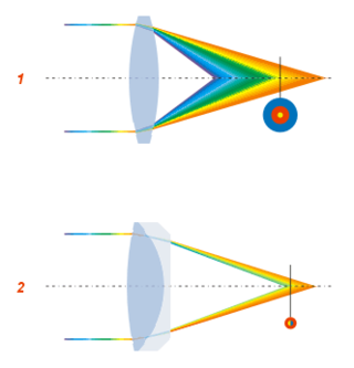
In optics, chromatic aberration (CA), also called chromatic distortion and spherochromatism, is a failure of a lens to focus all colors to the same point. It is caused by dispersion: the refractive index of the lens elements varies with the wavelength of light. The refractive index of most transparent materials decreases with increasing wavelength. Since the focal length of a lens depends on the refractive index, this variation in refractive index affects focusing. Chromatic aberration manifests itself as "fringes" of color along boundaries that separate dark and bright parts of the image.

<span class="mw-page-title-main">Optical microscope</span> Microscope that uses visible light

The optical microscope, also referred to as a light microscope, is a type of microscope that commonly uses visible light and a system of lenses to generate magnified images of small objects. Optical microscopes are the oldest design of microscope and were possibly invented in their present compound form in the 17th century. Basic optical microscopes can be very simple, although many complex designs aim to improve resolution and sample contrast.

<span class="mw-page-title-main">Transmission electron microscopy</span> Imaging and diffraction using electrons that pass through samples

Transmission electron microscopy (TEM) is a microscopy technique in which a beam of electrons is transmitted through a specimen to form an image. The specimen is most often an ultrathin section less than 100 nm thick or a suspension on a grid. An image is formed from the interaction of the electrons with the sample as the beam is transmitted through the specimen. The image is then magnified and focused onto an imaging device, such as a fluorescent screen, a layer of photographic film, or a detector such as a scintillator attached to a charge-coupled device or a direct electron detector.

<span class="mw-page-title-main">Angular resolution</span> Ability of any image-forming device to distinguish small details of an object

Angular resolution describes the ability of any image-forming device such as an optical or radio telescope, a microscope, a camera, or an eye, to distinguish small details of an object, thereby making it a major determinant of image resolution. It is used in optics applied to light waves, in antenna theory applied to radio waves, and in acoustics applied to sound waves. The colloquial use of the term "resolution" sometimes causes confusion; when an optical system is said to have a high resolution or high angular resolution, it means that the perceived distance, or actual angular distance, between resolved neighboring objects is small. The value that quantifies this property, θ, which is given by the Rayleigh criterion, is low for a system with a high resolution. The closely related term spatial resolution refers to the precision of a measurement with respect to space, which is directly connected to angular resolution in imaging instruments. The Rayleigh criterion shows that the minimum angular spread that can be resolved by an image forming system is limited by diffraction to the ratio of the wavelength of the waves to the aperture width. For this reason, high resolution imaging systems such as astronomical telescopes, long distance telephoto camera lenses and radio telescopes have large apertures.

<span class="mw-page-title-main">Refracting telescope</span> Type of optical telescope

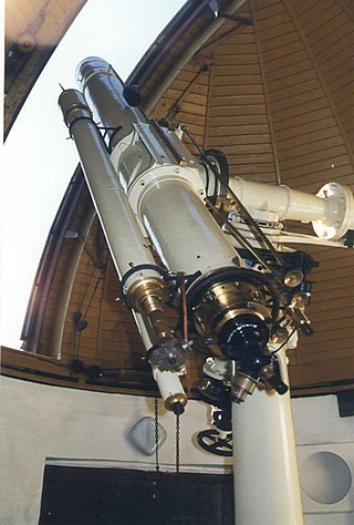
A refracting telescope is a type of optical telescope that uses a lens as its objective to form an image. The refracting telescope design was originally used in spyglasses and astronomical telescopes but is also used for long-focus camera lenses. Although large refracting telescopes were very popular in the second half of the 19th century, for most research purposes, the refracting telescope has been superseded by the reflecting telescope, which allows larger apertures. A refractor's magnification is calculated by dividing the focal length of the objective lens by that of the eyepiece.

<span class="mw-page-title-main">Diffraction-limited system</span> Optical system with resolution performance at the instruments theoretical limit

In optics, any optical instrument or system – a microscope, telescope, or camera – has a principal limit to its resolution due to the physics of diffraction. An optical instrument is said to be diffraction-limited if it has reached this limit of resolution performance. Other factors may affect an optical system's performance, such as lens imperfections or aberrations, but these are caused by errors in the manufacture or calculation of a lens, whereas the diffraction limit is the maximum resolution possible for a theoretically perfect, or ideal, optical system.

<span class="mw-page-title-main">Objective (optics)</span> Lens or mirror in optical instruments

In optical engineering, an objective is an optical element that gathers light from an object being observed and focuses the light rays from it to produce a real image of the object. Objectives can be a single lens or mirror, or combinations of several optical elements. They are used in microscopes, binoculars, telescopes, cameras, slide projectors, CD players and many other optical instruments. Objectives are also called object lenses, object glasses, or objective glasses.

<span class="mw-page-title-main">Eyepiece</span> Type of lens attached to a variety of optical devices such as telescopes and microscopes

An eyepiece, or ocular lens, is a type of lens that is attached to a variety of optical devices such as telescopes and microscopes. It is named because it is usually the lens that is closest to the eye when someone looks through an optical device to observe an object or sample. The objective lens or mirror collects light from an object or sample and brings it to focus creating an image of the object. The eyepiece is placed near the focal point of the objective to magnify this image to the eyes. The amount of magnification depends on the focal length of the eyepiece.

A total internal reflection fluorescence microscope (TIRFM) is a type of microscope with which a thin region of a specimen, usually less than 200 nanometers can be observed.

A solid immersion lens (SIL) has higher magnification and higher numerical aperture than common lenses by filling the object space with a high-refractive-index solid material. SIL was originally developed for enhancing the spatial resolution of optical microscopy. There are two types of SIL:

<span class="mw-page-title-main">Dark-field microscopy</span> Laboratory technique

Dark-field microscopy describes microscopy methods, in both light and electron microscopy, which exclude the unscattered beam from the image. Consequently, the field around the specimen is generally dark.

<span class="mw-page-title-main">Bright-field microscopy</span> Optical microscopy illumination technique

Bright-field microscopy (BF) is the simplest of all the optical microscopy illumination techniques. Sample illumination is transmitted white light, and contrast in the sample is caused by attenuation of the transmitted light in dense areas of the sample. Bright-field microscopy is the simplest of a range of techniques used for illumination of samples in light microscopes, and its simplicity makes it a popular technique. The typical appearance of a bright-field microscopy image is a dark sample on a bright background, hence the name.

Köhler illumination is a method of specimen illumination used for transmitted and reflected light optical microscopy. Köhler illumination acts to generate an even illumination of the sample and ensures that an image of the illumination source is not visible in the resulting image. Köhler illumination is the predominant technique for sample illumination in modern scientific light microscopy. It requires additional optical elements which are more expensive and may not be present in more basic light microscopes.

<span class="mw-page-title-main">Oil immersion</span> Light microscopy technique

In light microscopy, oil immersion is a technique used to increase the resolving power of a microscope. This is achieved by immersing both the objective lens and the specimen in a transparent oil of high refractive index, thereby increasing the numerical aperture of the objective lens.

In light microscopy, a water immersion objective is a specially designed objective lens used to increase the resolution of the microscope. This is achieved by immersing both the lens and the specimen in water which has a higher refractive index than air, thereby increasing the numerical aperture of the objective lens.

The design of photographic lenses for use in still or cine cameras is intended to produce a lens that yields the most acceptable rendition of the subject being photographed within a range of constraints that include cost, weight and materials. For many other optical devices such as telescopes, microscopes and theodolites where the visual image is observed but often not recorded the design can often be significantly simpler than is the case in a camera where every image is captured on film or image sensor and can be subject to detailed scrutiny at a later stage. Photographic lenses also include those used in enlargers and projectors.

<span class="mw-page-title-main">Optical sectioning</span> Imaging of focal planes within a thick sample

Optical sectioning is the process by which a suitably designed microscope can produce clear images of focal planes deep within a thick sample. This is used to reduce the need for thin sectioning using instruments such as the microtome. Many different techniques for optical sectioning are used and several microscopy techniques are specifically designed to improve the quality of optical sectioning.

References

  1. Royal Microscopical Society,"Journal of the Royal Microscopical Society", Williams and Norgate, London (1882), p.411-2
  2. Chamot, E.M., "Elementary chemical microscopy", John Wiley and Sons, London (1916), p.36
  3. "The Evolution of the Microscope". Bradbury. S, Pergamon Press, (1967)

Bibliography

General