Cooperativity

Last updated

Cooperativity is a phenomenon displayed by systems involving identical or near-identical elements, which act dependently of each other, relative to a hypothetical standard non-interacting system in which the individual elements are acting independently. One manifestation of this is enzymes or receptors that have multiple binding sites where the affinity of the binding sites for a ligand is apparently increased, positive cooperativity, or decreased, negative cooperativity, upon the binding of a ligand to a binding site. For example, when an oxygen atom binds to one of hemoglobin's four binding sites, the affinity to oxygen of the three remaining available binding sites increases; i.e. oxygen is more likely to bind to a hemoglobin bound to one oxygen than to an unbound hemoglobin. This is referred to as cooperative binding. [1]

Contents

We also see cooperativity in large chain molecules made of many identical (or nearly identical) subunits (such as DNA, proteins, and phospholipids), when such molecules undergo phase transitions such as melting, unfolding or unwinding. This is referred to as subunit cooperativity. However, the definition of cooperativity based on apparent increase or decrease in affinity to successive ligand binding steps is problematic, as the concept of "energy" must always be defined relative to a standard state. When we say that the affinity is increased upon binding of one ligand, it is empirically unclear what we mean since a non-cooperative binding curve is required to rigorously define binding energy and hence also affinity. A much more general and useful definition of positive cooperativity is: A process involving multiple identical incremental steps, in which intermediate states are statistically underrepresented relative to a hypothetical standard system (null hypothesis) where the steps occur independently of each other.

Likewise, a definition of negative cooperativity would be a process involving multiple identical incremental steps, in which the intermediate states are overrepresented relative to a hypothetical standard state in which individual steps occur independently. [2] These latter definitions for positive and negative cooperativity easily encompass all processes which we call "cooperative", including conformational transitions in large molecules (such as proteins) and even psychological phenomena of large numbers of people (which can act independently of each other, or in a co-operative fashion).

Cooperative binding

When a substrate binds to one enzymatic subunit, the rest of the subunits are stimulated and become active. Ligands can either have positive cooperativity, negative cooperativity, or non-cooperativity.

The sigmoidal shape of hemoglobin's oxygen-dissociation curve results from cooperative binding of oxygen to hemoglobin. Hemoglobin saturation curve.svg
The sigmoidal shape of hemoglobin's oxygen-dissociation curve results from cooperative binding of oxygen to hemoglobin.

An example of positive cooperativity is the binding of oxygen to hemoglobin. One oxygen molecule can bind to the ferrous iron of a heme molecule in each of the four chains of a hemoglobin molecule. Deoxy-hemoglobin has a relatively low affinity for oxygen, but when one molecule binds to a single heme, the oxygen affinity increases, allowing the second molecule to bind more easily, and the third and fourth even more easily. The oxygen affinity of 3-oxy-hemoglobin is ~300 times greater than that of deoxy-hemoglobin. This behavior leads the affinity curve of hemoglobin to be sigmoidal, rather than hyperbolic as with the monomeric myoglobin. By the same process, the ability for hemoglobin to lose oxygen increases as fewer oxygen molecules are bound. [1] See also Oxygen-hemoglobin dissociation curve.

Negative cooperativity means that the opposite will be true; as ligands bind to the protein, the protein's affinity for the ligand will decrease, i.e. it becomes less likely for the ligand to bind to the protein. An example of this occurring is the relationship between glyceraldehyde-3-phosphate and the enzyme glyceraldehyde-3-phosphate dehydrogenase.

Homotropic cooperativity refers to the fact that the molecule causing the cooperativity is the one that will be affected by it. Heterotropic cooperativity is where a third party substance causes the change in affinity. Homotropic or heterotropic cooperativity could be of both positives as well as negative types depend upon whether it support or oppose further binding of the ligand molecules to the enzymes. [3]

Subunit cooperativity

Cooperativity is not only a phenomenon of ligand binding, but also applies anytime energetic interactions make it easier or more difficult for something to happen involving multiple units as opposed to with single units. (That is, easier or more difficult compared with what is expected when only accounting for the addition of multiple units). For example, unwinding of DNA involves cooperativity: Portions of DNA must unwind in order for DNA to carry out replication, transcription and recombination. Positive cooperativity among adjacent DNA nucleotides makes it easier to unwind a whole group of adjacent nucleotides than it is to unwind the same number of nucleotides spread out along the DNA chain. The cooperative unit size is the number of adjacent bases that tend to unwind as a single unit due to the effects of positive cooperativity. This phenomenon applies to other types of chain molecules as well, such as the folding and unfolding of proteins and in the "melting" of phospholipid chains that make up the membranes of cells. Subunit cooperativity is measured on the relative scale known as Hill's Constant.

Hill equation

A simple and widely used model for molecular interactions is the Hill equation, which provides a way to quantify cooperative binding by describing the fraction of saturated ligand binding sites as a function of the ligand concentration.

Hill coefficient

The Hill coefficient is a measure of ultrasensitivity (i.e. how steep is the response curve).

From an operational point of view the Hill coefficient can be estimated as:

.

where and are the input values needed to produce the 10% and 90% of the maximal response, respectively.

Response coefficient

Global sensitivity measures such as the Hill coefficient do not characterise the local behaviours of the s-shaped curves. Instead, these features are well captured by the response coefficient measure [4] defined as:

In systems biology, such responses are referred to as elasticities.

Altszyler et al. (2017) have shown that these ultrasensitivity measures can be linked by the following equation: [5]

where denoted the mean value of the variable x over the range [a,b].

Ultrasensitivity in function composition

Consider two coupled ultrasensitive modules, disregarding effects of sequestration of molecular components between layers. In this case, the expression for the system's dose-response curve, F, results from the mathematical composition of the functions, , which describe the input/output relationship of isolated modules :

Brown et al. (1997) [6] [5] have shown that the local ultrasensitivity of the different layers combines multiplicatively:

.

In connection with this result, Ferrell et al. (1997) [7] showed, for Hill-type modules, that the overall cascade global ultrasensitivity had to be less than or equal to the product of the global ultrasensitivity estimations of each cascade's layer, [5]

,

where and are the Hill coefficient of modules 1 and 2 respectively.

Altszyler et al. (2017) [5] have shown that the cascade's global ultrasensitivity can be analytically calculated:

where and delimited the Hill input's working range of the composite system, i.e. the input values for the i-layer so that the last layer (corresponding to in this case) reached the 10% and 90% of it maximal output level. It followed this equation that the system's Hill coefficient n could be written as the product of two factors, and , which characterized local average sensitivities over the relevant input region for each layer: , with in this case.

For the more general case of a cascade of N modules, the Hill coefficient can be expressed as:

,

Supramultiplicativity

Several authors have reported the existence of supramultiplicative behavior in signaling cascades [8] [9] (i.e. the ultrasensitivity of the combination of layers is higher than the product of individual ultrasensitivities), but in many cases the ultimate origin of supramultiplicativity remained elusive. Altszyler et al. (2017) [5] framework naturally suggested a general scenario where supramultiplicative behavior could take place. This could occur when, for a given module, the corresponding Hill's input working range was located in an input region with local ultrasensitivities higher than the global ultrasensitivity of the respective dose-response curve.

Related Research Articles

In chemistry, biochemistry, and pharmacology, a dissociation constant (KD) is a specific type of equilibrium constant that measures the propensity of a larger object to separate (dissociate) reversibly into smaller components, as when a complex falls apart into its component molecules, or when a salt splits up into its component ions. The dissociation constant is the inverse of the association constant. In the special case of salts, the dissociation constant can also be called an ionization constant. For a general reaction:

<span class="mw-page-title-main">Hemoglobin</span> Metalloprotein that binds with oxygen

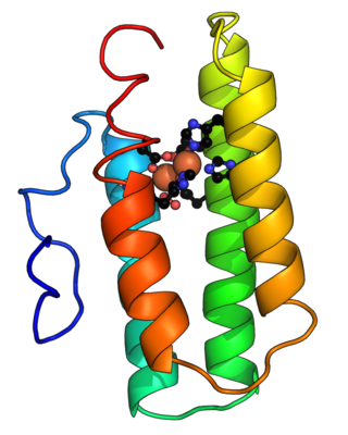
Hemoglobin is a protein containing iron that facilitates the transport of oxygen in red blood cells. Almost all vertebrates contain hemoglobin, with the exception of the fish family Channichthyidae. Hemoglobin in the blood carries oxygen from the respiratory organs to the other tissues of the body, where it releases the oxygen to enable aerobic respiration which powers the animal's metabolism. A healthy human has 12 to 20 grams of hemoglobin in every 100 mL of blood. Hemoglobin is a metalloprotein, a chromoprotein, and globulin.

<span class="mw-page-title-main">Hemocyanin</span> Proteins that transport oxygen throughout the bodies of some invertebrate animals

Hemocyanins (also spelled haemocyanins and abbreviated Hc) are proteins that transport oxygen throughout the bodies of some invertebrate animals. These metalloproteins contain two copper atoms that reversibly bind a single oxygen molecule (O2). They are second only to hemoglobin in frequency of use as an oxygen transport molecule. Unlike the hemoglobin in red blood cells found in vertebrates, hemocyanins are not confined in blood cells but are instead suspended directly in the hemolymph. Oxygenation causes a color change between the colorless Cu(I) deoxygenated form and the blue Cu(II) oxygenated form.

<span class="mw-page-title-main">Hemerythrin</span> InterPro Family

Hemerythrin (also spelled haemerythrin; Ancient Greek: αἷμα, romanized: haîma, lit. 'blood', Ancient Greek: ἐρυθρός, romanized: erythrós, lit. 'red') is an oligomeric protein responsible for oxygen (O2) transport in the marine invertebrate phyla of sipunculids, priapulids, brachiopods, and in a single annelid worm genus, Magelona. Myohemerythrin is a monomeric O2-binding protein found in the muscles of marine invertebrates. Hemerythrin and myohemerythrin are essentially colorless when deoxygenated, but turn a violet-pink in the oxygenated state.

Cooperative binding occurs in molecular binding systems containing more than one type, or species, of molecule and in which one of the partners is not mono-valent and can bind more than one molecule of the other species. In general, molecular binding is an interaction between molecules that results in a stable physical association between those molecules.

<span class="mw-page-title-main">Allosteric regulation</span> Regulation of enzyme activity

In biochemistry, allosteric regulation is the regulation of an enzyme by binding an effector molecule at a site other than the enzyme's active site.

<span class="mw-page-title-main">Solvation shell</span> Solvent interface of a solute

A solvation shell or solvation sheath is the solvent interface of any chemical compound or biomolecule that constitutes the solute in a solution. When the solvent is water it is called a hydration shell or hydration sphere. The number of solvent molecules surrounding each unit of solute is called the hydration number of the solute.

<span class="mw-page-title-main">Binding site</span> Molecule-specific coordinate bonding area in biological systems

In biochemistry and molecular biology, a binding site is a region on a macromolecule such as a protein that binds to another molecule with specificity. The binding partner of the macromolecule is often referred to as a ligand. Ligands may include other proteins, enzyme substrates, second messengers, hormones, or allosteric modulators. The binding event is often, but not always, accompanied by a conformational change that alters the protein's function. Binding to protein binding sites is most often reversible, but can also be covalent reversible or irreversible.

<span class="mw-page-title-main">Bohr effect</span> Concept in physiology

The Bohr effect is a phenomenon first described in 1904 by the Danish physiologist Christian Bohr. Hemoglobin's oxygen binding affinity (see oxygen–haemoglobin dissociation curve) is inversely related both to acidity and to the concentration of carbon dioxide. That is, the Bohr effect refers to the shift in the oxygen dissociation curve caused by changes in the concentration of carbon dioxide or the pH of the environment. Since carbon dioxide reacts with water to form carbonic acid, an increase in CO2 results in a decrease in blood pH, resulting in hemoglobin proteins releasing their load of oxygen. Conversely, a decrease in carbon dioxide provokes an increase in pH, which results in hemoglobin picking up more oxygen.

<span class="mw-page-title-main">Host–guest chemistry</span> Supramolecular structures held together other than by covalent bonds

In supramolecular chemistry, host–guest chemistry describes complexes that are composed of two or more molecules or ions that are held together in unique structural relationships by forces other than those of full covalent bonds. Host–guest chemistry encompasses the idea of molecular recognition and interactions through non-covalent bonding. Non-covalent bonding is critical in maintaining the 3D structure of large molecules, such as proteins and is involved in many biological processes in which large molecules bind specifically but transiently to one another.

<span class="mw-page-title-main">Hill equation (biochemistry)</span> Diagram showing the proportion of a receptor bound to a ligand

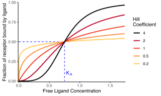
In biochemistry and pharmacology, the Hill equation refers to two closely related equations that reflect the binding of ligands to macromolecules, as a function of the ligand concentration. A ligand is "a substance that forms a complex with a biomolecule to serve a biological purpose", and a macromolecule is a very large molecule, such as a protein, with a complex structure of components. Protein-ligand binding typically changes the structure of the target protein, thereby changing its function in a cell.

<span class="mw-page-title-main">Monod–Wyman–Changeux model</span> Biochemical model of protein transitions

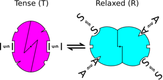
In biochemistry, the Monod–Wyman–Changeux model describes allosteric transitions of proteins made up of identical subunits. It was proposed by Jean-Pierre Changeux in his PhD thesis, and described by Jacques Monod, Jeffries Wyman, and Jean-Pierre Changeux. It contrasts with the sequential model and substrate presentation.

<span class="mw-page-title-main">Oxygen–hemoglobin dissociation curve</span> Visual tool used to understand how human blood carries and releases oxygen

The oxygen–hemoglobin dissociation curve, also called the oxyhemoglobin dissociation curve or oxygen dissociation curve (ODC), is a curve that plots the proportion of hemoglobin in its saturated (oxygen-laden) form on the vertical axis against the prevailing oxygen tension on the horizontal axis. This curve is an important tool for understanding how our blood carries and releases oxygen. Specifically, the oxyhemoglobin dissociation curve relates oxygen saturation (SO2) and partial pressure of oxygen in the blood (PO2), and is determined by what is called "hemoglobin affinity for oxygen"; that is, how readily hemoglobin acquires and releases oxygen molecules into the fluid that surrounds it.

EC<sub>50</sub> Concentration of a compound where 50% of its maximal effect is observed

Half maximal effective concentration (EC50) is a measure of the concentration of a drug, antibody or toxicant which induces a biological response halfway between the baseline and maximum after a specified exposure time. More simply, EC50 can be defined as the concentration required to obtain a 50% [...] effect and may be also written as [A]50. It is commonly used as a measure of a drug's potency, although the use of EC50 is preferred over that of 'potency', which has been criticised for its vagueness. EC50 is a measure of concentration, expressed in molar units (M), where 1 M is equivalent to 1 mol/L.

Iron-binding proteins are carrier proteins and metalloproteins that are important in iron metabolism and the immune response. Iron is required for life.

<span class="mw-page-title-main">Sequential model</span> Type of model used in biochemistry

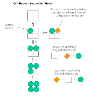
The sequential model is a theory that describes cooperativity of protein subunits. It postulates that a protein's conformation changes with each binding of a ligand, thus sequentially changing its affinity for the ligand at neighboring binding sites. It gives one explanation for cooperative binding.

Molecular binding is an attractive interaction between two molecules that results in a stable association in which the molecules are in close proximity to each other. It is formed when atoms or molecules bind together by sharing of electrons. It often, but not always, involves some chemical bonding.

<span class="mw-page-title-main">Goldbeter–Koshland kinetics</span> Steady-state solution for a 2-state biological system

The Goldbeter–Koshland kinetics describe a steady-state solution for a 2-state biological system. In this system, the interconversion between these two states is performed by two enzymes with opposing effect. One example would be a protein Z that exists in a phosphorylated form ZP and in an unphosphorylated form Z; the corresponding kinase Y and phosphatase X interconvert the two forms. In this case we would be interested in the equilibrium concentration of the protein Z. It has many applications in the description of biological systems.

In chemistry, binding selectivity is defined with respect to the binding of ligands to a substrate forming a complex. Binding selectivity describes how a ligand may bind more preferentially to one receptor than another. A selectivity coefficient is the equilibrium constant for the reaction of displacement by one ligand of another ligand in a complex with the substrate. Binding selectivity is of major importance in biochemistry and in chemical separation processes.

<span class="mw-page-title-main">Ultrasensitivity</span>

In molecular biology, ultrasensitivity describes an output response that is more sensitive to stimulus change than the hyperbolic Michaelis-Menten response. Ultrasensitivity is one of the biochemical switches in the cell cycle and has been implicated in a number of important cellular events, including exiting G2 cell cycle arrests in Xenopus laevis oocytes, a stage to which the cell or organism would not want to return.

References

  1. 1 2 Whitford D (2005). Proteins: structure and function. John Wiley & Sons. pp. 66–74.
  2. Abeliovich H (July 2005). "An empirical extremum principle for the hill coefficient in ligand-protein interactions showing negative cooperativity". Biophysical Journal. 89 (1): 76–9. Bibcode:2005BpJ....89...76A. doi:10.1529/biophysj.105.060194. PMC   1366580 . PMID   15834004.
  3. Hussain R, Kumari I, Sharma S, Ahmed M, Khan TA, Akhter Y (December 2017). "Catalytic diversity and homotropic allostery of two Cytochrome P450 monooxygenase like proteins from Trichoderma brevicompactum". Journal of Biological Inorganic Chemistry. 22 (8): 1197–1209. doi:10.1007/s00775-017-1496-6. PMID   29018974. S2CID   25685603.
  4. Kholodenko BN, Hoek JB, Westerhoff HV, Brown GC (September 1997). "Quantification of information transfer via cellular signal transduction pathways". FEBS Letters. 414 (2): 430–4. doi: 10.1016/S0014-5793(97)01018-1 . PMID   9315734. S2CID   19466336.
  5. 1 2 3 4 5 Altszyler E, Ventura AC, Colman-Lerner A, Chernomoretz A (29 June 2017). "Ultrasensitivity in signaling cascades revisited: Linking local and global ultrasensitivity estimations". PLOS ONE. 12 (6): e0180083. arXiv: 1608.08007 . Bibcode:2017PLoSO..1280083A. doi: 10.1371/journal.pone.0180083 . PMC   5491127 . PMID   28662096. CC-BY icon.svg This article contains quotations from this source, which is available under the Creative Commons Attribution 4.0 International (CC BY 4.0) license.
  6. Brown GC, Hoek JB, Kholodenko BN (August 1997). "Why do protein kinase cascades have more than one level?". Trends in Biochemical Sciences. 22 (8): 288. doi:10.1016/s0968-0004(97)82216-5. PMID   9270298.
  7. Ferrell JE (August 1997). "How responses get more switch-like as you move down a protein kinase cascade". Trends in Biochemical Sciences. 22 (8): 288–9. doi:10.1016/s0968-0004(97)82217-7. PMID   9270299.
  8. Altszyler E, Ventura A, Colman-Lerner A, Chernomoretz A (October 2014). "Impact of upstream and downstream constraints on a signaling module's ultrasensitivity". Physical Biology. 11 (6): 066003. Bibcode:2014PhBio..11f6003A. doi:10.1088/1478-3975/11/6/066003. PMC   4233326 . PMID   25313165.
  9. Rácz E, Slepchenko BM (July 2008). "On sensitivity amplification in intracellular signaling cascades". Physical Biology. 5 (3): 036004. Bibcode:2008PhBio...5c6004R. doi:10.1088/1478-3975/5/3/036004. PMC   2675913 . PMID   18663279.