Cell biology is a branch of biology that studies the structure, function, and behavior of cells. All living organisms are made of cells. A cell is the basic unit of life that is responsible for the living and functioning of organisms. Cell biology is the study of the structural and functional units of cells. Cell biology encompasses both prokaryotic and eukaryotic cells and has many subtopics which may include the study of cell metabolism, cell communication, cell cycle, biochemistry, and cell composition. The study of cells is performed using several microscopy techniques, cell culture, and cell fractionation. These have allowed for and are currently being used for discoveries and research pertaining to how cells function, ultimately giving insight into understanding larger organisms. Knowing the components of cells and how cells work is fundamental to all biological sciences while also being essential for research in biomedical fields such as cancer, and other diseases. Research in cell biology is interconnected to other fields such as genetics, molecular genetics, molecular biology, medical microbiology, immunology, and cytochemistry.

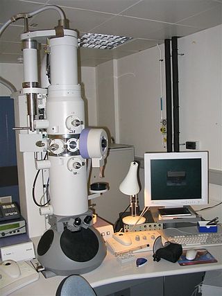
An electron microscope is a microscope that uses a beam of electrons as a source of illumination. They use electron optics that are analogous to the glass lenses of an optical light microscope to control the electron beam, for instance focusing them to produce magnified images or electron diffraction patterns. As the wavelength of an electron can be up to 100,000 times smaller than that of visible light, electron microscopes have a much higher resolution of about 0.1 nm, which compares to about 200 nm for light microscopes. Electron microscope may refer to:

Histology, also known as microscopic anatomy or microanatomy, is the branch of biology that studies the microscopic anatomy of biological tissues. Histology is the microscopic counterpart to gross anatomy, which looks at larger structures visible without a microscope. Although one may divide microscopic anatomy into organology, the study of organs, histology, the study of tissues, and cytology, the study of cells, modern usage places all of these topics under the field of histology. In medicine, histopathology is the branch of histology that includes the microscopic identification and study of diseased tissue. In the field of paleontology, the term paleohistology refers to the histology of fossil organisms.

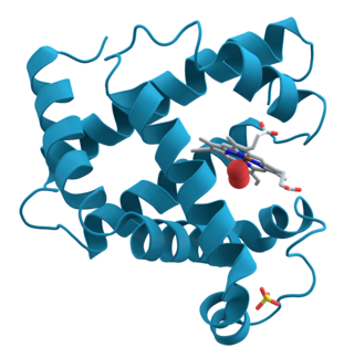
Proteins are large biomolecules and macromolecules that comprise one or more long chains of amino acid residues. Proteins perform a vast array of functions within organisms, including catalysing metabolic reactions, DNA replication, responding to stimuli, providing structure to cells and organisms, and transporting molecules from one location to another. Proteins differ from one another primarily in their sequence of amino acids, which is dictated by the nucleotide sequence of their genes, and which usually results in protein folding into a specific 3D structure that determines its activity.

In biochemistry, immunostaining is any use of an antibody-based method to detect a specific protein in a sample. The term "immunostaining" was originally used to refer to the immunohistochemical staining of tissue sections, as first described by Albert Coons in 1941. However, immunostaining now encompasses a broad range of techniques used in histology, cell biology, and molecular biology that use antibody-based staining methods.

Phalloidin belongs to a class of toxins called phallotoxins, which are found in the death cap mushroom (Amanita phalloides). It is a rigid bicyclic heptapeptide that is lethal after a few days when injected into the bloodstream. The major symptom of phalloidin poisoning is acute hunger due to the destruction of liver cells. It functions by binding and stabilizing filamentous actin (F-actin) and effectively prevents the depolymerization of actin fibers. Due to its tight and selective binding to F-actin, derivatives of phalloidin containing fluorescent tags are used widely in microscopy to visualize F-actin in biomedical research.
In cell biology, a granule is a small particle. It can be any structure barely visible by light microscopy. The term is most often used to describe a secretory vesicle.

The glycocalyx, also known as the pericellular matrix and sometime cell coat, is a glycoprotein and glycolipid covering that surrounds the cell membranes of bacteria, epithelial cells, and other cells. It was described in a review article in 1970.

Hybridoma technology is a method for producing large numbers of identical antibodies. This process starts by injecting a mouse with an antigen that provokes an immune response. A type of white blood cell, the B cell, produces antibodies that bind to the injected antigen. These antibody producing B-cells are then harvested from the mouse and, in turn, fused with immortal B cell cancer cells, a myeloma, to produce a hybrid cell line called a hybridoma, which has both the antibody-producing ability of the B-cell and the longevity and reproductivity of the myeloma. The hybridomas can be grown in culture, each culture starting with one viable hybridoma cell, producing cultures each of which consists of genetically identical hybridomas which produce one antibody per culture (monoclonal) rather than mixtures of different antibodies (polyclonal). The myeloma cell line that is used in this process is selected for its ability to grow in tissue culture and for an absence of antibody synthesis. In contrast to polyclonal antibodies, which are mixtures of many different antibody molecules, the monoclonal antibodies produced by each hybridoma line are all chemically identical.

A fluorescence microscope is an optical microscope that uses fluorescence instead of, or in addition to, scattering, reflection, and attenuation or absorption, to study the properties of organic or inorganic substances. "Fluorescence microscope" refers to any microscope that uses fluorescence to generate an image, whether it is a simple set up like an epifluorescence microscope or a more complicated design such as a confocal microscope, which uses optical sectioning to get better resolution of the fluorescence image.

Immunocytochemistry (ICC) is a common laboratory technique that is used to anatomically visualize the localization of a specific protein or antigen in cells by use of a specific primary antibody that binds to it. The primary antibody allows visualization of the protein under a fluorescence microscope when it is bound by a secondary antibody that has a conjugated fluorophore. ICC allows researchers to evaluate whether or not cells in a particular sample express the antigen in question. In cases where an immunopositive signal is found, ICC also allows researchers to determine which sub-cellular compartments are expressing the antigen.
Nanovid microscopy, from "nanometer video-enhanced microscopy", is a microscopic technique aimed at visualizing colloidal gold particles of 20–40 nm diameter as dynamic markers at the light-microscopic level. The nanogold particles as such are smaller than the diffraction limit of light, but can be visualized by using video-enhanced differential interference contrast (VEDIC). The technique is based on the use of contrast enhancement by video techniques and digital image processing. Nanovid microscopy, by combining small colloidal gold probes with video-enhanced quantitative microscopy, allows studying the intracellular dynamics of specific proteins in living cells.

John E. Heuser is an American Professor of Biophysics in the department of Cell Biology and Physiology at the Washington University School of Medicine as well as a Professor at the Institute for Integrated Cell-Material Sciences (iCeMS) at Kyoto University.

Immunolabeling is a biochemical process that enables the detection and localization of an antigen to a particular site within a cell, tissue, or organ. Antigens are organic molecules, usually proteins, capable of binding to an antibody. These antigens can be visualized using a combination of antigen-specific antibody as well as a means of detection, called a tag, that is covalently linked to the antibody. If the immunolabeling process is meant to reveal information about a cell or its substructures, the process is called immunocytochemistry. Immunolabeling of larger structures is called immunohistochemistry.
Alex Benjamin Novikoff was a Russian Empire-born American biologist who is recognized for his pioneering works in the discoveries of cell organelles. A victim of American Cold War antagonism to communism that he supported, he is also recognized as a public figure of the mid-20th century at the height of McCarthyism in America. As his original discoveries such as cell organelles and autophagy earned other scientists Nobel Prizes, he is regarded as one of the overlooked scientists to get Nobel Prize.
Standards for the identification of cell death have changed. Cell death used to be defined and described based on morphology. Now there is a switch in classifying it basing on molecular and genetic definitions. This description is more functional and applies to both in vitro and in vivo, so cell death subroutines are now described by a series of precise, measurable, biochemical features. A set of recommendations for describing the terminology of cell death was proposed by the Nomenclature Committee on Cell Death (NCCD) in 2009, because misusing words and concepts may slow down progress in the area of cell death research.

The Golgi matrix is a collection of proteins involved in the structure and function of the Golgi apparatus. The matrix was first isolated in 1994 as an amorphous collection of 12 proteins that remained associated together in the presence of detergent and 150 mM NaCl. Treatment with a protease enzyme removed the matrix, which confirmed the importance of proteins for the matrix structure. Modern freeze etch electron microscopy (EM) clearly shows a mesh connecting Golgi cisternae and associated vesicles. Further support for the existence of a matrix comes from EM images showing that ribosomes are excluded from regions between and near Golgi cisternae.

Cell unroofing is any of various methods to isolate and expose the cell membrane of cells. Differently from the more common membrane extraction protocols performed with multiple steps of centrifugation, in cell unroofing the aim is to tear and preserve patches of the plasma membrane in order to perform in situ experiments using.

Convergent beam electron diffraction (CBED) is an electron diffraction technique where a convergent or divergent beam of electrons is used to study materials.
Lucas Andrew Staehelin was a retired Swiss-American cell biologist. He was professor emeritus at the University of Colorado Boulder.


