Developmental biology is the study of the process by which animals and plants grow and develop. Developmental biology also encompasses the biology of regeneration, asexual reproduction, metamorphosis, and the growth and differentiation of stem cells in the adult organism.

A zygote is an eukaryotic cell formed by a fertilization event between two gametes. The zygote's genome is a combination of the DNA in each gamete, and contains all of the genetic information necessary to form a new individual. In multicellular organisms, the zygote is the earliest developmental stage. In single-celled organisms, the zygote can divide asexually by mitosis to produce identical offspring.
A somatic cell, or vegetal cell, is any biological cell forming the body of an organism; that is, in a multicellular organism, any cell other than a gamete, germ cell, gametocyte or undifferentiated stem cell.
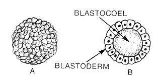
Blastulation is the stage in early animal embryonic development that produces the blastula. The blastula (from Greek βλαστός is a hollow sphere of cells surrounding an inner fluid-filled cavity. Embryonic development begins with a sperm fertilizing an egg cell to become a zygote, which undergoes many cleavages to develop into a ball of cells called a morula. Only when the blastocoel is formed does the early embryo become a blastula. The blastula precedes the formation of the gastrula in which the germ layers of the embryo form.

The blastocyst is a structure formed in the early development of mammals. It possesses an inner cell mass (ICM) which subsequently forms the embryo. The outer layer of the blastocyst consists of cells collectively called the trophoblast. This layer surrounds the inner cell mass and a fluid-filled cavity known as the blastocoel. The trophoblast gives rise to the placenta. The name "blastocyst" arises from the Greek βλαστός blastos and κύστις kystis. In other animals this is called a blastula.
An oocyte, oöcyte, ovocyte, or rarely ocyte, is a female gametocyte or germ cell involved in reproduction. In other words, it is an immature ovum, or egg cell. An oocyte is produced in the ovary during female gametogenesis. The female germ cells produce a primordial germ cell (PGC), which then undergoes mitosis, forming oogonia. During oogenesis, the oogonia become primary oocytes. An oocyte is a form of genetic material that can be collected for cryoconservation. Cryoconservation of animal genetic resources has been put into action as a means of conserving traditional livestock.
In biology, a blastomere is a type of cell produced by cleavage of the zygote after fertilization and is an essential part of blastula formation.

A blastocoel, also spelled blastocoele and blastocele, and also called blastocyst cavity is a fluid-filled cavity that forms in the blastula (blastocyst) of early amphibian and echinoderm embryos, or between the epiblast and hypoblast of avian, reptilian, and mammalian blastoderm-stage embryos.
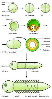
In developmental biology, embryonic development, also known as embryogenesis, is the development of an animal or plant embryo. Embryonic development starts with the fertilization of an egg cell (ovum) by a sperm cell, (spermatozoon). Once fertilized, the ovum becomes a single diploid cell known as a zygote. The zygote undergoes mitotic divisions with no significant growth and cellular differentiation, leading to development of a multicellular embryo after passing through an organizational checkpoint during mid-embryogenesis. In mammals, the term refers chiefly to the early stages of prenatal development, whereas the terms fetus and fetal development describe later stages.
In developmental biology, cleavage is the division of cells in the early embryo. The process follows fertilization, with the transfer being triggered by the activation of a cyclin-dependent kinase complex. The zygotes of many species undergo rapid cell cycles with no significant overall growth, producing a cluster of cells the same size as the original zygote. The different cells derived from cleavage are called blastomeres and form a compact mass called the morula. Cleavage ends with the formation of the blastula.
In early embryogenesis of most eutherian mammals, the inner cell mass is the mass of cells inside the primordial embryo that will eventually give rise to the definitive structures of the fetus. This structure forms in the earliest steps of development, before implantation into the endometrium of the uterus has occurred. The ICM lies within the blastocoele and is entirely surrounded by the single layer of cells called trophoblast.
An asymmetric cell division produces two daughter cells with different cellular fates. This is in contrast to symmetric cell divisions which give rise to daughter cells of equivalent fates. Notably, stem cells divide asymmetrically to give rise to two distinct daughter cells: one copy of the original stem cell as well as a second daughter programmed to differentiate into a non-stem cell fate.
Within the field of developmental biology, one goal is to understand how a particular cell develops into a final cell type, known as fate determination. Within an embryo, several processes play out at the cellular and tissue level to create an organism. These processes include cell proliferation, differentiation, cellular movement and programmed cell death. Each cell in an embryo receives molecular signals from neighboring cells in the form of proteins, RNAs and even surface interactions. Almost all animals undergo a similar sequence of events during very early development, a conserved process known as embryogenesis. During embryogenesis, cells exist in three germ layers, and undergo gastrulation. While embryogenesis has been studied for more than a century, it was only recently that scientists discovered that a basic set of the same proteins and mRNAs are involved in embryogenesis. Evolutionary conservation is one of the reasons that model systems such as the fly, the mouse, and other organisms are used as models to study embryogenesis and developmental biology. Studying model organisms provides information relevant to other animals, including humans. New discoveries and investigations includen how RNAs and proteins are expressed differentially between cells types, temporally and spatially; and how they are responsible for cell fate determination contributing to the vast diversity of organisms.
In the field of developmental biology, regional differentiation is the process by which different areas are identified in the development of the early embryo. The process by which the cells become specified differs between organisms.

The development of fishes is unique in some specific aspects compared to the development of other animals.

Human embryonic development, or human embryogenesis, refers to the development and formation of the human embryo. It is characterised by the processes of cell division and cellular differentiation of the embryo that occurs during the early stages of development. In biological terms, the development of the human body entails growth from a one-celled zygote to an adult human being. Fertilisation occurs when the sperm cell successfully enters and fuses with an egg cell (ovum). The genetic material of the sperm and egg then combine to form a single cell called a zygote and the germinal stage of development commences. Embryonic development in the human, covers the first eight weeks of development; at the beginning of the ninth week the embryo is termed a fetus. Human embryology is the study of this development during the first eight weeks after fertilisation. The normal period of gestation (pregnancy) is about nine months or 40 weeks.
Embryomics is the identification, characterization and study of the diverse cell types which arise during embryogenesis, especially as this relates to the location and developmental history of cells in the embryo. Cell type may be determined according to several criteria: location in the developing embryo, gene expression as indicated by protein and nucleic acid markers and surface antigens, and also position on the embryogenic tree.

Cell potency is a cell's ability to differentiate into other cell types. The more cell types a cell can differentiate into, the greater its potency. Potency is also described as the gene activation potential within a cell, which like a continuum, begins with totipotency to designate a cell with the most differentiation potential, pluripotency, multipotency, oligopotency, and finally unipotency.
Leech embryogenesis is the process by which the embryo of the leech forms and develops. The embryonic development of the larva occurs as a series of stages. During stage 1, the first cleavage occurs, which gives rise to an AB and a CD blastomere, and is in the interphase of this cell division when a yolk-free cytoplasm called teloplasm is formed. The teloplasm is known to be a determinant for the specification of the D cell fate. In stage 3, during the second cleavage, an unequal division occurs in the CD blastomere. As a consequence, it creates a large D cell on the left and a smaller C cell to the right. This unequal division process is dependent on actomyosin, and by the end of stage 3 the AB cell divides. On stage 4 of development, the micromeres and teloblast stem cells are formed and subsequently, the D quadrant divides to form the DM and the DNOPQ teloblast precursor cells. By the end stage 6, the zygote contains a set of 25 micromeres, 3 macromeres and 10 teloblasts derived from the D quadrant.