D-peptide

Last updated

A D-peptide is a small sequence of D-amino acids. Since ribosomes are specific to L-amino acids, D-peptides rarely occur naturally in organisms and are not easily digested or degraded. D-peptide peptidomimetics are D-peptides designed to mimic natural L-peptides that commonly have therapeutic properties. A peptide with secondary structure cannot be mimicked by its retro-inverse, as linking in the reverse order breaks many backbone interactions essential for the secondary structure. [1] An approach to mimicking these peptides is by searching for similar (sidechain) structures in a mirrored copy of the Protein Data Bank for the structured elements, and then linking the sections by retro-inversed versions of the loops found in the original protein. [2]

Figure 2. D-peptides assume the mirror image conformation of their L-peptide analogues. Many D-proteins and other D-peptides when placed in a nonchiral solvent like water, assume the mirror image conformation of their L-peptide counterpart. Pictured is an L-peptide (1) fragment with the sequence Asp-Val-Ser and the D-peptide Asp-Val-Ser (2) shown from C-terminus to N-terminus. L-Peptide-D-PeptideMirrorImages.png
Figure 2. D-peptides assume the mirror image conformation of their L-peptide analogues. Many D-proteins and other D-peptides when placed in a nonchiral solvent like water, assume the mirror image conformation of their L-peptide counterpart. Pictured is an L-peptide (1) fragment with the sequence Asp-Val-Ser and the D-peptide Asp-Val-Ser (2) shown from C-terminus to N-terminus.

When placed in a nonchiral solvent like water, D-peptides, as well as the larger polypeptide D-proteins, have similar but mirrored properties to the L-peptides and L-proteins with identical sequences. If an L-protein does not require a chaperone or a structural cofactor to fold, its D-enantiomer protein should have a mirror image conformation with respect to the L-protein (Figure 2). A D-enzyme should act on substrates of reverse chirality compared to the L-enzyme with the same sequence. Similarly, if an L-peptide binds to an L-protein, their D-peptide and D-protein counterparts should bind together in a mirrored way. [3]

D-peptides also have properties that make them attractive as drugs. D-peptides are less susceptible to be degraded in the stomach or inside cells by proteolysis. D-peptide drugs can, therefore, be taken orally and are effective for a longer period of time. D-peptides are easy to synthesize when compared to many other drugs. In some cases, D-peptides can have a low immunogenic response. [4]

Ret design

An L-peptide has three analogue sequences (Figure 3) built from L and D amino acids: the D-enantiomer or inverso-peptide with the same sequence, but composed of D-amino acids and a mirror conformation; the retro-peptide, consisting of the same sequence of L amino acids but in reverse order; and the retro-inverso or D-retro-enantiomer peptide, consisting of D-amino acids in the reversed sequence. [5] [6]

While the L-peptide and its D-enantiomer are mirror structures of each other, the L-retro-peptide is the mirror image of the D-retro-inverso-peptide. On the other hand, the L-peptide and the D-retro-inverso-peptide share a similar arrangement of side-chains, although their carboxyl and amino groups point in opposing directions. For small peptides that do not depend on a secondary structure for binding, an L-peptide and its D-retro-inverso-peptide is likely to have a similar binding affinity with a target L-protein.

Figure 3. An L-peptide and its analogues. An L-peptide (1) sequence has three analogues: the D-enantiomer (3) with the same sequence, the retro L-peptide (4) with the inverted sequence, and the retro-inverso D-peptide(2), with all D-amino acids and the inverted sequence. In this image (1) and (3) are shown from C-terminus on the left to N-terminus on the right, while (2) and (4) are shown from N-terminus to C-terminus. Note that (1) and (2) have similar side chain positions; one is the retro-inverso sequence of the other. The same applies to (3) and (4). L-peptideD-peptideAnalogues.png
Figure 3. An L-peptide and its analogues. An L-peptide (1) sequence has three analogues: the D-enantiomer (3) with the same sequence, the retro L-peptide (4) with the inverted sequence, and the retro-inverso D-peptide(2), with all D-amino acids and the inverted sequence. In this image (1) and (3) are shown from C-terminus on the left to N-terminus on the right, while (2) and (4) are shown from N-terminus to C-terminus. Note that (1) and (2) have similar side chain positions; one is the retro-inverso sequence of the other. The same applies to (3) and (4).

Mirror-image phage display

Phage display is a technique to screen large libraries of peptides for binding to a target protein. In phage display, the DNA sequence that codes the potential drug-peptide is fused to the gene of the protein coat of bacteriophages and introduced into a vector. Diversity can be introduced to the peptide by mutagenesis. The protein coats peptides are then expressed and purified, and applied to a surface of immobilized protein targets. The surface is then washed away to remove non-binding peptides, while the remaining binding peptides are eluted. [7]

Mirror-image phage display is a similar method that can be used to screen large libraries of D-peptides that bind to target L-proteins. More precisely, since D-peptides can not be expressed in bacteriophages, mirror-image phage display screens L-peptides that bind to immobilized D-proteins that are previously chemically synthesized. Because of the mirror properties of D-peptides, the D-enantiomer of an L-peptide that binds to a D-protein will bind to the L-protein.

Mirror-image phage display, however, has two disadvantages when compared to phage display. Target D-proteins must be chemically synthesized, which is normally an expensive and time-consuming process. Also, the target protein must not require a cofactor or a chaperone to fold, otherwise the chemically synthesized D-protein will not fold to the target, mirror structure.

Figure 4. Mirror image phage display. (A) A target L-amino acid protein (L-protein) for which an L-peptide inhibitor might be available is selected. The D-enantiomer protein (D-protein) is chemically synthesized from the same sequence using D-amino acids. If the target L-protein does not require a chaperone or co-factor to fold, the D-protein will mirror the conformation and properties of the L-protein, but the L-peptide inhibitor will most likely have little binding affinity towards it. (B) The synthesized D-proteins are fixed to a surface and exposed to phage displaying many different L-peptides. (C) L-peptides that do not bind well to the surface are washed away. The remaining L-peptides are sequenced. (D) The D-enantiomer peptides (D-peptides) of the binding L-peptides are synthesized using the same sequence and tested against the L-proteins. The D-peptide will bind the L-protein but most likely not bind the D-protein mirror. MirrorImagePhageDisplay.png
Figure 4. Mirror image phage display. (A) A target L-amino acid protein (L-protein) for which an L-peptide inhibitor might be available is selected. The D-enantiomer protein (D-protein) is chemically synthesized from the same sequence using D-amino acids. If the target L-protein does not require a chaperone or co-factor to fold, the D-protein will mirror the conformation and properties of the L-protein, but the L-peptide inhibitor will most likely have little binding affinity towards it. (B) The synthesized D-proteins are fixed to a surface and exposed to phage displaying many different L-peptides. (C) L-peptides that do not bind well to the surface are washed away. The remaining L-peptides are sequenced. (D) The D-enantiomer peptides (D-peptides) of the binding L-peptides are synthesized using the same sequence and tested against the L-proteins. The D-peptide will bind the L-protein but most likely not bind the D-protein mirror.

Related Research Articles

<span class="mw-page-title-main">Zinc finger</span> Small structural protein motif found mostly in transcriptional proteins

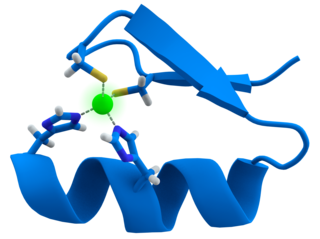
A zinc finger is a small protein structural motif that is characterized by the coordination of one or more zinc ions (Zn2+) which stabilizes the fold. It was originally coined to describe the finger-like appearance of a hypothesized structure from the African clawed frog (Xenopus laevis) transcription factor IIIA. However, it has been found to encompass a wide variety of differing protein structures in eukaryotic cells. Xenopus laevis TFIIIA was originally demonstrated to contain zinc and require the metal for function in 1983, the first such reported zinc requirement for a gene regulatory protein followed soon thereafter by the Krüppel factor in Drosophila. It often appears as a metal-binding domain in multi-domain proteins.

<span class="mw-page-title-main">Peptidomimetic</span> Class of compounds designed to mimic features of peptides

A peptidomimetic is a small protein-like chain designed to mimic a peptide. They typically arise either from modification of an existing peptide, or by designing similar systems that mimic peptides, such as peptoids and β-peptides. Irrespective of the approach, the altered chemical structure is designed to advantageously adjust the molecular properties such as stability or biological activity. This can have a role in the development of drug-like compounds from existing peptides. Peptidomimetics can be prepared by cyclization of linear peptides or coupling of stable unnatural amino acids. These modifications involve changes to the peptide that will not occur naturally. Unnatural amino acids can be generated from their native analogs via modifications such as amine alkylation, side chain substitution, structural bond extension cyclization, and isosteric replacements within the amino acid backbone. Based on their similarity with the precursor peptide, peptidomimetics can be grouped into four classes where A features the most and D the least similarities. Classes A and B involve peptide-like scaffolds, while classes C and D include small molecules.

<span class="mw-page-title-main">Phage display</span> Biological technique to evolve proteins using bacteriophages

Phage display is a laboratory technique for the study of protein–protein, protein–peptide, and protein–DNA interactions that uses bacteriophages to connect proteins with the genetic information that encodes them. In this technique, a gene encoding a protein of interest is inserted into a phage coat protein gene, causing the phage to "display" the protein on its outside while containing the gene for the protein on its inside, resulting in a connection between genotype and phenotype. The proteins that the phages are displaying can then be screened against other proteins, peptides or DNA sequences, in order to detect interaction between the displayed protein and those of other molecules. In this way, large libraries of proteins can be screened and amplified in a process called in vitro selection, which is analogous to natural selection.

<span class="mw-page-title-main">Dermorphin</span> Opioid agonist peptide compound

Dermorphin is a hepta-peptide first isolated from the skin of South American frogs belonging to the genus Phyllomedusa. The peptide is a natural opioid that binds as an agonist with high potency and selectivity to mu opioid receptors. Dermorphin is about 30–40 times more potent than morphine, but theoretically may be less likely to produce drug tolerance and addiction due to its high potency. The amino acid sequence of dermorphin is H-Tyr-D-Ala-Phe-Gly-Tyr-Pro-Ser-NH2.

<span class="mw-page-title-main">Chemical biology</span> Scientific discipline

Chemical biology is a scientific discipline between the fields of chemistry and biology. The discipline involves the application of chemical techniques, analysis, and often small molecules produced through synthetic chemistry, to the study and manipulation of biological systems. Although often confused with biochemistry, which studies the chemistry of biomolecules and regulation of biochemical pathways within and between cells, chemical biology remains distinct by focusing on the application of chemical tools to address biological questions.

Ribosome display is a technique used to perform in vitro protein evolution to create proteins that can bind to a desired ligand. The process results in translated proteins that are associated with their mRNA progenitor which is used, as a complex, to bind to an immobilized ligand in a selection step. The mRNA-protein hybrids that bind well are then reverse transcribed to cDNA and their sequence amplified via PCR. The result is a nucleotide sequence that can be used to create tightly binding proteins.

<span class="mw-page-title-main">Aptamer</span> Oligonucleotide or peptide molecules that bind specific targets

Aptamers are short sequences of artificial DNA, RNA, XNA, or peptide that bind a specific target molecule, or family of target molecules. They exhibit a range of affinities, with variable levels of off-target binding and are sometimes classified as chemical antibodies. Aptamers and antibodies can be used in many of the same applications, but the nucleic acid-based structure of aptamers, which are mostly oligonucleotides, is very different from the amino acid-based structure of antibodies, which are proteins. This difference can make aptamers a better choice than antibodies for some purposes.

<span class="mw-page-title-main">Epitope mapping</span> Identifying the binding site of an antibody on its target antigen

In immunology, epitope mapping is the process of experimentally identifying the binding site, or epitope, of an antibody on its target antigen. Identification and characterization of antibody binding sites aid in the discovery and development of new therapeutics, vaccines, and diagnostics. Epitope characterization can also help elucidate the binding mechanism of an antibody and can strengthen intellectual property (patent) protection. Experimental epitope mapping data can be incorporated into robust algorithms to facilitate in silico prediction of B-cell epitopes based on sequence and/or structural data.

<span class="mw-page-title-main">Single-domain antibody</span> Antibody fragment

A single-domain antibody (sdAb), also known as a Nanobody, is an antibody fragment consisting of a single monomeric variable antibody domain. Like a whole antibody, it is able to bind selectively to a specific antigen. With a molecular weight of only 12–15 kDa, single-domain antibodies are much smaller than common antibodies which are composed of two heavy protein chains and two light chains, and even smaller than Fab fragments and single-chain variable fragments.

<i>trp</i> operon Operon that codes for the components for production of tryptophan

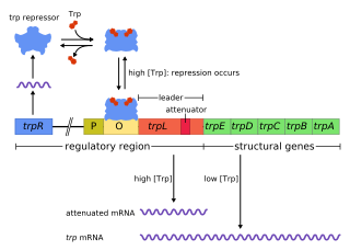
The trp operon is a group of genes that are transcribed together, encoding the enzymes that produce the amino acid tryptophan in bacteria. The trp operon was first characterized in Escherichia coli, and it has since been discovered in many other bacteria. The operon is regulated so that, when tryptophan is present in the environment, the genes for tryptophan synthesis are repressed.

mRNA display

mRNA display is a display technique used for in vitro protein, and/or peptide evolution to create molecules that can bind to a desired target. The process results in translated peptides or proteins that are associated with their mRNA progenitor via a puromycin linkage. The complex then binds to an immobilized target in a selection step. The mRNA-protein fusions that bind well are then reverse transcribed to cDNA and their sequence amplified via a polymerase chain reaction. The result is a nucleotide sequence that encodes a peptide with high affinity for the molecule of interest.

<span class="mw-page-title-main">Lysin</span>

Lysins, also known as endolysins or murein hydrolases, are hydrolytic enzymes produced by bacteriophages in order to cleave the host's cell wall during the final stage of the lytic cycle. Lysins are highly evolved enzymes that are able to target one of the five bonds in peptidoglycan (murein), the main component of bacterial cell walls, which allows the release of progeny virions from the lysed cell. Cell-wall-containing Archaea are also lysed by specialized pseudomurein-cleaving lysins, while most archaeal viruses employ alternative mechanisms. Similarly, not all bacteriophages synthesize lysins: some small single-stranded DNA and RNA phages produce membrane proteins that activate the host's autolytic mechanisms such as autolysins.

Zinc finger protein chimera are chimeric proteins composed of a DNA-binding zinc finger protein domain and another domain through which the protein exerts its effect. The effector domain may be a transcriptional activator (A) or repressor (R), a methylation domain (M) or a nuclease (N).

Imperatoxin I (IpTx) is a peptide toxin derived from the venom of the African scorpion Pandinus imperator.

<span class="mw-page-title-main">Racemic crystallography</span>

Racemic crystallography is a technique used in structural biology where crystals of a protein molecule are developed from an equimolar mixture of an L-protein molecule of natural chirality and its D-protein mirror image. L-protein molecules consist of 'left-handed' L-amino acids and the achiral amino acid glycine, whereas the mirror image D-protein molecules consist of 'right-handed' D-amino acids and glycine. Typically, both the L-protein and the D-protein are prepared by total chemical synthesis.

DNA-encoded chemical libraries (DECL) is a technology for the synthesis and screening on an unprecedented scale of collections of small molecule compounds. DECL is used in medicinal chemistry to bridge the fields of combinatorial chemistry and molecular biology. The aim of DECL technology is to accelerate the drug discovery process and in particular early phase discovery activities such as target validation and hit identification.

Affibody molecules are small, robust proteins engineered to bind to a large number of target proteins or peptides with high affinity, imitating monoclonal antibodies, and are therefore a member of the family of antibody mimetics. Affibody molecules are used in biochemical research and are being developed as potential new biopharmaceutical drugs. These molecules can be used for molecular recognition in diagnostic and therapeutic applications.

DARPins are genetically engineered antibody mimetic proteins typically exhibiting highly specific and high-affinity target protein binding. They are derived from natural ankyrin repeat proteins, one of the most common classes of binding proteins in nature, which are responsible for diverse functions such as cell signaling, regulation and structural integrity of the cell. DARPins consist of at least three, repeat motifs or modules, of which the most N- and the most C-terminal modules are referred to as "caps", since they shield the hydrophobic core of the protein. The number of internal modules is indicated as number while the caps are indicated with "N" or "C", respectively. The molecular mass of e.g. 14 or 18 kDa (kilodaltons) for four- (N2C) or five- (N3C) repeat DARPins is rather small for a biologic.

Avimers are artificial proteins that are able to specifically bind to certain antigens via multiple binding sites. Since they are not structurally related to antibodies, they are classified as a type of antibody mimetic. Avimers have been developed by the biotechnology company Avidia, now part of Amgen, as potential new pharmaceutical drugs.

<small>L</small>-Ribonucleic acid aptamer RNA-like molecule

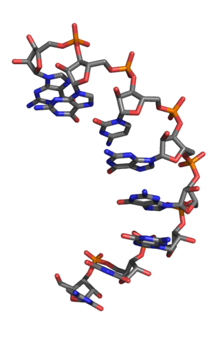
An L-ribonucleic acid aptamer is an RNA-like molecule built from L-ribose units. It is an artificial oligonucleotide named for being a mirror image of natural oligonucleotides. L-RNA aptamers are a form of aptamers. Due to their L-nucleotides, they are highly resistant to degradation by nucleases. L-RNA aptamers are considered potential drugs and are currently being tested in clinical trials.

References

  1. Lacroix E, Viguera AR, Serrano L (April 1998). "Reading protein sequences backwards". Folding & Design. 3 (2): 79–85. doi:10.1016/S1359-0278(98)00013-3. PMID   9565752.
  2. Garton M, Nim S, Stone TA, Wang KE, Deber CM, Kim PM (February 2018). "Method to generate highly stable D-amino acid analogs of bioactive helical peptides using a mirror image of the entire PDB". Proceedings of the National Academy of Sciences of the United States of America. 115 (7): 1505–1510. Bibcode:2018PNAS..115.1505G. doi: 10.1073/pnas.1711837115 . PMC   5816147 . PMID   29378946.
  3. Milton RC, Milton SC, Kent SB (1992). "Total chemical synthesis of a D-enzyme: the enantiomers of HIV-1 protease show demonstration of reciprocal chiral substrate specificity". Science. 256 (5062): 1445–1448. doi:10.1126/science.1604320. PMID   1604320.
  4. Welch BD, VanDemark AP, Heroux A, Hill CP, Kay MS (October 2007). "Potent D-peptide inhibitors of HIV-1 entry". Proceedings of the National Academy of Sciences of the United States of America. 104 (43): 16828–33. Bibcode:2007PNAS..10416828W. doi: 10.1073/pnas.0708109104 . PMC   2040420 . PMID   17942675.
  5. Guichard G, Benkirane N, Zeder-Lutz G, van Regenmortel MH, Briand JP, Muller S (October 1994). "Antigenic mimicry of natural L-peptides with retro-inverso-peptidomimetics". Proceedings of the National Academy of Sciences of the United States of America. 91 (21): 9765–9. Bibcode:1994PNAS...91.9765G. doi: 10.1073/pnas.91.21.9765 . PMC   44897 . PMID   7937888.
  6. Cardó-Vila M, Giordano RJ, Sidman RL, Bronk LF, Fan Z, Mendelsohn J, Arap W, Pasqualini R (March 2010). "From combinatorial peptide selection to drug prototype (II): targeting the epidermal growth factor receptor pathway". Proceedings of the National Academy of Sciences of the United States of America. 107 (11): 5118–23. Bibcode:2010PNAS..107.5118C. doi: 10.1073/pnas.0915146107 . PMC   2841862 . PMID   20190183.
  7. Wiesehan K, Willbold D (September 2003). "Mirror-image Phage Display: Aiming at the Mirror". ChemBioChem. 4 (9): 811–5. doi:10.1002/cbic.200300570. PMID   12964153. S2CID   34014107.