Warts are non-cancerous viral growths usually occurring on the hands and feet but can also affect other locations, such as the genitals or face. One or many warts may appear. They are distinguished from cancerous tumors as they are caused by a viral infection, such as a human papillomavirus, rather than a cancerous growth.
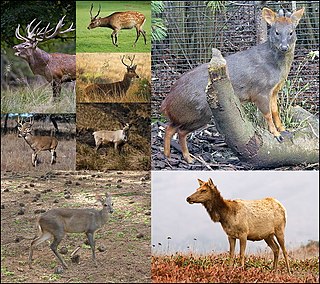
A deer or true deer is a hoofed ruminant mammal of the family Cervidae. The two main groups of deer are the Cervinae, including muntjac, elk (wapiti), red deer, and fallow deer; and the Capreolinae, including reindeer (caribou), white-tailed deer, roe deer, and moose. Male deer of all species, as well as female reindeer, grow and shed new antlers each year. In this, they differ from permanently horned antelope, which are part of a different family (Bovidae) within the same order of even-toed ungulates (Artiodactyla).

Myxomatosis is a disease caused by Myxoma virus, a poxvirus in the genus Leporipoxvirus. The natural hosts are tapeti in South and Central America, and brush rabbits in North America. The myxoma virus causes only a mild disease in these species, but causes a severe and usually fatal disease in European rabbits.

Chronic wasting disease (CWD), sometimes called zombie deer disease, is a transmissible spongiform encephalopathy (TSE) affecting deer. TSEs are a family of diseases thought to be caused by misfolded proteins called prions and include similar diseases such as BSE in cattle, Creutzfeldt-Jakob disease (CJD) in humans and scrapie in sheep. In the United States, CWD affects mule deer, white-tailed deer, red deer, sika deer, elk, caribou, and moose. Natural infection causing CWD affects members of the deer family. Experimental transmission of CWD to other species such as squirrel monkeys and genetically modified mice has been shown.

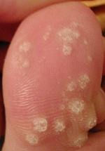
A plantar wart, or verruca vulgaris, is a wart occurring on the bottom of the foot or toes. Its color is typically similar to that of the skin. Small black dots often occur on the surface. One or more may occur in an area. They may result in pain with pressure such that walking is difficult.
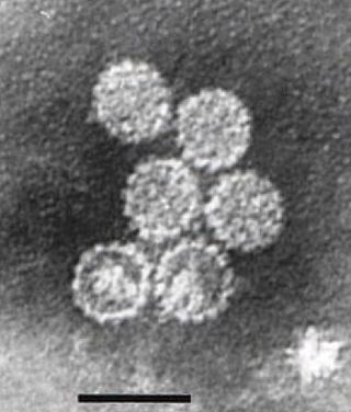
Papillomaviridae is a family of non-enveloped DNA viruses whose members are known as papillomaviruses. Several hundred species of papillomaviruses, traditionally referred to as "types", have been identified infecting all carefully inspected mammals, but also other vertebrates such as birds, snakes, turtles and fish. Infection by most papillomavirus types, depending on the type, is either asymptomatic or causes small benign tumors, known as papillomas or warts. Papillomas caused by some types, however, such as human papillomaviruses 16 and 18, carry a risk of becoming cancerous.
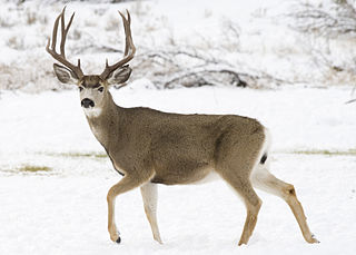
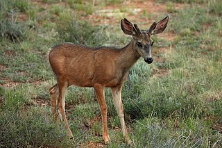
The mule deer is a deer indigenous to western North America; it is named for its ears, which are large like those of the mule. Two subspecies of mule deer are grouped into the black-tailed deer.

The term fibromatosis refers to a group of soft tissue tumors which have certain characteristics in common, including absence of cytologic and clinical malignant features, a histology consistent with proliferation of well-differentiated fibroblasts, an infiltrative growth pattern, and aggressive clinical behavior with frequent local recurrence. It is classed by the World Health Organization as an intermediate soft tissue tumor related to the sarcoma family. Arthur Purdy Stout coined the term fibromatosis, in 1954.
Parelaphostrongylus tenuis is a neurotropic nematode parasite common to white-tailed deer, Odocoileus virginianus, which causes damage to the central nervous system. Moose, elk, caribou, mule deer, and others are also susceptible to the parasite, but are aberrant hosts and are infected in neurological instead of meningeal tissue. The frequency of infection in these species increases dramatically when their ranges overlap high densities of white-tailed deer.

Bovine papillomaviruses (BPV) are a paraphyletic group of DNA viruses of the subfamily Firstpapillomavirinae of Papillomaviridae that are common in cattle. All BPVs have a circular double-stranded DNA genome. Infection causes warts of the skin and alimentary tract, and more rarely cancers of the alimentary tract and urinary bladder. They are also thought to cause the skin tumour equine sarcoid in horses and donkeys.
Elaeophora schneideri is a nematode which infests several mammalian hosts in North America. It is transmitted by horse-flies. Infection in the normal definitive hosts, mule deer or black-tailed deer, seldom produces clinical symptoms. In other hosts, such as sheep, elk, moose, and goats, infection with E. schneideri leads to elaeophorosis. Symptoms of elaeophorosis include necrosis of the muzzle, ears, and optic nerves; lack of coordination (ataxia); facial or lower limb dermatitis; horn deformities; blindness; and death.
Epizootic hemorrhagic disease (EHD) is a hemorrhagic disease of white-tailed deer caused by an infection of a virus from the genus Orbivirus subsequently called Epizootic hemorrhagic disease virus (EHDV). It is an infectious, and sometimes fatal, virus that is characterized by extensive hemorrhages, and is found throughout the United States. Large-scale outbreaks in wild ruminants affect livestock and the production industry. EHD has been found in some domestic ruminants and many species of deer including white-tailed deer, mule deer, elk, and pronghorn antelope. Seropositive black-tailed deer, fallow deer, red deer, wapiti, and roe deer have also been found, which essentially means that they were exposed to the disease at some time in the past but may not be involved in transmission. Outbreaks of EHD have been reported in cattle, although they rarely develop disease or die. Sheep may develop clinical signs, but this is also rare. EHD is often called bluetongue, but this is incorrect. Bluetongue virus is closely related to EHDV, and has similar clinical signs, but it is a different disease. Bluetongue is a serious disease in cattle, as well as other ruminants, and can have a significant effect on international trade. Testing at animal health laboratories is necessary to distinguish between the viruses that cause bluetongue and EHD.

The Shope papilloma virus (SPV), also known as cottontail rabbit papilloma virus (CRPV) or Kappapapillomavirus 2, is a papillomavirus which infects certain leporids, causing keratinous carcinomas resembling horns, typically on or near the animal's head. The carcinomas can metastasize or become large enough to interfere with the host's ability to eat, causing starvation. Richard E. Shope investigated the horns and discovered the virus in 1933, an important breakthrough in the study of oncoviruses. The virus was originally discovered in cottontail rabbits in the Midwestern U.S. but can also infect brush rabbits, black-tailed jackrabbits, snowshoe hares, European rabbits, and domestic rabbits.
The Capreolinae, Odocoileinae, or the New World deer are a subfamily of deer. Alternatively, they are known as the telemetacarpal deer, due to their bone structure being different from the plesiometacarpal deer subfamily Cervinae. The telemetacarpal deer maintain their distal lateral metacarpals, while the plesiometacarpal deer maintain only their proximal lateral metacarpals. The Capreolinae are believed to have originated in the Middle Miocene, between 7.7 and 11.5 million years ago, in Central Asia.

Cutaneous horns, also known by the Latin name cornu cutaneum, are unusual keratinous skin tumors with the appearance of horns, or sometimes of wood or coral. Formally, this is a clinical diagnosis for a "conical projection above the surface of the skin." They are usually small and localized but can, in very rare cases, be much larger. Although often benign, they can also be malignant or premalignant.

Angiofibroma (AGF) is a descriptive term for a wide range of benign skin or mucous membrane lesions in which individuals have:
- benign papules, i.e. pinhead-sized elevations that lack visible evidence of containing fluid;
- nodules, i.e. small firm lumps usually >0.1 cm in diameter; and/or
- tumors, i.e. masses often regarded as ~0.8 cm or larger.
Infantile digital fibromatosis (IDF), also termed inclusion body fibromatosis, Reye tumor, or Reye's tumor, usually occurs as a single, small, asymptomatic, nodule in the dermis on a finger or toe of infants and young children. IMF is a rare disorder with approximately 200 cases reported in the medical literature as of 2021. The World Health Organization in 2020 classified these nodules as a specific benign tumor type in the category of fibroblastic and myofibroblastic tumors. IDF was first described by the Australian pathologist, Douglas Reye, in 1965.
Fibroblastic and myofibroblastic tumors (FMTs) develop from the mesenchymal stem cells which differentiate into fibroblasts and/or the myocytes/myoblasts that differentiate into muscle cells. FMTs are a heterogeneous group of soft tissue neoplasms. The World Health Organization (2020) defined tumors as being FMTs based on their morphology and, more importantly, newly discovered abnormalities in the expression levels of key gene products made by these tumors' neoplastic cells. Histopathologically, FMTs consist of neoplastic connective tissue cells which have differented into cells that have microscopic appearances resembling fibroblasts and/or myofibroblasts. The fibroblastic cells are characterized as spindle-shaped cells with inconspicuous nucleoli that express vimentin, an intracellular protein typically found in mesenchymal cells, and CD34, a cell surface membrane glycoprotein. Myofibroblastic cells are plumper with more abundant cytoplasm and more prominent nucleoli; they express smooth muscle marker proteins such as smooth muscle actins, desmin, and caldesmon. The World Health Organization further classified FMTs into four tumor forms based on their varying levels of aggressiveness: benign, intermediate, intermediate, and malignant.
Gardner fibroma (GF) is a benign fibroblastic tumor. GF tumors typically develop in the dermis and adjacent subcutaneous tissue lying just below the dermis. These tumors typically occur on the back, abdomen, and other superficial sites but in rare cases have been diagnoses in internal sites such as the retroperitoneum and around the large blood vessels in the upper thoracic cavity. The World Health Organization, 2020, classified Gardner fibroma as a benign tumor in the category of fibroblastic and myofibroblastic tumors.
