Digital pathology

Last updated
Digital pathology is a major part of pathology informatics, and encompasses topics including slide scanning, digital imaging, image analysis and telepathology. Major topics of pathology informatics.png
Digital pathology is a major part of pathology informatics, and encompasses topics including slide scanning, digital imaging, image analysis and telepathology.

Digital pathology is a sub-field of pathology that focuses on data management based on information generated from digitized specimen slides. Through the use of computer-based technology, digital pathology utilizes virtual microscopy. [1] Glass slides are converted into digital slides that can be viewed, managed, shared and analyzed on a computer monitor. With the practice of whole-slide imaging (WSI), which is another name for virtual microscopy, [2] the field of digital pathology is growing and has applications in diagnostic medicine, with the goal of achieving efficient and cheaper diagnoses, prognosis, and prediction of diseases due to the success in machine learning and artificial intelligence in healthcare. [3]

Contents

History

The roots of digital pathology go back to the 1960s, when first telepathology experiments took place. Later in the 1990s the principle of virtual microscopy [4] appeared in several life science research areas. At the turn of the century the scientific community more and more agreed on the term "digital pathology" to denote digitization efforts in pathology. However, in 2000, the technical requirements (scanner, storage, network) were still a limiting factor for a broad dissemination of digital pathology concepts. This changed as new powerful and affordable scanner technology as well as mass / cloud storage technologies appeared on the market. The field of radiology has undergone the digital transformation almost 15 years ago, not because radiology is more advanced, but there are fundamental differences between digital images in radiology and digital pathology: The image source in radiology is the (alive) patient, and today in most cases, the image is even primarily captured in digital format. In pathology the scanning is done from preserved and processed specimens, for retrospective studies even from slides stored in a biobank. Besides this difference in pre-analytics and metadata content, the required storage in digital pathology is two to three orders of magnitude higher than in radiology. However, the advantages anticipated through digital pathology are similar to those in radiology:

Digital pathology is today widely used for educational purposes [5] in telepathology and teleconsultation as well as in research projects. Digital pathology allows to share and annotate slides in a much easier way and to download annotated lecture sets generates new opportunities for e-learning and knowledge sharing in pathology. Digital pathology in diagnostics is an emerging and upcoming field.

Environment

Scan

A microscopy slide scanner. Leica microscopy slide scanner, annotated.jpg
A microscopy slide scanner.
Whole slide image quality comparison, with a slide scanned with a 20x objective and about 0.8 gigabytes (GB) in size to the left, and a 40x objective and approximately 1.2 GB in size to the right. Each image shows a red blood cell. Whole slide image quality comparison.png
Whole slide image quality comparison, with a slide scanned with a 20x objective and about 0.8 gigabytes (GB) in size to the left, and a 40x objective and approximately 1.2 GB in size to the right. Each image shows a red blood cell.

Digital slides are created from glass slides using specialized scanning machines. All high quality scans must be free of dust, scratches, and other obstructions. There are two common methods for digital slide scanning, tile-based scanning and line-based scanning. [6] Both technologies use an integrated camera and a motorized stage to move the slide around while parts of the tissue are imaged. Tile scanners capture square field-of-view images covering the entire tissue area on the slide, while line-scanners capture images of the tissue in long, uninterrupted stripes rather than tiles. In both cases, software associated with the scanner stitch the tiles or lines together into a single, seamless image.

Z-stacking is the scanning of a slide at multiple focal planes along the vertical z-axis. [7]

View

Digital slides are accessible for viewing via a computer monitor and viewing software either locally or remotely via the Internet. An example of an open-source, web-based viewer for this purpose implemented in pure JavaScript, for desktop and mobile, is the OpenSeadragon [8] viewer. QuPath [9] is another such open source software, which is often used for digital pathology applications because it offers a powerful set of tools for working with whole slide images. OpenSlide, [10] on the other hand is a C library (Python and Java bindings are also available) that provides a simple interface to read and view whole-slide images.

Manage

Digital slides are maintained in an information management system that allows for archival and intelligent retrieval.

Network

Digital slides are often stored and delivered over the Internet or private networks, for viewing and consultation.

Analyze

Image analysis tools are used to derive objective quantification measures from digital slides. Image segmentation and classification algorithms, often implemented using deep learning neural networks, are used to identify medically significant regions and objects on digital slides. A GPU acceleration software for pathology imaging analysis, cross-comparing spatial boundaries of a huge amount of segmented micro-anatomic objects has been developed. [11] The core algorithm of PixelBox in this software has been adopted in Fixstars' Geometric Performance Primitives (GPP) library [12] as a part of NVIDIA Developer, which is a production geometry engine for advanced graphical information systems, electronic design automation, computer vision and motion planning solutions. [13]

Integrate

Digital pathology workflow is integrated into the institution's overall operational environment. Slide digitization is expected to reduce the number of routine, manually reviewed slides, maximizing workload efficiency.

Sharing

Digital pathology also allows internet information sharing for education, diagnostics, publication and research. This may take the form of publicly available datasets or open source access to machine learning algorithms.

Challenges

Bone tissue is particularly prone to folding artifacts. In this micrograph, the automatic camera is focused on a fold (left in image), resulting in defocus aberration (blur) of the surrounding tissue (right in image). Folding artifact on whole slide imaging of bone.png
Bone tissue is particularly prone to folding artifacts. In this micrograph, the automatic camera is focused on a fold (left in image), resulting in defocus aberration (blur) of the surrounding tissue (right in image).
In this case, there is no clear distinction between tumor cells and surrounding large stromal cells, requiring delimitation before applying automatic stain quantification tools. CD117 (c-kit) stain of mixed malignant germ cell tumor - crop.png
In this case, there is no clear distinction between tumor cells and surrounding large stromal cells, requiring delimitation before applying automatic stain quantification tools.

Digital pathology has been approved by the FDA for primary diagnosis. [16] The approval was based on a multi-center study of 1,992 cases in which whole-slide imaging (WSI) was shown to be non-inferior to microscopy across a wide range of surgical pathology specimens, sample types and stains. [17] While there are advantages to WSI when creating digital data from glass slides, when it comes to real-time telepathology applications, WSI is not a strong choice for discussion and collaboration between multiple remote pathologists. [18] Furthermore, unlike digital radiology where the elimination of film made return on investment (ROI) clear, the ROI on digital pathology equipment is less obvious. The strongest ROI justification includes improved quality of healthcare, increased efficiency for pathologists, and reduced costs in handling glass slides. [19]

Validation

Validation of a digital microscopy workflow in a specific environment (see above) is important to ensure high diagnostic performance of pathologists when evaluating digital whole-slide images. There are different methods that can be used for this validation process. [20] The College of American Pathologists has published a guideline with minimal requirements for validation of whole slide imaging systems for diagnostic purposes in human pathology. [21]

Potential

Trained pathologists traditionally view tissue slides under a microscope. These tissue slides may be stained to highlight cellular structures. When slides are digitized, they are able to be shared through tele-pathology and are numerically analyzed using computer algorithms. Algorithms can be used to automate the manual counting of structures, or for classifying the condition of tissue such as is used in grading tumors. They can additionally be used for feature detection of mitotic figures, epithelial cells, or tissue specific structures such as lung cancer nodules, glomeruli, or vessels, or estimation of molecular biomarkers such as mutated genes, tumor mutational burden, or transcriptional changes. [22] [23] [24] This has the potential to reduce human error and improve accuracy of diagnoses. Digital slides can be easily shared, increasing the potential for data usage in education as well as in consultations between expert pathologists. Multiplexed imaging (staining multiple markers on the same slide) allows pathologists to understand finer distribution of cell-types and their relative locations. [25] An understanding of the spatial distribution of cell-types or markers and pathways they express, can allow for prescription of targeted drugs or build combinational therapies in a personalized manner.

See also

Related Research Articles

<span class="mw-page-title-main">Magnetic resonance imaging</span> Medical imaging technique

Magnetic resonance imaging (MRI) is a medical imaging technique used in radiology to form pictures of the anatomy and the physiological processes inside the body. MRI scanners use strong magnetic fields, magnetic field gradients, and radio waves to generate images of the organs in the body. MRI does not involve X-rays or the use of ionizing radiation, which distinguishes it from computed tomography (CT) and positron emission tomography (PET) scans. MRI is a medical application of nuclear magnetic resonance (NMR) which can also be used for imaging in other NMR applications, such as NMR spectroscopy.

<span class="mw-page-title-main">Pathology</span> Study of the causes and effects of disease or injury, and how they arise

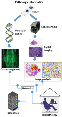
Pathology is the study of disease and injury. The word pathology also refers to the study of disease in general, incorporating a wide range of biology research fields and medical practices. However, when used in the context of modern medical treatment, the term is often used in a narrower fashion to refer to processes and tests that fall within the contemporary medical field of "general pathology", an area that includes a number of distinct but inter-related medical specialties that diagnose disease, mostly through analysis of tissue and human cell samples. Idiomatically, "a pathology" may also refer to the predicted or actual progression of particular diseases, and the affix pathy is sometimes used to indicate a state of disease in cases of both physical ailment and psychological conditions. A physician practicing pathology is called a pathologist.

<span class="mw-page-title-main">CT scan</span> Medical imaging procedure using X-rays to produce cross-sectional images

A computed tomography scan is a medical imaging technique used to obtain detailed internal images of the body. The personnel that perform CT scans are called radiographers or radiology technologists.

<span class="mw-page-title-main">Medical imaging</span> Technique and process of creating visual representations of the interior of a body

Medical imaging is the technique and process of imaging the interior of a body for clinical analysis and medical intervention, as well as visual representation of the function of some organs or tissues (physiology). Medical imaging seeks to reveal internal structures hidden by the skin and bones, as well as to diagnose and treat disease. Medical imaging also establishes a database of normal anatomy and physiology to make it possible to identify abnormalities. Although imaging of removed organs and tissues can be performed for medical reasons, such procedures are usually considered part of pathology instead of medical imaging.

<span class="mw-page-title-main">Virtual colonoscopy</span> Medical imaging of the colon

Virtual colonoscopy is the use of CT scanning or magnetic resonance imaging (MRI) to produce two- and three-dimensional images of the colon, from the lowest part, the rectum, to the lower end of the small intestine, and to display the images on an electronic display device. The procedure is used to screen for colon cancer and polyps, and may detect diverticulosis. A virtual colonoscopy can provide 3D reconstructed endoluminal views of the bowel. VC provides a secondary benefit of revealing diseases or abnormalities outside the colon.

<span class="mw-page-title-main">Computer-aided diagnosis</span> Type of diagnosis assisted by computers

Computer-aided detection (CADe), also called computer-aided diagnosis (CADx), are systems that assist doctors in the interpretation of medical images. Imaging techniques in X-ray, MRI, Endoscopy, and ultrasound diagnostics yield a great deal of information that the radiologist or other medical professional has to analyze and evaluate comprehensively in a short time. CAD systems process digital images or videos for typical appearances and to highlight conspicuous sections, such as possible diseases, in order to offer input to support a decision taken by the professional.

<span class="mw-page-title-main">Virtual microscopy</span>

Virtual microscopy is a method of posting microscope images on, and transmitting them over, computer networks. This allows independent viewing of images by large numbers of people in diverse locations. It involves a synthesis of microscopy technologies and digital technologies. The use of virtual microscopes can transform traditional teaching methods by removing the reliance on physical space, equipment, and specimens to a model that is solely dependent upon computer-internet access. This increases the convenience of accessing the slide sets and making the slides available to a broader audience. Digitized slides can have a high resolution and are resistant to being damaged or broken over time.

<span class="mw-page-title-main">Surgical pathology</span> Area of practice for anatomical pathologists

Surgical pathology is the most significant and time-consuming area of practice for most anatomical pathologists. Surgical pathology involves gross and microscopic examination of surgical specimens, as well as biopsies submitted by surgeons and non-surgeons such as general internists, medical subspecialists, dermatologists, and interventional radiologists.

<span class="mw-page-title-main">High-resolution computed tomography</span> Diagnostic imaging test

High-resolution computed tomography (HRCT) is a type of computed tomography (CT) with specific techniques to enhance image resolution. It is used in the diagnosis of various health problems, though most commonly for lung disease, by assessing the lung parenchyma. On the other hand, HRCT of the temporal bone is used to diagnose various middle ear diseases such as otitis media, cholesteatoma, and evaluations after ear operations.

A virtual slide is created when glass slides are digitally scanned in their entirety to provide a high resolution digital image using a digital scanning system for the purpose of medical digital image analysis. Digital slides can be retrieved from a storage system, and viewed on a computer screen, by running image management software on a standard web browser, and assessed in exactly the same way as on a microscope. Digital slides can be used as an alternative to traditional viewing for the purpose of teleconsultation.

<span class="mw-page-title-main">Automated tissue image analysis</span>

Automated tissue image analysis or histopathology image analysis (HIMA) is a process by which computer-controlled automatic test equipment is used to evaluate tissue samples, using computations to derive quantitative measurements from an image to avoid subjective errors.

<span class="mw-page-title-main">Telepathology</span>

Telepathology is the practice of pathology at a distance. It uses telecommunications technology to facilitate the transfer of image-rich pathology data between distant locations for the purposes of diagnosis, education, and research. Performance of telepathology requires that a pathologist selects the video images for analysis and the rendering of diagnoses. The use of "television microscopy", the forerunner of telepathology, did not require that a pathologist have physical or virtual "hands-on" involvement in the selection of microscopic fields-of-view for analysis and diagnosis.

<span class="mw-page-title-main">Cone beam computed tomography</span> Medical imaging technique

Cone beam computed tomography is a medical imaging technique consisting of X-ray computed tomography where the X-rays are divergent, forming a cone.

<span class="mw-page-title-main">CellNetix</span>

CellNetix Pathology & Laboratories, LLC, headquartered in Tukwila, Washington, is a premier anatomic pathology provider in the Pacific Northwest, with 60 physicians and more than 300 total staff. Services include cytology, histology, fine needle aspiration (FNA) services, flow cytometry, immunohistochemistry, immunofluorescence, UroVysion™, and molecular diagnostics.

A digital autopsy is a non-invasive autopsy in which digital imaging technology, such as with computerized tomography (CT) or magnetic resonance imaging (MRI) scans, is used to develop three-dimensional images for a virtual exploration of a human body.

<span class="mw-page-title-main">Artificial intelligence in healthcare</span> Overview of the use of artificial intelligence in healthcare

Artificial intelligence in healthcare is a term used to describe the use of machine-learning algorithms and software, or artificial intelligence (AI), to copy human cognition in the analysis, presentation, and understanding of complex medical and health care data, or to exceed human capabilities by providing new ways to diagnose, treat, or prevent disease. Specifically, AI is the ability of computer algorithms to arrive at approximate conclusions based solely on input data.

<span class="mw-page-title-main">Ronald S. Weinstein</span> American pathologist (1938–2021)

Ronald S. Weinstein was an American pathologist. He was a professor at the University of Arizona College of Medicine-Tucson. Weinstein served for 32 years as an academic pathology department chair, in Chicago, Illinois and then Tucson, Arizona, while also serving as a serial entrepreneur engaged in university technology transfer.

Microscopy with UV Surface Excitation (MUSE) is a novel microscopy method that utilizes the shallow penetration of UV photons excitation. Compared to conventional microscopes, which usually require sectioning to exclude blurred signals from outside of the focal plane, MUSE's low penetration depth limits the excitation volume to a thin layer, and removes the tissue sectioning requirement. The entire signal collected is the desired light, and all photons collected contribute to the image formation.

Elizabeth Anne Krupinski is a Professor and Vice Chair for Research of Radiology & Imaging Sciences at Emory University. She works on the perception of medical images and decision-making. Dr. Krupinski is a Fellow of SPIE, Fellow of the Society Imaging Informatics in Medicine, Fellow of the American Telemedicine Association(ATA) and Fellow of the American Institute for Medical & Biological Engineering (AIMBE). She has previously served as Chair for the SPIE Medical Imaging Conference, Chair of SIIM, President of the American Telemedicine Association, President of the Medical Image Perception Society and Vice President of the Society for Education and the Advancement of Research in Connected Health.

Tissue image cytometry or tissue cytometry is a method of digital histopathology and combines classical digital pathology and computational pathology into one integrated approach with solutions for all kinds of diseases, tissue and cell types as well as molecular markers and corresponding staining methods to visualize these markers. Tissue cytometry uses virtual slides as they can be generated by multiple, commercially available slide scanners, as well as dedicated image analysis software – preferentially including machine and deep learning algorithms. Tissue cytometry enables cellular analysis within thick tissues, retaining morphological and contextual information, including spatial information on defined cellular subpopulations. In this process, a tissue sample, either formalin-fixed paraffin-embedded (FFPE) or frozen tissue section, also referred to as “cryocut”, is labelled with either immunohistochemistry(IHC) or immunofluorescent markers, scanned with high-throughput slide scanners and the data gathered from virtual slides is processed and analyzed using software that is able to identify individual cells in tissue context automatically and distinguish between nucleus and cytoplasm for each cell. Additional algorithms can identify cellular membranes, subcellular structures and/or multicellular tissue structures.

References

  1. Pantanowitz L (2018). "Twenty Years of Digital Pathology: An Overview of the Road Travelled, What is on the Horizon, and the Emergence of Vendor-Neutral Archives". Journal of Pathology Informatics. 9: 40. doi: 10.4103/jpi.jpi_69_18 . PMC   6289005 . PMID   30607307.
  2. "Whole Slide Imaging | MBF Bioscience". www.mbfbioscience.com. Retrieved 2019-12-02.
  3. Saillard, Charlie; Dubois, Rémy; Tchita, Oussama; Loiseau, Nicolas; Garcia, Thierry; Adriansen, Aurélie; Carpentier, Séverine; Reyre, Joelle; Enea, Diana; von Loga, Katharina; Kamoun, Aurélie; Rossat, Stéphane; Wiscart, Corentin; Sefta, Meriem; Auffret, Michaël (2023-11-06). "Validation of MSIntuit as an AI-based pre-screening tool for MSI detection from colorectal cancer histology slides". Nature Communications. 14 (1): 6695. doi:10.1038/s41467-023-42453-6. ISSN   2041-1723. PMID   37932267.
  4. Ferreira, R; Moon, J; Humphries, J; Sussman, A; Saltz, J; Miller, R; Demarzo, A (1997). "The virtual microscope". Romanian Journal of Morphology and Embryology. 45: 449–453. PMC   2233368 . PMID   9357666.
  5. Hamilton, Peter W.; Wang, Yinhai; McCullough, Stephen J.; Sussman (2012). "Virtual microscopy and digital pathology in training and education". APMIS . 120 (4): 305–315. doi: 10.1111/j.1600-0463.2011.02869.x . PMID   22429213. S2CID   20599493.
  6. "Informatics, digital & computational pathology".
  7. Evans AJ, Salama ME, Henricks WH, Pantanowitz L (2017). "Implementation of Whole Slide Imaging for Clinical Purposes: Issues to Consider From the Perspective of Early Adopters". Arch Pathol Lab Med. 141 (7): 944–959. doi: 10.5858/arpa.2016-0074-OA . PMID   28440660.
  8. OpenSeadragon on GitHub
  9. QuPath on GitHub
  10. OpenSlide website
  11. Kaibo Wang; Yin Huai; Rubao Lee; Fusheng Wang; Xiaodong Zhang; Joel H. Saltz (2012). "Accelerating Pathology Image Data Cross-Comparison on CPU-GPU Hybrid Systems". Proceedings VLDB Endowment. Proceedings of the VLDB Endowment. International Conference on Very Large Data Bases. Vol. 5, no. 11. pp. 1543–1554. PMC   3553551 . PMID   23355955.
  12. "Geometric Performance Primitives (GPP)". NVIDIA Developer.
  13. "Letter to Kaibo Wang" (PDF).
  14. Lourenço BC, Guimarães-Teixeira C, Flores BCT, Miranda-Gonçalves V, Guimarães R, Cantante M, et al. (2022). "Ki67 and LSD1 Expression in Testicular Germ Cell Tumors Is Not Associated with Patient Outcome: Investigation Using a Digital Pathology Algorithm". Life. 12 (2): 264. doi: 10.3390/life12020264 . PMC   8875543 . PMID   35207551.
    - Figure 2 - available via license: Creative Commons Attribution 4.0 International
  15. Cruz-Roa A, Gilmore H, Basavanhally A, Feldman M, Ganesan S, Shih NNC, et al. (2017). "Accurate and reproducible invasive breast cancer detection in whole-slide images: A Deep Learning approach for quantifying tumor extent". Sci Rep. 7: 46450. doi:10.1038/srep46450. PMC   5394452 . PMID   28418027.
    "This work is licensed under a Creative Commons Attribution 4.0 International License."
  16. "FDA allows marketing of first whole slide imaging system for digital pathology" (Press release). FDA. April 12, 2017. Retrieved May 24, 2017.
  17. Mukhopadhyay, Sanjay; Feldman, Michael; Abels, Esther (2017). "Whole slide imaging versus microscopy for primary diagnosis in surgical pathology: a multicenter randomized blinded noninferiority study of 1992 cases (pivotal study)". American Journal of Surgical Pathology. 42 (1): 39–52. doi:10.1097/PAS.0000000000000948. PMC   5737464 . PMID   28961557.
  18. Siegel, Gabriel; Regelman, Dan; Maronpot, Robert; Rosenstock, Moti; Hayashi, Shim-mo; Nyska, Abraham (Oct 2018). "Utilizing novel telepathology system in preclinical studies and peer review". Journal of Toxicologic Pathology. 31 (4): 315–319. doi:10.1293/tox.2018-0032. PMC   6206289 . PMID   30393436.
  19. "How to Build a Business Case to Justify the Investment in Digital Pathology". Sectra Medical Systems. Retrieved April 26, 2015.
  20. Bertram, Christof A; Stathonikos, Nikolas; Donovan, Taryn A; Bartel, Alexander; Fuchs-Baumgartinger, Andrea; Lipnik, Karoline; can Diest, Paul J; Bonsembiante, Federico; Klopfleisch, Robert (September 2021). "Validation of digital microscopy: Review of validation methods and sources of bias". Veterinary Pathology. 59 (1): 26–38. doi:10.1177/03009858211040476. PMC   8761960 . PMID   34433345.
  21. Evans, Andrew J; Brown, Richard; Bui, Marilyn (2021). "Validating whole slide imaging systems for diagnostic purposes in pathology: guideline update from the College of American Pathologists in collaboration with the American Society for Clinical Pathology and the Association for Pathology Informatics". Arch Pathol Lab Med. 146 (4): 440–450. doi: 10.5858/arpa.2020-0723-CP . PMID   34003251.
  22. Aeffner, Famke; Zarella, Mark D.; Buchbinder, Nathan; Bui, Marilyn M.; Goodman, Matthew R.; Hartman, Douglas J.; Lujan, Giovanni M.; Molani, Mariam A.; Parwani, Anil V.; Lillard, Kate; Turner, Oliver C. (2019-03-08). "Introduction to Digital Image Analysis in Whole-slide Imaging: A White Paper from the Digital Pathology Association". Journal of Pathology Informatics. 10: 9. doi: 10.4103/jpi.jpi_82_18 . ISSN   2229-5089. PMC   6437786 . PMID   30984469.
  23. Jain, Mika S.; Massoud, Tarik F. (2020). "Predicting tumour mutational burden from histopathological images using multiscale deep learning". Nature Machine Intelligence. 2 (6): 356–362. doi:10.1038/s42256-020-0190-5. ISSN   2522-5839. S2CID   220510782.
  24. Serag, Ahmed; Ion-Margineanu, Adrian; Qureshi, Hammad; McMillan, Ryan; Saint Martin, Marie-Judith; Diamond, Jim; O'Reilly, Paul; Hamilton, Peter (2019). "Translational AI and Deep Learning in Diagnostic Pathology". Frontiers in Medicine. 6: 185. doi: 10.3389/fmed.2019.00185 . ISSN   2296-858X. PMC   6779702 . PMID   31632973.
  25. Nirmal, Ajit J.; Maliga, Zoltan; Vallius, Tuulia; Quattrochi, Brian; Chen, Alyce A.; Jacobson, Connor A.; Pelletier, Roxanne J.; Yapp, Clarence; Arias-Camison, Raquel; Chen, Yu-An; Lian, Christine G. (2022-04-11). "The spatial landscape of progression and immunoediting in primary melanoma at single cell resolution". Cancer Discovery. 12 (6): 1518–1541. doi:10.1158/2159-8290.CD-21-1357. ISSN   2159-8290. PMC   9167783 . PMID   35404441.

Further reading