Related Research Articles
Digital signal processing (DSP) is the use of digital processing, such as by computers or more specialized digital signal processors, to perform a wide variety of signal processing operations. The digital signals processed in this manner are a sequence of numbers that represent samples of a continuous variable in a domain such as time, space, or frequency. In digital electronics, a digital signal is represented as a pulse train, which is typically generated by the switching of a transistor.

A wavelet is a wave-like oscillation with an amplitude that begins at zero, increases or decreases, and then returns to zero one or more times. Wavelets are termed a "brief oscillation". A taxonomy of wavelets has been established, based on the number and direction of its pulses. Wavelets are imbued with specific properties that make them useful for signal processing.

Magnetoencephalography (MEG) is a functional neuroimaging technique for mapping brain activity by recording magnetic fields produced by electrical currents occurring naturally in the brain, using very sensitive magnetometers. Arrays of SQUIDs are currently the most common magnetometer, while the SERF magnetometer is being investigated for future machines. Applications of MEG include basic research into perceptual and cognitive brain processes, localizing regions affected by pathology before surgical removal, determining the function of various parts of the brain, and neurofeedback. This can be applied in a clinical setting to find locations of abnormalities as well as in an experimental setting to simply measure brain activity.

In mathematics, the Morlet wavelet is a wavelet composed of a complex exponential (carrier) multiplied by a Gaussian window (envelope). This wavelet is closely related to human perception, both hearing and vision.

Neurofeedback is a form of biofeedback that uses electrical potentials in the brain to reinforce desired brain states through operant conditioning. This process is non-invasive and typically collects brain activity data using electroencephalography (EEG). Several neurofeedback protocols exist, with potential additional benefit from use of quantitative electroencephalography (QEEG) or functional magnetic resonance imaging (fMRI) to localize and personalize treatment. Related technologies include functional near-infrared spectroscopy-mediated (fNIRS) neurofeedback, hemoencephalography biofeedback (HEG), and fMRI biofeedback.

A brain–computer interface (BCI), sometimes called a brain–machine interface (BMI) or smartbrain, is a direct communication pathway between the brain's electrical activity and an external device, most commonly a computer or robotic limb. BCIs are often directed at researching, mapping, assisting, augmenting, or repairing human cognitive or sensory-motor functions. They are often conceptualized as a human–machine interface that skips the intermediary component of the physical movement of body parts, although they also raise the possibility of the erasure of the discreteness of brain and machine. Implementations of BCIs range from non-invasive and partially invasive to invasive, based on how close electrodes get to brain tissue.

In mathematics, the continuous wavelet transform (CWT) is a formal tool that provides an overcomplete representation of a signal by letting the translation and scale parameter of the wavelets vary continuously.
A gamma wave or gamma rhythm is a pattern of neural oscillation in humans with a frequency between 25 and 140 Hz, the 40 Hz point being of particular interest. Gamma rhythms are correlated with large-scale brain network activity and cognitive phenomena such as working memory, attention, and perceptual grouping, and can be increased in amplitude via meditation or neurostimulation. Altered gamma activity has been observed in many mood and cognitive disorders such as Alzheimer's disease, epilepsy, and schizophrenia.

Neural oscillations, or brainwaves, are rhythmic or repetitive patterns of neural activity in the central nervous system. Neural tissue can generate oscillatory activity in many ways, driven either by mechanisms within individual neurons or by interactions between neurons. In individual neurons, oscillations can appear either as oscillations in membrane potential or as rhythmic patterns of action potentials, which then produce oscillatory activation of post-synaptic neurons. At the level of neural ensembles, synchronized activity of large numbers of neurons can give rise to macroscopic oscillations, which can be observed in an electroencephalogram. Oscillatory activity in groups of neurons generally arises from feedback connections between the neurons that result in the synchronization of their firing patterns. The interaction between neurons can give rise to oscillations at a different frequency than the firing frequency of individual neurons. A well-known example of macroscopic neural oscillations is alpha activity.
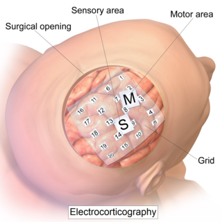
Electrocorticography (ECoG), a type of intracranial electroencephalography (iEEG), is a type of electrophysiological monitoring that uses electrodes placed directly on the exposed surface of the brain to record electrical activity from the cerebral cortex. In contrast, conventional electroencephalography (EEG) electrodes monitor this activity from outside the skull. ECoG may be performed either in the operating room during surgery or outside of surgery. Because a craniotomy is required to implant the electrode grid, ECoG is an invasive procedure.

The sensorimotor mu rhythm, also known as mu wave, comb or wicket rhythms or arciform rhythms, are synchronized patterns of electrical activity involving large numbers of neurons, probably of the pyramidal type, in the part of the brain that controls voluntary movement. These patterns as measured by electroencephalography (EEG), magnetoencephalography (MEG), or electrocorticography (ECoG), repeat at a frequency of 7.5–12.5 Hz, and are most prominent when the body is physically at rest. Unlike the alpha wave, which occurs at a similar frequency over the resting visual cortex at the back of the scalp, the mu rhythm is found over the motor cortex, in a band approximately from ear to ear. People suppress mu rhythms when they perform motor actions or, with practice, when they visualize performing motor actions. This suppression is called desynchronization of the wave because EEG wave forms are caused by large numbers of neurons firing in synchrony. The mu rhythm is even suppressed when one observes another person performing a motor action or an abstract motion with biological characteristics. Researchers such as V. S. Ramachandran and colleagues have suggested that this is a sign that the mirror neuron system is involved in mu rhythm suppression, although others disagree.
Synaptic noise refers to the constant bombardment of synaptic activity in neurons. This occurs in the background of a cell when potentials are produced without the nerve stimulation of an action potential, and are due to the inherently random nature of synapses. These random potentials have similar time courses as excitatory postsynaptic potentials (EPSPs) and inhibitory postsynaptic potentials (IPSPs), yet they lead to variable neuronal responses. The variability is due to differences in the discharge times of action potentials.
Fault detection, isolation, and recovery (FDIR) is a subfield of control engineering which concerns itself with monitoring a system, identifying when a fault has occurred, and pinpointing the type of fault and its location. Two approaches can be distinguished: A direct pattern recognition of sensor readings that indicate a fault and an analysis of the discrepancy between the sensor readings and expected values, derived from some model. In the latter case, it is typical that a fault is said to be detected if the discrepancy or residual goes above a certain threshold. It is then the task of fault isolation to categorize the type of fault and its location in the machinery. Fault detection and isolation (FDI) techniques can be broadly classified into two categories. These include model-based FDI and signal processing based FDI.

Electroencephalography (EEG) is a method to record an electrogram of the spontaneous electrical activity of the brain. The biosignals detected by EEG have been shown to represent the postsynaptic potentials of pyramidal neurons in the neocortex and allocortex. It is typically non-invasive, with the EEG electrodes placed along the scalp using the International 10–20 system, or variations of it. Electrocorticography, involving surgical placement of electrodes, is sometimes called "intracranial EEG". Clinical interpretation of EEG recordings is most often performed by visual inspection of the tracing or quantitative EEG analysis.
Quantitative electroencephalography is a field concerned with the numerical analysis of electroencephalography (EEG) data and associated behavioral correlates.
Brain connectivity estimators represent patterns of links in the brain. Connectivity can be considered at different levels of the brain's organisation: from neurons, to neural assemblies and brain structures. Brain connectivity involves different concepts such as: neuroanatomical or structural connectivity, functional connectivity and effective connectivity.

Burst suppression is an electroencephalography (EEG) pattern that is characterized by periods of high-voltage electrical activity alternating with periods of no activity in the brain. The pattern is found in patients with inactivated brain states, such as from general anesthesia, coma, or hypothermia. This pattern can be physiological, as during early development, or pathological, as in diseases such as Ohtahara syndrome.
Corticocortical coherence is referred to the synchrony in the neural activity of different cortical brain areas. The neural activities are picked up by electrophysiological recordings from the brain. It is a method to study the brain's neural communication and function at rest or during functional tasks.
Neural dust is a hypothetical class of nanometer-sized devices operated as wirelessly powered nerve sensors; it is a type of brain–computer interface. The sensors may be used to study, monitor, or control the nerves and muscles and to remotely monitor neural activity. In practice, a medical treatment could introduce thousands of neural dust devices into human brains. The term is derived from "smart dust", as the sensors used as neural dust may also be defined by this concept.

Dimitri Van De Ville is a Swiss and Belgian computer scientist and neuroscientist specialized in dynamical and network aspects of brain activity. He is a professor of bioengineering at EPFL and the head of the Medical Image Processing Laboratory at EPFL's School of Engineering.
References
- ↑ Pardey, J.; Roberts, S.; Tarassenko, L. (January 1996). "A review of parametric modelling techniques for EEG analysis". Medical Engineering & Physics. 18 (1): 2–11. CiteSeerX 10.1.1.51.9271 . doi:10.1016/1350-4533(95)00024-0. ISSN 1350-4533. PMID 8771033.
- ↑ Acharya, U. Rajendra; Vinitha Sree, S.; Swapna, G.; Martis, Roshan Joy; Suri, Jasjit S. (June 2013). "Automated EEG analysis of epilepsy: A review". Knowledge-Based Systems. 45: 147–165. doi:10.1016/j.knosys.2013.02.014. ISSN 0950-7051.
- ↑ Acharya, U. Rajendra; Vinitha Sree, S.; Swapna, G.; Martis, Roshan Joy; Suri, Jasjit S. (June 2013). "Automated EEG analysis of epilepsy: A review". Knowledge-Based Systems. 45: 147–165. doi:10.1016/j.knosys.2013.02.014. ISSN 0950-7051.
- ↑ Dressler, O.; Schneider, G.; Stockmanns, G.; Kochs, E.F. (December 2004). "Awareness and the EEG power spectrum: analysis of frequencies". British Journal of Anaesthesia. 93 (6): 806–809. doi: 10.1093/bja/aeh270 . ISSN 0007-0912. PMID 15377585.
- ↑ Ogilvie, Robert D.; Simons, Iain A.; Kuderian, Roxanne H.; MacDonald, Thomas; Rustenburg, John (January 1991). "Behavioral, Event‐Related Potential, and EEG/FFT Changes at Sleep Onset". Psychophysiology. 28 (1): 54–64. doi:10.1111/j.1469-8986.1991.tb03386.x. ISSN 0048-5772.
- ↑ Acharya, U. Rajendra; Vinitha Sree, S.; Swapna, G.; Martis, Roshan Joy; Suri, Jasjit S. (June 2013). "Automated EEG analysis of epilepsy: A review". Knowledge-Based Systems. 45: 147–165. doi:10.1016/j.knosys.2013.02.014. ISSN 0950-7051.
- ↑ Hjorth, Bo (September 1970). "EEG analysis based on time domain properties". Electroencephalography and Clinical Neurophysiology. 29 (3): 306–310. doi:10.1016/0013-4694(70)90143-4. ISSN 0013-4694. PMID 4195653.
- ↑ Hjorth, Bo (September 1970). "EEG analysis based on time domain properties". Electroencephalography and Clinical Neurophysiology. 29 (3): 306–310. doi:10.1016/0013-4694(70)90143-4. ISSN 0013-4694. PMID 4195653.
- ↑ Zhang, Yong; Liu, Bo; Ji, Xiaomin; Huang, Dan (April 2017). "Classification of EEG Signals Based on Autoregressive Model and Wavelet Packet Decomposition". Neural Processing Letters. 45 (2): 365–378. doi:10.1007/s11063-016-9530-1. ISSN 1370-4621.
- ↑ Adeli, Hojjat; Zhou, Ziqin; Dadmehr, Nahid (February 2003). "Analysis of EEG records in an epileptic patient using wavelet transform". Journal of Neuroscience Methods. 123 (1): 69–87. doi:10.1016/s0165-0270(02)00340-0. ISSN 0165-0270. PMID 12581851. S2CID 30980416.
- ↑ Adeli, Hojjat; Zhou, Ziqin; Dadmehr, Nahid (February 2003). "Analysis of EEG records in an epileptic patient using wavelet transform". Journal of Neuroscience Methods. 123 (1): 69–87. doi:10.1016/s0165-0270(02)00340-0. ISSN 0165-0270. PMID 12581851. S2CID 30980416.
- ↑ Hazarika, N.; Chen, J.Z.; Ah Chung Tsoi; Sergejew, A. (1997). "Classification of EEG signals using the wavelet transform". Proceedings of 13th International Conference on Digital Signal Processing. Vol. 1. IEEE. pp. 89–92. doi:10.1109/icdsp.1997.627975. ISBN 978-0780341371.
- ↑ Acharya, U. Rajendra; Vinitha Sree, S.; Swapna, G.; Martis, Roshan Joy; Suri, Jasjit S. (June 2013). "Automated EEG analysis of epilepsy: A review". Knowledge-Based Systems. 45: 147–165. doi:10.1016/j.knosys.2013.02.014. ISSN 0950-7051.
- ↑ Pigorini, Andrea; Casali, Adenauer G.; Casarotto, Silvia; Ferrarelli, Fabio; Baselli, Giuseppe; Mariotti, Maurizio; Massimini, Marcello; Rosanova, Mario (June 2011). "Time–frequency spectral analysis of TMS-evoked EEG oscillations by means of Hilbert–Huang transform". Journal of Neuroscience Methods. 198 (2): 236–245. doi:10.1016/j.jneumeth.2011.04.013. ISSN 0165-0270. PMID 21524665. S2CID 11151845.
- ↑ Stam, C.J. (October 2005). "Nonlinear dynamical analysis of EEG and MEG: Review of an emerging field". Clinical Neurophysiology. 116 (10): 2266–2301. CiteSeerX 10.1.1.126.4927 . doi:10.1016/j.clinph.2005.06.011. ISSN 1388-2457. PMID 16115797. S2CID 15359405.
- ↑ Acharya, U. Rajendra; Vinitha Sree, S.; Swapna, G.; Martis, Roshan Joy; Suri, Jasjit S. (June 2013). "Automated EEG analysis of epilepsy: A review". Knowledge-Based Systems. 45: 147–165. doi:10.1016/j.knosys.2013.02.014. ISSN 0950-7051.
- ↑ Stam, C.J. (October 2005). "Nonlinear dynamical analysis of EEG and MEG: Review of an emerging field". Clinical Neurophysiology. 116 (10): 2266–2301. CiteSeerX 10.1.1.126.4927 . doi:10.1016/j.clinph.2005.06.011. ISSN 1388-2457. PMID 16115797. S2CID 15359405.
- ↑ Petrosian, Arthur; Prokhorov, Danil; Homan, Richard; Dasheiff, Richard; Wunsch, Donald (January 2000). "Recurrent neural network based prediction of epileptic seizures in intra- and extracranial EEG". Neurocomputing. 30 (1–4): 201–218. doi:10.1016/s0925-2312(99)00126-5. ISSN 0925-2312.
- ↑ Subasi, Abdulhamit; Erçelebi, Ergun (May 2005). "Classification of EEG signals using neural network and logistic regression". Computer Methods and Programs in Biomedicine. 78 (2): 87–99. doi:10.1016/j.cmpb.2004.10.009. ISSN 0169-2607. PMID 15848265.
- ↑ Subasi, Abdulhamit; Erçelebi, Ergun (May 2005). "Classification of EEG signals using neural network and logistic regression". Computer Methods and Programs in Biomedicine. 78 (2): 87–99. doi:10.1016/j.cmpb.2004.10.009. ISSN 0169-2607. PMID 15848265.
- ↑ Übeyli, Elif Derya (January 2009). "Analysis of EEG signals by implementing eigenvector methods/recurrent neural networks". Digital Signal Processing. 19 (1): 134–143. doi:10.1016/j.dsp.2008.07.007. ISSN 1051-2004.
- ↑ Schirrmeister, R.; Gemein, L.; Eggensperger, K.; Hutter, F.; Ball, T. (December 2017). "Deep learning with convolutional neural networks for decoding and visualization of EEG pathology". 2017 IEEE Signal Processing in Medicine and Biology Symposium (SPMB). IEEE. pp. 1–7. arXiv: 1708.08012 . doi:10.1109/spmb.2017.8257015. ISBN 9781538648735. S2CID 5692066.
- ↑ Hosseini, Mohammad-Parsa; Soltanian-Zadeh, Hamid; Elisevich, Kost; Pompili, Dario (December 2016). "Cloud-based deep learning of big EEG data for epileptic seizure prediction". 2016 IEEE Global Conference on Signal and Information Processing (GlobalSIP). IEEE. pp. 1151–1155. arXiv: 1702.05192 . doi:10.1109/globalsip.2016.7906022. ISBN 9781509045457. S2CID 2675362.
- ↑ Biasiucci, Andrea; Franceschiello, Benedetta; Murray, Micah M. (February 2019). "Electroencephalography". Current Biology. 29 (3): R80–R85. doi: 10.1016/j.cub.2018.11.052 . ISSN 0960-9822.
- ↑ McFarland, D. J.; Wolpaw, J. R. (2017-12-01). "EEG-based brain–computer interfaces". Current Opinion in Biomedical Engineering. Synthetic Biology and Biomedical Engineering / Neural Engineering. 4: 194–200. doi:10.1016/j.cobme.2017.11.004. ISSN 2468-4511. PMC 5839510 . PMID 29527584.
- ↑ Subasi, Abdulhamit; Erçelebi, Ergun (May 2005). "Classification of EEG signals using neural network and logistic regression". Computer Methods and Programs in Biomedicine. 78 (2): 87–99. doi:10.1016/j.cmpb.2004.10.009. ISSN 0169-2607. PMID 15848265.
- ↑ Jeong, Jaeseung; Gore, John C; Peterson, Bradley S (May 2001). "Mutual information analysis of the EEG in patients with Alzheimer's disease". Clinical Neurophysiology. 112 (5): 827–835. doi:10.1016/s1388-2457(01)00513-2. ISSN 1388-2457. PMID 11336898. S2CID 9851741.
- ↑ Guger, C.; Ramoser, H.; Pfurtscheller, G. (2000). "Real-time EEG analysis with subject-specific spatial patterns for a brain-computer interface (BCI)". IEEE Transactions on Rehabilitation Engineering. 8 (4): 447–456. doi:10.1109/86.895947. ISSN 1063-6528. PMID 11204035. S2CID 9504054.
- ↑ Biasiucci, Andrea; Franceschiello, Benedetta; Murray, Micah M. (February 2019). "Electroencephalography". Current Biology. 29 (3): R80–R85. doi: 10.1016/j.cub.2018.11.052 . ISSN 0960-9822.
- ↑ "Introduction - Brainstorm". neuroimage.usc.edu. Retrieved 2018-12-16.
- ↑ Soleymani, Mohammad; Asghari-Esfeden, Sadjad; Pantic, Maja; Fu, Yun (July 2014). "Continuous emotion detection using EEG signals and facial expressions". 2014 IEEE International Conference on Multimedia and Expo (ICME). IEEE. pp. 1–6. CiteSeerX 10.1.1.649.3590 . doi:10.1109/icme.2014.6890301. ISBN 9781479947614. S2CID 16028962.