Epigenetic clock

Last updated

An epigenetic clock is a biochemical test that can be used to measure age. The test is based on DNA methylation levels, measuring the accumulation of methyl groups to one's DNA molecules.

Contents

History

The strong effects of age on DNA methylation levels have been known since the late 1960s. [1] A vast literature describes sets of CpGs whose DNA methylation levels correlate with age. [2] [3] [4] [5] [6] The first robust demonstration that DNA methylation levels in saliva could generate age predictors with an average accuracy of 5.2 years was published by a UCLA team including Sven Bocklandt, Steve Horvath, and Eric Vilain in 2011 (Bocklandt et al. 2011). [7] [8] The laboratories of Trey Ideker and Kang Zhang at the University of California, San Diego published the Hannum epigenetic clock (Hannum 2013), [9] which consisted of 71 markers that accurately estimate age based on blood methylation levels. The first multi-tissue epigenetic clock, Horvath's epigenetic clock, was developed by Steve Horvath, a professor of human genetics and biostatistics at UCLA (Horvath 2013). [10] [11] Horvath spent over 4 years collecting publicly available Illumina DNA methylation data and identifying suitable statistical methods. [12]

The personal story behind the discovery was featured in Nature. [13] The age estimator was developed using 8,000 samples from 82 Illumina DNA methylation array datasets, encompassing 51 healthy tissues and cell types. The major innovation of Horvath's epigenetic clock lies in its wide applicability: the same set of 353 CpGs and the same prediction algorithm is used irrespective of the DNA source within the organism, i.e. it does not require any adjustments or offsets. [10] This property allows one to compare the ages of different areas of the human body using the same aging clock. Shortly afterwards, a derivation of Horvath's clock, the IEAA (Intrinsic Epigenetic Age Acceleration), an estimator based on the cellular composition of the blood, was developed.

A second generation of epigenetic clocks emerged a few years later and improved on the first in age estimation. This was thanks to the incorporation not only of epigenetic variants such as DNA methylation but also environmental variants such as smoking or chronological age. Among these clocks, the PhenoAge and GrimAge clocks stand out. PhenoAge is an epigenetic clock that takes chronological age into account, and GrimAge uses the mortality risks of age together with the smoking variant among others as a risk factor. Taking into account environmental variants allows GrimAge to outperform any other epigenetic clock in "predicting death".

Third-generation epigenetic clocks are designed to be applicable across multiple species simultaneously. Specifically, pan-mammalian epigenetic clocks determine the age of tissues from all mammalian species by analyzing cytosine methylation in DNA regions that are highly conserved. [14]

New age estimation tools have been developed continuously, which also facilitate the prognosis of certain diseases.

Relationship to a cause of biological aging

It is not yet known what exactly is measured by DNA methylation age. Horvath hypothesized that DNA methylation age measures the cumulative effect of an epigenetic maintenance system but details are unknown. The fact that DNA methylation age of blood predicts all-cause mortality in later life [15] [16] [17] [18] has been used to argue that it relates to a process that causes aging. [15] However, if a particular CpG played a direct causal role in the aging process, the mortality it created would make it less likely to be observed in older individuals, making the site less likely to have been chosen as a predictor; the 353 clock CpGs therefore likely have no causal effect whatsoever. [19] Rather, the epigenetic clock captures an emergent property of the epigenome.

Epigenetic clock theory of aging

In 2010, a new unifying model of aging and the development of complex diseases was proposed, incorporating classical aging theories and epigenetics. [20] [21] Horvath and Raj [22] extended this theory, proposing an epigenetic clock theory of aging with the following tenets:

Motivation for biological clocks

In general, biological aging clocks and biomarkers of aging are expected to find many uses in biological research since age is a fundamental characteristic of most organisms. Accurate measures of biological age (biological aging clocks) could be useful for

Overall, biological clocks are expected to be useful for studying what causes aging and what can be done against it. However, they can only capture the effects of interventions that affect the rate of future aging, i.e. the slope of the Gompertz curve by which mortality increases with age, and not that of interventions that act at one moment in time, e.g. to lower mortality across all ages, i.e. the intercept of the Gompertz curve. [19]

Properties of Horvath's clock

The clock is defined as an age estimation method based on 353 epigenetic markers on the DNA. The 353 markers measure DNA methylation of CpG dinucleotides. Estimated age ("predicted age" in mathematical usage), also referred to as DNA methylation age, has the following properties: first, it is close to zero for embryonic and induced pluripotent stem cells; second, it correlates with cell passage number; third, it gives rise to a highly heritable measure of age acceleration; and, fourth, it is applicable to chimpanzee tissues (which are used as human analogs for biological testing purposes). Organismal growth (and concomitant cell division) leads to a high ticking rate of the epigenetic clock that slows down to a constant ticking rate (linear dependence) after adulthood (age 20). [10] The fact that DNA methylation age of blood predicts all-cause mortality in later life even after adjusting for known risk factors [15] [16] is compatible with a variety of causal relationships, e.g. a common cause for both. Similarly, markers of physical and mental fitness are associated with the epigenetic clock (lower abilities associated with age acceleration). [23] It systematically underestimates age from older individuals. [24]

Salient features of Horvath's epigenetic clock include its applicability to a broad spectrum of tissues and cell types. Since it allows one to contrast the ages of different tissues from the same subject, it can be used to identify tissues that show evidence of accelerated age due to disease.

Genetic estimators in the Horvath clock

The Horvath clock, specifically the IEAA variant, is associated with several ageing-related genes:14

Statistical approach

The basic approach is to form a weighted average of the 353 clock CpGs, which is then transformed to DNAm age using a calibration function. The calibration function reveals that the epigenetic clock has a high ticking rate until adulthood, after which it slows to a constant ticking rate. Using the training data sets, Horvath used a penalized regression model (Elastic net regularization) to regress a calibrated version of chronological age on 21,369 CpG probes that were present both on the Illumina 450K and 27K platform and had fewer than 10 missing values. DNAm age is defined as estimated ("predicted") age. The elastic net predictor automatically selected 353 CpGs. 193 of the 353 CpGs correlate positively with age while the remaining 160 CpGs correlate negatively with age. R software and a freely available web-based tool can be found at the following webpage. [26]

Accuracy

The median error of estimated age is 3.6 years across a wide spectrum of tissues and cell types, [10] although this increases for older individuals [24] The epigenetic clock performs well in heterogeneous tissues (for example, whole blood, peripheral blood mononuclear cells, cerebellar samples, occipital cortex, buccal epithelium, colon, adipose, kidney, liver, lung, saliva, uterine cervix, epidermis, muscle) as well as in individual cell types such as CD4 T cells, CD14 monocytes, glial cells, neurons, immortalized B cells, mesenchymal stromal cells. [10] However, accuracy depends to some extent on the source of the DNA.

Comparison with other biological clocks

The epigenetic clock leads to a chronological age prediction that has a Pearson correlation coefficient of r = 0.96 with chronological age (Figure 2 in [10] ). Thus the age correlation is close to its maximum possible correlation value of 1. Other biological clocks are based on a) telomere length, b) p16INK4a expression levels (also known as INK4a/ARF locus), [27] and c) microsatellite mutations. [28] The correlation between chronological age and telomere length is r = −0.51 in women and r = −0.55 in men. [29] The correlation between chronological age and expression levels of p16INK4a in T cells is r = 0.56. [30]

Applications of Horvath's clock

By contrasting DNA methylation age (estimated age) with chronological age, one can define measures of age acceleration. Age acceleration can be defined as the difference between DNA methylation age and chronological age. Alternatively, it can be defined as the residual that results from regressing DNAm age on chronological age. The latter measure is attractive because it does not correlate with chronological age. A positive/negative value of epigenetic age acceleration suggests that the underlying tissue ages faster/slower than expected.

Genetic studies of epigenetic age acceleration

The broad sense heritability (defined via Falconer's formula) of age acceleration of blood from older subjects is around 40% but it appears to be much higher in newborns. [10] Similarly, the age acceleration of brain tissue (prefrontal cortex) was found to be 41% in older subjects. [31] Genome-wide association studies (GWAS) of epigenetic age acceleration in postmortem brain samples have identified several SNPs at a genomewide significance level. [32] [33] GWAS of age acceleration in blood have identified several genome-wide significant genetic loci including the telomerase reverse transcriptase gene (TERT) locus. [34] Genetic variants associated with longer leukocyte telomere length in TERT gene paradoxically confer higher epigenetic age acceleration in blood. [34]

Lifestyle factors

In general, lifestyle factors have only weak associations with epigenetic age acceleration in blood. [35] [36] [37] Cross sectional studies of extrinsic epigenetic aging rates in blood show reduced epigenetic aging correlates with higher education, eating a high plant diet with lean meats, moderate alcohol consumption, and physical activity [36] and the risks associated with metabolic syndrome. However, studies suggest that high levels of alcohol consumption are associated with accelerated aging of certain epigenetic clocks. [37]

Obesity and metabolic syndrome

The epigenetic clock was used to study the relationship between high body mass index (BMI) and the DNA methylation ages of human blood, liver, muscle and adipose tissue. [38] A significant correlation (r = 0.42) between BMI and epigenetic age acceleration could be observed for the liver. A much larger sample size (n = 4200 blood samples) revealed a weak but statistically significant correlation (r = 0.09) between BMI and intrinsic age acceleration of blood. [35] The same large study found that various biomarkers of metabolic syndrome (glucose-, insulin-, triglyceride levels, C-reactive protein, waist-to-hip ratio) were associated with epigenetic age acceleration in blood. [35] Conversely, high levels of HDL cholesterol were associated with a lower epigenetic aging rate of blood. [35] Other research suggests very strong associations between higher body mass index, waist-to-hip ratio, and waist circumference and accelerated epigenetic clocks, with evidence that physical activity may lessen these effects. [36]

Female breast tissue is older than expected

DNAm age is higher than chronological age in female breast tissue that is adjacent to breast cancer tissue. [10] Since normal tissue which is adjacent to other cancer types does not exhibit a similar age acceleration effect, this finding suggests that normal female breast tissue ages faster than other parts of the body. [10] Similarly, normal breast tissue samples from women without cancer have been found to be substantially older than blood samples collected from the same women at the same time. [39]

Female breast cancer

In a study of three epigenetic clocks and breast cancer risk, DNAm age was found to be accelerated in blood samples of cancer-free women, years before diagnosis. [40]

Cancer tissue

Cancer tissues show both positive and negative age acceleration effects. For most tumor types, no significant relationship can be observed between age acceleration and tumor morphology (grade/stage). [10] [41] On average, cancer tissues with mutated TP53 have a lower age acceleration than those without it. [10] Further, cancer tissues with high age acceleration tend to have fewer somatic mutations than those with low age acceleration. [10] [41] Age acceleration is highly related to various genomic aberrations in cancer tissues. Somatic mutations in estrogen receptors or progesterone receptors are associated with accelerated DNAm age in breast cancer. [10] Colorectal cancer samples with a BRAF (V600E) mutation or promoter hypermethylation of the mismatch repair gene MLH1 are associated with an increased age acceleration. [10] Age acceleration in glioblastoma multiforme samples is highly significantly associated with certain mutations in H3F3A. [10] One study suggests that the epigenetic age of blood tissue may be prognostic of lung cancer incidence. [42]

Trisomy 21 (Down syndrome)

Down syndrome entails an increased risk of many chronic diseases that are typically associated with older age. The clinical manifestations of accelerated aging suggest that trisomy 21 increases the biological age of tissues, but molecular evidence for this hypothesis has been sparse. According to the epigenetic clock, trisomy 21 significantly increases the age of blood and brain tissue (on average by 6.6 years). [43]

Epigenetic age acceleration of the human prefrontal cortex was found to be correlated with several neuropathological measurements that play a role in Alzheimer's disease [31] Further, it was found to be associated with a decline in global cognitive functioning, and memory functioning among individuals with Alzheimer's disease. [31] The epigenetic age of blood relates to cognitive functioning in the elderly. [23] Overall, these results strongly suggest that the epigenetic clock lends itself for measuring the biological age of the brain.

Cerebellum ages slowly

It has been difficult to identify tissues that seem to evade aging due to the lack of biomarkers of tissue age that allow one to contrast compare the ages of different tissues. An application of epigenetic clock to 30 anatomic sites from six centenarians and younger subjects revealed that the cerebellum ages slowly: it is about 15 years younger than expected in a centenarian. [44] This finding might explain why the cerebellum exhibits fewer neuropathological hallmarks of age related dementias compared to other brain regions. In younger subjects (e.g. younger than 70), brain regions and brain cells appear to have roughly the same age. [10] [44] Several SNPs and genes have been identified that relate to the epigenetic age of the cerebellum. [32]

Huntington's disease

Huntington's disease has been found to increase the epigenetic aging rates of several human brain regions. [45]

Centenarians age slowly

The offspring of semi-supercentenarians (subjects who reached an age of 105–109 years) have a lower epigenetic age than age-matched controls (age difference = 5.1 years in blood) and centenarians are younger (8.6 years) than expected based on their chronological age. [18]

HIV infection

Infection with the Human Immunodeficiency Virus-1 (HIV) is associated with clinical symptoms of accelerated aging, as evidenced by increased incidence and diversity of age-related illnesses at relatively young ages. But it has been difficult to detect an accelerated aging effect on a molecular level. An epigenetic clock analysis of human DNA from HIV+ subjects and controls detected a significant age acceleration effect in brain (7.4 years) and blood (5.2 years) tissue due to HIV-1 infection. [46] These results are consistent with an independent study that also found an age advancement of 5 years in blood of HIV patients and a strong effect of the HLA locus. [47]

Parkinson's disease

A large-scale study suggests that the blood of Parkinson's disease subjects, in particular, their granulocyte ratio, exhibits (relatively weak) accelerated aging effects. [48]

Developmental disorder: syndrome X

Children with a very rare disorder known as syndrome X maintain the façade of persistent toddler-like features while aging from birth to adulthood. Since the physical development of these children is dramatically delayed, these children appear to be a toddler or at best a preschooler. According to an epigenetic clock analysis, blood tissue from syndrome X cases is not younger than expected. [49]

Menopause accelerates epigenetic aging

The following results strongly suggest that the loss of female hormones resulting from menopause accelerates the epigenetic aging rate of blood and possibly that of other tissues. [50] First, early menopause has been found to be associated with an increased epigenetic age acceleration of blood. [50] Second, surgical menopause (due to bilateral oophorectomy) is associated with epigenetic age acceleration in blood and saliva. Third, menopausal hormone therapy, which mitigates hormonal loss, is associated with a negative age acceleration of buccal cells (but not of blood cells). [50] Fourth, genetic markers that are associated with early menopause are also associated with increased epigenetic age acceleration in blood. [50]

Cellular senescence versus epigenetic aging

A confounding aspect of biological aging is the nature and role of senescent cells. It is unclear whether the three major types of cellular senescence, namely replicative senescence, oncogene-induced senescence and DNA damage-induced senescence are descriptions of the same phenomenon instigated by different sources, or if each of these is distinct, and how they are associated with epigenetic aging. Induction of replicative senescence (RS) and oncogene-induced senescence (OIS) were found to be accompanied by epigenetic aging of primary cells but senescence induced by DNA damage was not, even though RS and OIS activate the cellular DNA damage response pathway. [51] These results highlight the independence of cellular senescence from epigenetic aging. Consistent with this, telomerase-immortalised cells continued to age (according to the epigenetic clock) without having been treated with any senescence inducers or DNA-damaging agents, re-affirming the independence of the process of epigenetic ageing from telomeres, cellular senescence, and the DNA damage response pathway. Although the uncoupling of senescence from cellular aging appears at first sight to be inconsistent with the fact that senescent cells contribute to the physical manifestation of organism ageing, as demonstrated by Baker et al., where removal of senescent cells slowed down aging. [52]

The epigenetic clock analysis of senescence, however, suggests that cellular senescence is a state that cells are forced into as a result of external pressures such as DNA damage, ectopic oncogene expression and exhaustive proliferation of cells to replenish those eliminated by external/environmental factors. [51] These senescent cells, in sufficient numbers, will probably cause the deterioration of tissues, which is interpreted as organism ageing. However, at the cellular level, aging, as measured by the epigenetic clock, is distinct from senescence. It is an intrinsic mechanism that exists from the birth of the cell and continues. This implies that if cells are not shunted into senescence by the external pressures described above, they would still continue to age. This is consistent with the fact that mice with naturally long telomeres still age and eventually die even though their telomere lengths are far longer than the critical limit, and they age prematurely when their telomeres are forcibly shortened, due to replicative senescence. Therefore, cellular senescence is a route by which cells exit prematurely from the natural course of cellular aging. [51]

Effect of sex and race/ethnicity

Men age faster than women according to epigenetic age acceleration in blood, brain, saliva, but it depends on the structure being researched and the lifestyle. [53] The epigenetic clock method applies to all examined racial/ethnic groups in the sense that DNAm age is highly correlated with chronological age. But ethnicity can be associated with epigenetic age acceleration. [53] For example, the blood of Hispanics and the Tsimané ages more slowly than that of other populations which might explain the Hispanic mortality paradox. [53]

Rejuvenation effect due to stem cell transplantation in blood

Hematopoietic stem cell transplantation, which transplants these cells from a young donor to an older recipient, rejuvenates the epigenetic age of blood to that of the donor. However, graft-versus-host disease is associated with increased DNA methylation age. [54]

Progeria

Adult progeria also known as Werner syndrome is associated with epigenetic age acceleration in blood. [55] Fibroblast samples from children with Hutchinson-Gilford Progeria exhibit accelerated epigenetic aging effects according to the "skin & blood" epigenetic clock but not according to the original pan tissue clock from Horvath. [56]

Biological mechanism behind the epigenetic clock

Possible explanation 1: Epigenomic maintenance system

Horvath hypothesized that his clock arises from a methylation footprint left by an epigenomic maintenance system. [10]

Possible explanation 2: Unrepaired DNA damages

Endogenous DNA damages occur frequently including about 50 double-strand DNA breaks per cell cycle [57] and about 10,000 oxidative damages per day (see DNA damage (naturally occurring)). During repair of double-strand breaks many epigenetic alterations are introduced, and in a percentage of cases epigenetic alterations remain after repair is completed, including increased methylation of CpG island promoters. [58] [59] [60] Similar, but usually transient epigenetic alterations were recently found during repair of oxidative damages caused by H2O2, and it was suggested that occasionally these epigenetic alterations may also remain after repair. [61] These accumulated epigenetic alterations may contribute to the epigenetic clock. Accumulation of epigenetic alterations may parallel the accumulation of un-repaired DNA damages that are proposed to cause aging (see DNA damage theory of aging). In line with stochastic DNA damage accumulation, age-related alterations in DNA methylation have been observed to predominantly undergo stochastic changes as individuals age. [62] This accumulation of stochastic variation has demonstrated sufficient capacity to build aging clocks, further supporting the notion that epigenetic changes may be driven by the gradual accrual of unprogrammed stochastic damage. [63]

Related Research Articles

<span class="mw-page-title-main">Epigenetics</span> Study of DNA modifications that do not change its sequence

In biology, epigenetics is the study of heritable traits, or a stable change of cell function, that happen without changes to the DNA sequence. The Greek prefix epi- in epigenetics implies features that are "on top of" or "in addition to" the traditional genetic mechanism of inheritance. Epigenetics usually involves a change that is not erased by cell division, and affects the regulation of gene expression. Such effects on cellular and physiological phenotypic traits may result from environmental factors, or be part of normal development. They can lead to cancer.

<span class="mw-page-title-main">5-Methylcytosine</span> Chemical compound which is a modified DNA base

5-Methylcytosine is a methylated form of the DNA base cytosine (C) that regulates gene transcription and takes several other biological roles. When cytosine is methylated, the DNA maintains the same sequence, but the expression of methylated genes can be altered. 5-Methylcytosine is incorporated in the nucleoside 5-methylcytidine.

Senescence or biological aging is the gradual deterioration of functional characteristics in living organisms. Whole organism senescence involves an increase in death rates and/or a decrease in fecundity with increasing age, at least in the later part of an organism's life cycle. However, the resulting effects of senescence can be delayed. The 1934 discovery that calorie restriction can extend lifespans by 50% in rats, the existence of species having negligible senescence, and the existence of potentially immortal organisms such as members of the genus Hydra have motivated research into delaying senescence and thus age-related diseases. Rare human mutations can cause accelerated aging diseases.

<span class="mw-page-title-main">CpG site</span> Region of often-methylated DNA with a cytosine followed by a guanine

The CpG sites or CG sites are regions of DNA where a cytosine nucleotide is followed by a guanine nucleotide in the linear sequence of bases along its 5' → 3' direction. CpG sites occur with high frequency in genomic regions called CpG islands.

<span class="mw-page-title-main">Biogerontology</span> Sub-field of gerontology

Biogerontology is the sub-field of gerontology concerned with the biological aging process, its evolutionary origins, and potential means to intervene in the process. The term "biogerontology" was coined by S. Rattan, and came in regular use with the start of the journal BIOGERONTOLOGY in 2000. It involves interdisciplinary research on the causes, effects, and mechanisms of biological aging. Biogerontologist Leonard Hayflick has said that the natural average lifespan for a human is around 92 years and, if humans do not invent new approaches to treat aging, they will be stuck with this lifespan. James Vaupel has predicted that life expectancy in industrialized countries will reach 100 for children born after the year 2000. Many surveyed biogerontologists have predicted life expectancies of more than three centuries for people born after the year 2100. Other scientists, more controversially, suggest the possibility of unlimited lifespans for those currently living. For example, Aubrey de Grey offers the "tentative timeframe" that with adequate funding of research to develop interventions in aging such as strategies for engineered negligible senescence, "we have a 50/50 chance of developing technology within about 25 to 30 years from now that will, under reasonable assumptions about the rate of subsequent improvements in that technology, allow us to stop people from dying of aging at any age". The idea of this approach is to use presently available technology to extend lifespans of currently living humans long enough for future technological progress to resolve any remaining aging-related issues. This concept has been referred to as longevity escape velocity.

<span class="mw-page-title-main">DNA repair</span> Cellular mechanism

DNA repair is a collection of processes by which a cell identifies and corrects damage to the DNA molecules that encode its genome. In human cells, both normal metabolic activities and environmental factors such as radiation can cause DNA damage, resulting in tens of thousands of individual molecular lesions per cell per day. Many of these lesions cause structural damage to the DNA molecule and can alter or eliminate the cell's ability to transcribe the gene that the affected DNA encodes. Other lesions induce potentially harmful mutations in the cell's genome, which affect the survival of its daughter cells after it undergoes mitosis. As a consequence, the DNA repair process is constantly active as it responds to damage in the DNA structure. When normal repair processes fail, and when cellular apoptosis does not occur, irreparable DNA damage may occur. This can eventually lead to malignant tumors, or cancer as per the two-hit hypothesis.

<span class="mw-page-title-main">Neoplasm</span> Tumor or other abnormal growth of tissue

A neoplasm is a type of abnormal and excessive growth of tissue. The process that occurs to form or produce a neoplasm is called neoplasia. The growth of a neoplasm is uncoordinated with that of the normal surrounding tissue, and persists in growing abnormally, even if the original trigger is removed. This abnormal growth usually forms a mass, which may be called a tumour or tumor.

p16 Mammalian protein found in Homo sapiens

p16, is a protein that slows cell division by slowing the progression of the cell cycle from the G1 phase to the S phase, thereby acting as a tumor suppressor. It is encoded by the CDKN2A gene. A deletion in this gene can result in insufficient or non-functional p16, accelerating the cell cycle and resulting in many types of cancer.

Chromatin remodeling is the dynamic modification of chromatin architecture to allow access of condensed genomic DNA to the regulatory transcription machinery proteins, and thereby control gene expression. Such remodeling is principally carried out by 1) covalent histone modifications by specific enzymes, e.g., histone acetyltransferases (HATs), deacetylases, methyltransferases, and kinases, and 2) ATP-dependent chromatin remodeling complexes which either move, eject or restructure nucleosomes. Besides actively regulating gene expression, dynamic remodeling of chromatin imparts an epigenetic regulatory role in several key biological processes, egg cells DNA replication and repair; apoptosis; chromosome segregation as well as development and pluripotency. Aberrations in chromatin remodeling proteins are found to be associated with human diseases, including cancer. Targeting chromatin remodeling pathways is currently evolving as a major therapeutic strategy in the treatment of several cancers.

<span class="mw-page-title-main">Cellular senescence</span> Phenomenon characterized by the cessation of cell division

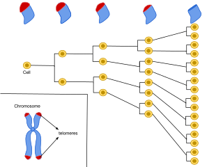
Cellular senescence is a phenomenon characterized by the cessation of cell division. In their experiments during the early 1960s, Leonard Hayflick and Paul Moorhead found that normal human fetal fibroblasts in culture reach a maximum of approximately 50 cell population doublings before becoming senescent. This process is known as "replicative senescence", or the Hayflick limit. Hayflick's discovery of mortal cells paved the path for the discovery and understanding of cellular aging molecular pathways. Cellular senescence can be initiated by a wide variety of stress inducing factors. These stress factors include both environmental and internal damaging events, abnormal cellular growth, oxidative stress, autophagy factors, among many other things.

The DNA damage theory of aging proposes that aging is a consequence of unrepaired accumulation of naturally occurring DNA damage. Damage in this context is a DNA alteration that has an abnormal structure. Although both mitochondrial and nuclear DNA damage can contribute to aging, nuclear DNA is the main subject of this analysis. Nuclear DNA damage can contribute to aging either indirectly or directly.

<span class="mw-page-title-main">YPEL3</span> Protein-coding gene in humans

Yippee-like 3 (Drosophila) is a protein that in humans is encoded by the YPEL3 gene. YPEL3 has growth inhibitory effects in normal and tumor cell lines. One of five family members (YPEL1-5), YPEL3 was named in reference to its Drosophila melanogaster orthologue. Initially discovered in a gene expression profiling assay of p53 activated MCF7 cells, induction of YPEL3 has been shown to trigger permanent growth arrest or cellular senescence in certain human normal and tumor cell types. DNA methylation of a CpG island near the YPEL3 promoter as well as histone acetylation may represent possible epigenetic mechanisms leading to decreased gene expression in human tumors.

miR-137

In molecular biology, miR-137 is a short non-coding RNA molecule that functions to regulate the expression levels of other genes by various mechanisms. miR-137 is located on human chromosome 1p22 and has been implicated to act as a tumor suppressor in several cancer types including colorectal cancer, squamous cell carcinoma and melanoma via cell cycle control.

<span class="mw-page-title-main">Biomarkers of aging</span> Type of biomarkers

Biomarkers of aging are biomarkers that could predict functional capacity at some later age better than chronological age. Stated another way, biomarkers of aging would give the true "biological age", which may be different from the chronological age.

<span class="mw-page-title-main">Cancer epigenetics</span> Field of study in cancer research

Cancer epigenetics is the study of epigenetic modifications to the DNA of cancer cells that do not involve a change in the nucleotide sequence, but instead involve a change in the way the genetic code is expressed. Epigenetic mechanisms are necessary to maintain normal sequences of tissue specific gene expression and are crucial for normal development. They may be just as important, if not even more important, than genetic mutations in a cell's transformation to cancer. The disturbance of epigenetic processes in cancers, can lead to a loss of expression of genes that occurs about 10 times more frequently by transcription silencing than by mutations. As Vogelstein et al. points out, in a colorectal cancer there are usually about 3 to 6 driver mutations and 33 to 66 hitchhiker or passenger mutations. However, in colon tumors compared to adjacent normal-appearing colonic mucosa, there are about 600 to 800 heavily methylated CpG islands in the promoters of genes in the tumors while these CpG islands are not methylated in the adjacent mucosa. Manipulation of epigenetic alterations holds great promise for cancer prevention, detection, and therapy. In different types of cancer, a variety of epigenetic mechanisms can be perturbed, such as the silencing of tumor suppressor genes and activation of oncogenes by altered CpG island methylation patterns, histone modifications, and dysregulation of DNA binding proteins. There are several medications which have epigenetic impact, that are now used in a number of these diseases.

Epigenetic therapy refers to the use of drugs or other interventions to modify gene expression patterns, potentially treating diseases by targeting epigenetic mechanisms such as DNA methylation and histone modifications.

<span class="mw-page-title-main">Epigenome-wide association study</span>

An epigenome-wide association study (EWAS) is an examination of a genome-wide set of quantifiable epigenetic marks, such as DNA methylation, in different individuals to derive associations between epigenetic variation and a particular identifiable phenotype/trait. When patterns change such as DNA methylation at specific loci, discriminating the phenotypically affected cases from control individuals, this is considered an indication that epigenetic perturbation has taken place that is associated, causally or consequentially, with the phenotype.

<span class="mw-page-title-main">Steve Horvath</span> German–American aging researcher, geneticist and biostatistician

Steve Horvath is a German–American aging researcher, geneticist, and biostatistician. He is a professor at the University of California, Los Angeles known for developing the Horvath aging clock, which is a highly accurate molecular biomarker of aging, and for developing weighted correlation network analysis. His work on the genomic biomarkers of aging, the aging process, and many age related diseases/conditions has earned him several research awards. Horvath is a principal investigator at the anti-aging startup Altos Labs and co-founder of nonprofit Clock Foundation.

Aging is characterized by a progressive loss of physiological integrity, leading to impaired function and increased vulnerability to death. The hallmarks of aging are the types of biochemical changes that occur in all organisms that experience biological aging and lead to a progressive loss of physiological integrity, impaired function and, eventually, death. They were first listed in a landmark paper in 2013 to conceptualize the essence of biological aging and its underlying mechanisms.

<span class="mw-page-title-main">Epiphenotyping</span> Epiphenotyping is the use of DNA methylation patterns to predict phenotypes.

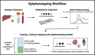
Epiphenotyping involves studying the relationship between DNA methylation patterns and phenotypic traits in individuals and populations to be able to predict a phenotype from a DNA methylation profile. In the following sections, the background of epiphenotyping, an overview of a general methodology, its applications, advantages, and limitations are covered.

References

  1. Berdyshev GD, Korotaev GK, Boiarskikh GV, Vaniushin BF (1967). "[Nucleotide composition of DNA and RNA from somatic tissues of humpback and its changes during spawning]". Biokhimiia. 32 (5): 988–993. PMID   5628601.
  2. Rakyan VK, Down TA, Maslau S, Andrew T, Yang TP, Beyan H, et al. (April 2010). "Human aging-associated DNA hypermethylation occurs preferentially at bivalent chromatin domains". Genome Research. 20 (4): 434–439. doi:10.1101/gr.103101.109. PMC   2847746 . PMID   20219945.
  3. Teschendorff AE, Menon U, Gentry-Maharaj A, Ramus SJ, Weisenberger DJ, Shen H, et al. (April 2010). "Age-dependent DNA methylation of genes that are suppressed in stem cells is a hallmark of cancer". Genome Research. 20 (4): 440–446. doi:10.1101/gr.103606.109. PMC   2847747 . PMID   20219944.
  4. Koch CM, Wagner W (October 2011). "Epigenetic-aging-signature to determine age in different tissues". Aging. 3 (10): 1018–1027. doi:10.18632/aging.100395. PMC   3229965 . PMID   22067257.
  5. Horvath S, Zhang Y, Langfelder P, Kahn RS, Boks MP, van Eijk K, et al. (October 2012). "Aging effects on DNA methylation modules in human brain and blood tissue". Genome Biology. 13 (10): R97. doi: 10.1186/gb-2012-13-10-r97 . PMC   4053733 . PMID   23034122.
  6. Bell JT, Tsai PC, Yang TP, Pidsley R, Nisbet J, Glass D, et al. (2012). "Epigenome-wide scans identify differentially methylated regions for age and age-related phenotypes in a healthy ageing population". PLOS Genetics. 8 (4): e1002629. doi: 10.1371/journal.pgen.1002629 . PMC   3330116 . PMID   22532803.
  7. "Scientists discover new biological clock with age-measuring potential". Forbes. 21 October 2013. Retrieved 21 October 2013.
  8. Bocklandt S, Lin W, Sehl ME, Sánchez FJ, Sinsheimer JS, Horvath S, Vilain E (2011). "Epigenetic predictor of age". PLOS ONE. 6 (6): e14821. Bibcode:2011PLoSO...614821B. doi: 10.1371/journal.pone.0014821 . PMC   3120753 . PMID   21731603.
  9. Hannum G, Guinney J, Zhao L, Zhang L, Hughes G, Sadda S, et al. (January 2013). "Genome-wide methylation profiles reveal quantitative views of human aging rates". Molecular Cell. 49 (2): 359–367. doi:10.1016/j.molcel.2012.10.016. PMC   3780611 . PMID   23177740.
  10. 1 2 3 4 5 6 7 8 9 10 11 12 13 14 15 16 17 Horvath S (2013). "DNA methylation age of human tissues and cell types". Genome Biology. 14 (10): R115. doi: 10.1186/gb-2013-14-10-r115 . PMC   4015143 . PMID   24138928.
  11. "Scientist uncovers internal clock able to measure age of most human tissues; Women's breast tissue ages faster than rest of body". ScienceDaily. 20 October 2013. Retrieved 22 October 2013.
  12. "Novel epigenetic clock predicts tissue age". Biome. October 21, 2013. Archived from the original on December 31, 2013.
  13. Gibbs WW (April 2014). "Biomarkers and ageing: The clock-watcher". Nature. 508 (7495): 168–170. Bibcode:2014Natur.508..168G. doi: 10.1038/508168a . PMID   24717494.
  14. Lu AT, Fei Z, Haghani A, et al. (2023). "Universal DNA methylation age across mammalian tissues [published correction appears in Nat Aging. 2023 Sep 6;]". Nat Aging. 3 (9): 1144–1166. doi: 10.1038/s43587-023-00462-6 . hdl: 10023/28280 . PMC   10501909 . PMID   37563227.
  15. 1 2 3 Chen BH, Marioni RE, Colicino E, Peters MJ, Ward-Caviness CK, Tsai PC, et al. (September 2016). "DNA methylation-based measures of biological age: meta-analysis predicting time to death". Aging. 8 (9): 1844–1865. doi:10.18632/aging.101020. PMC   5076441 . PMID   27690265.
  16. 1 2 Marioni RE, Shah S, McRae AF, Chen BH, Colicino E, Harris SE, et al. (January 2015). "DNA methylation age of blood predicts all-cause mortality in later life". Genome Biology. 16 (1): 25. doi: 10.1186/s13059-015-0584-6 . PMC   4350614 . PMID   25633388.
  17. Christiansen L, Lenart A, Tan Q, Vaupel JW, Aviv A, McGue M, Christensen K (February 2016). "DNA methylation age is associated with mortality in a longitudinal Danish twin study". Aging Cell. 15 (1): 149–154. doi:10.1111/acel.12421. PMC   4717264 . PMID   26594032.
  18. 1 2 Horvath S, Pirazzini C, Bacalini MG, Gentilini D, Di Blasio AM, Delledonne M, et al. (December 2015). "Decreased epigenetic age of PBMCs from Italian semi-supercentenarians and their offspring". Aging. 7 (12): 1159–1170. doi:10.18632/aging.100861. PMC   4712339 . PMID   26678252.
  19. 1 2 Nelson PG, Promislow DE, Masel J (February 2020). "Biomarkers for Aging Identified in Cross-sectional Studies Tend to Be Non-causative". The Journals of Gerontology. Series A, Biological Sciences and Medical Sciences. 75 (3): 466–472. doi:10.1093/gerona/glz174. PMC   7457180 . PMID   31353411.
  20. Schumacher, Axel (2010). Trygve Tollefsbol, editors, Handbook of Epigenetics: The New Molecular and Medical Genetics. Elsevier. pp. 405–422. ISBN   978-0123757098.
  21. Schumacher, Axel (21 August 2017). Trygve Tollefsbol, editors, Handbook of Epigenetics: The New Molecular and Medical Genetics - 2nd Edition. Elsevier. p. Ch. 26. ISBN   9780128053881.
  22. Horvath S, Raj K (June 2018). "DNA methylation-based biomarkers and the epigenetic clock theory of ageing". Nature Reviews. Genetics. 19 (6): 371–384. doi:10.1038/s41576-018-0004-3. PMID   29643443. S2CID   4709691.
  23. 1 2 Marioni RE, Shah S, McRae AF, Ritchie SJ, Muniz-Terrera G, Harris SE, et al. (August 2015). "The epigenetic clock is correlated with physical and cognitive fitness in the Lothian Birth Cohort 1936". International Journal of Epidemiology. 44 (4): 1388–1396. doi:10.1093/ije/dyu277. PMC   4588858 . PMID   25617346.
  24. 1 2 El Khoury LY, Gorrie-Stone T, Smart M, Hughes A, Bao Y, Andrayas A, et al. (December 2019). "Systematic underestimation of the epigenetic clock and age acceleration in older subjects". Genome Biology. 20 (1): 283. doi: 10.1186/s13059-019-1810-4 . PMC   6915902 . PMID   31847916.
  25. McCartney DL, Min JL, Richmond RC, Lu AT, Sobczyk MK, Davies G, et al. (June 2021). "Genome-wide association studies identify 137 genetic loci for DNA methylation biomarkers of aging". Genome Biology. 22 (1): 194. doi: 10.1186/s13059-021-02398-9 . PMC   8243879 . PMID   34187551. CC-BY icon.svg Text was copied from this source, which is available under a Creative Commons Attribution 4.0 International License.
  26. DNA methylation age calculator
  27. Collado M, Blasco MA, Serrano M (July 2007). "Cellular senescence in cancer and aging". Cell. 130 (2): 223–233. doi: 10.1016/j.cell.2007.07.003 . PMID   17662938. S2CID   18689141.
  28. Forster P, Hohoff C, Dunkelmann B, Schürenkamp M, Pfeiffer H, Neuhuber F, Brinkmann B (March 2015). "Elevated germline mutation rate in teenage fathers". Proceedings. Biological Sciences. 282 (1803): 20142898. doi:10.1098/rspb.2014.2898. PMC   4345458 . PMID   25694621.
  29. Nordfjäll K, Svenson U, Norrback KF, Adolfsson R, Roos G (March 2010). "Large-scale parent-child comparison confirms a strong paternal influence on telomere length". European Journal of Human Genetics. 18 (3): 385–389. doi:10.1038/ejhg.2009.178. PMC   2987222 . PMID   19826452.
  30. Wang Y, Zang X, Wang Y, Chen P (2012). "High expression of p16INK4a and low expression of Bmi1 are associated with endothelial cellular senescence in the human cornea". Molecular Vision. 18: 803–815. PMC   3324359 . PMID   22509111.
  31. 1 2 3 Levine ME, Lu AT, Bennett DA, Horvath S (December 2015). "Epigenetic age of the pre-frontal cortex is associated with neuritic plaques, amyloid load, and Alzheimer's disease related cognitive functioning". Aging. 7 (12): 1198–1211. doi:10.18632/aging.100864. PMC   4712342 . PMID   26684672.
  32. 1 2 Lu AT, Hannon E, Levine ME, Hao K, Crimmins EM, Lunnon K, et al. (February 2016). "Genetic variants near MLST8 and DHX57 affect the epigenetic age of the cerebellum". Nature Communications. 7: 10561. Bibcode:2016NatCo...710561L. doi:10.1038/ncomms10561. PMC   4740877 . PMID   26830004.
  33. Lu AT, Hannon E, Levine ME, Crimmins EM, Lunnon K, Mill J, et al. (May 2017). "Genetic architecture of epigenetic and neuronal ageing rates in human brain regions". Nature Communications. 8 (15353): 15353. Bibcode:2017NatCo...815353L. doi:10.1038/ncomms15353. PMC   5454371 . PMID   28516910.
  34. 1 2 Lu AT, Xue L, Salfati EL, Chen BH, Ferrucci L, Levy D, et al. (January 2018). "GWAS of epigenetic aging rates in blood reveals a critical role for TERT". Nature Communications. 9 (1): 387. Bibcode:2018NatCo...9..387L. doi:10.1038/s41467-017-02697-5. PMC   5786029 . PMID   29374233.
  35. 1 2 3 4 Quach A, Levine ME, Tanaka T, Lu AT, Chen BH, Ferrucci L, et al. (February 2017). "Epigenetic clock analysis of diet, exercise, education, and lifestyle factors". Aging. 9 (2): 419–446. doi:10.18632/aging.101168. PMC   5361673 . PMID   28198702.
  36. 1 2 3 Kresovich JK, Garval EL, Martinez Lopez AM, Xu Z, Niehoff NM, White AJ, et al. (June 2021). "Associations of Body Composition and Physical Activity Level With Multiple Measures of Epigenetic Age Acceleration". American Journal of Epidemiology. 190 (6): 984–993. doi:10.1093/aje/kwaa251. PMC   8168202 . PMID   33693587.
  37. 1 2 Kresovich JK, Martinez Lopez AM, Garval EL, Xu Z, White AJ, Sandler DP, Taylor JA (November 2021). "Alcohol Consumption and Methylation-Based Measures of Biological Age". The Journals of Gerontology. Series A, Biological Sciences and Medical Sciences. 76 (12): 2107–2111. doi: 10.1093/gerona/glab149 . PMC   8599006 . PMID   34038541.
  38. Horvath S, Erhart W, Brosch M, Ammerpohl O, von Schönfels W, Ahrens M, et al. (October 2014). "Obesity accelerates epigenetic aging of human liver". Proceedings of the National Academy of Sciences of the United States of America. 111 (43): 15538–15543. Bibcode:2014PNAS..11115538H. doi: 10.1073/pnas.1412759111 . PMC   4217403 . PMID   25313081.
  39. Sehl ME, Henry JE, Storniolo AM, Ganz PA, Horvath S (July 2017). "DNA methylation age is elevated in breast tissue of healthy women". Breast Cancer Research and Treatment. 164 (1): 209–219. doi:10.1007/s10549-017-4218-4. PMC   5487725 . PMID   28364215.
  40. Kresovich JK, Xu Z, O'Brien KM, Weinberg CR, Sandler DP, Taylor JA (October 2019). "Methylation-Based Biological Age and Breast Cancer Risk". Journal of the National Cancer Institute. 111 (10): 1051–1058. doi:10.1093/jnci/djz020. PMC   6792078 . PMID   30794318.
  41. 1 2 Horvath S (May 2015). "Erratum to: DNA methylation age of human tissues and cell types". Genome Biology. 16 (1): 96. doi: 10.1186/s13059-015-0649-6 . PMC   4427927 . PMID   25968125.
  42. Levine ME, Hosgood HD, Chen B, Absher D, Assimes T, Horvath S (September 2015). "DNA methylation age of blood predicts future onset of lung cancer in the women's health initiative". Aging. 7 (9): 690–700. doi:10.18632/aging.100809. PMC   4600626 . PMID   26411804.
  43. Horvath S, Garagnani P, Bacalini MG, Pirazzini C, Salvioli S, Gentilini D, et al. (June 2015). "Accelerated epigenetic aging in Down syndrome". Aging Cell. 14 (3): 491–495. doi:10.1111/acel.12325. PMC   4406678 . PMID   25678027.
  44. 1 2 Horvath S, Mah V, Lu AT, Woo JS, Choi OW, Jasinska AJ, et al. (May 2015). "The cerebellum ages slowly according to the epigenetic clock". Aging. 7 (5): 294–306. doi:10.18632/aging.100742. PMC   4468311 . PMID   26000617.
  45. Horvath S, Langfelder P, Kwak S, Aaronson J, Rosinski J, Vogt TF, et al. (July 2016). "Huntington's disease accelerates epigenetic aging of human brain and disrupts DNA methylation levels". Aging. 8 (7): 1485–1512. doi:10.18632/aging.101005. PMC   4993344 . PMID   27479945.
  46. Horvath S, Levine AJ (November 2015). "HIV-1 Infection Accelerates Age According to the Epigenetic Clock". The Journal of Infectious Diseases. 212 (10): 1563–1573. doi:10.1093/infdis/jiv277. PMC   4621253 . PMID   25969563.
  47. Gross AM, Jaeger PA, Kreisberg JF, Licon K, Jepsen KL, Khosroheidari M, et al. (April 2016). "Methylome-wide Analysis of Chronic HIV Infection Reveals Five-Year Increase in Biological Age and Epigenetic Targeting of HLA". Molecular Cell. 62 (2): 157–168. doi:10.1016/j.molcel.2016.03.019. PMC   4995115 . PMID   27105112.
  48. Horvath S, Ritz BR (December 2015). "Increased epigenetic age and granulocyte counts in the blood of Parkinson's disease patients". Aging. 7 (12): 1130–1142. doi:10.18632/aging.100859. PMC   4712337 . PMID   26655927.
  49. Walker RF, Liu JS, Peters BA, Ritz BR, Wu T, Ophoff RA, Horvath S (May 2015). "Epigenetic age analysis of children who seem to evade aging". Aging. 7 (5): 334–339. doi:10.18632/aging.100744. PMC   4468314 . PMID   25991677.
  50. 1 2 3 4 Levine ME, Lu AT, Chen BH, Hernandez DG, Singleton AB, Ferrucci L, et al. (August 2016). "Menopause accelerates biological aging". Proceedings of the National Academy of Sciences of the United States of America. 113 (33): 9327–9332. doi: 10.1073/pnas.1604558113 . PMC   4995944 . PMID   27457926.
  51. 1 2 3 Lowe D, Horvath S, Raj K (February 2016). "Epigenetic clock analyses of cellular senescence and ageing". Oncotarget. 7 (8): 8524–8531. doi:10.18632/oncotarget.7383. PMC   4890984 . PMID   26885756.
  52. Baker DJ, Wijshake T, Tchkonia T, LeBrasseur NK, Childs BG, van de Sluis B, et al. (November 2011). "Clearance of p16Ink4a-positive senescent cells delays ageing-associated disorders". Nature. 479 (7372): 232–236. Bibcode:2011Natur.479..232B. doi:10.1038/nature10600. PMC   3468323 . PMID   22048312.
  53. 1 2 3 Horvath S, Gurven M, Levine ME, Trumble BC, Kaplan H, Allayee H, et al. (August 2016). "An epigenetic clock analysis of race/ethnicity, sex, and coronary heart disease". Genome Biology. 17 (1): 171. doi: 10.1186/s13059-016-1030-0 . PMC   4980791 . PMID   27511193.
  54. Stölzel F, Brosch M, Horvath S, Kramer M, Thiede C, von Bonin M, et al. (August 2017). "Dynamics of epigenetic age following hematopoietic stem cell transplantation". Haematologica. 102 (8): e321–e323. doi:10.3324/haematol.2016.160481. PMC   5541887 . PMID   28550187.
  55. Maierhofer A, Flunkert J, Oshima J, Martin GM, Haaf T, Horvath S (April 2017). "Accelerated epigenetic aging in Werner syndrome". Aging. 9 (4): 1143–1152. doi:10.18632/aging.101217. PMC   5425119 . PMID   28377537.
  56. Horvath S, Oshima J, Martin GM, Lu AT, Quach A, Cohen H, et al. (July 2018). "Epigenetic clock for skin and blood cells applied to Hutchinson Gilford Progeria Syndrome and ex vivo studies". Aging. 10 (7): 1758–1775. doi: 10.18632/aging.101508 . PMC   6075434 . PMID   30048243.
  57. Vilenchik MM, Knudson AG (October 2003). "Endogenous DNA double-strand breaks: production, fidelity of repair, and induction of cancer". Proceedings of the National Academy of Sciences of the United States of America. 100 (22): 12871–12876. Bibcode:2003PNAS..10012871V. doi: 10.1073/pnas.2135498100 . PMC   240711 . PMID   14566050.
  58. Cuozzo C, Porcellini A, Angrisano T, Morano A, Lee B, Di Pardo A, et al. (July 2007). "DNA damage, homology-directed repair, and DNA methylation". PLOS Genetics. 3 (7): e110. doi: 10.1371/journal.pgen.0030110 . PMC   1913100 . PMID   17616978.
  59. O'Hagan HM, Mohammad HP, Baylin SB (August 2008). "Double strand breaks can initiate gene silencing and SIRT1-dependent onset of DNA methylation in an exogenous promoter CpG island". PLOS Genetics. 4 (8): e1000155. doi: 10.1371/journal.pgen.1000155 . PMC   2491723 . PMID   18704159.
  60. Morano A, Angrisano T, Russo G, Landi R, Pezone A, Bartollino S, et al. (January 2014). "Targeted DNA methylation by homology-directed repair in mammalian cells. Transcription reshapes methylation on the repaired gene". Nucleic Acids Research. 42 (2): 804–821. doi:10.1093/nar/gkt920. PMC   3902918 . PMID   24137009.
  61. Ding N, Bonham EM, Hannon BE, Amick TR, Baylin SB, O'Hagan HM (June 2016). "Mismatch repair proteins recruit DNA methyltransferase 1 to sites of oxidative DNA damage". Journal of Molecular Cell Biology. 8 (3): 244–254. doi:10.1093/jmcb/mjv050. PMC   4937888 . PMID   26186941.
  62. Tarkhov, Andrei E.; Lindstrom-Vautrin, Thomas; Zhang, Sirui; Ying, Kejun; Moqri, Mahdi; Zhang, Bohan; Tyshkovskiy, Alexander; Levy, Orr; Gladyshev, Vadim N. (2024-05-09). "Nature of epigenetic aging from a single-cell perspective". Nature Aging: 1–17. doi:10.1038/s43587-024-00616-0. ISSN   2662-8465.
  63. Meyer, David H.; Schumacher, Björn (2024-05-09). "Aging clocks based on accumulating stochastic variation". Nature Aging: 1–15. doi:10.1038/s43587-024-00619-x. ISSN   2662-8465.

Further reading