Fibroblast

Last updated
Fibroblast
NIH 3T3.jpg
Details
Location Connective tissue
Function Extracellular matrix and collagen creation
Identifiers
Latin fibroblastus
MeSH D005347
TH H2.00.03.0.01002
FMA 63877
Anatomical terms of microanatomy

A fibroblast is a type of biological cell typically with a spindle shape [1] that synthesizes the extracellular matrix and collagen, [2] produces the structural framework (stroma) for animal tissues, and plays a critical role in wound healing. [3] Fibroblasts are the most common cells of connective tissue in animals.

Contents

Structure

Microfilaments (blue and red), mitochondria (yellow), and nuclei (green) in fibroblast cells Fibroblastid (BPAE).jpg
Microfilaments (blue and red), mitochondria (yellow), and nuclei (green) in fibroblast cells

Fibroblasts have a branched cytoplasm surrounding an elliptical, speckled nucleus having two or more nucleoli. Active fibroblasts can be recognized by their abundant rough endoplasmic reticulum (RER). Inactive fibroblasts, called 'fibrocytes', are smaller, spindle-shaped, and have less RER. Although disjointed and scattered when covering large spaces, fibroblasts often locally align in parallel clusters when crowded together.

Unlike the epithelial cells lining the body structures, fibroblasts do not form flat monolayers and are not restricted by a polarizing attachment to a basal lamina on one side, although they may contribute to basal lamina components in some situations (e.g. subepithelial myofibroblasts in intestine may secrete the α-2 chain-carrying component of the laminin, which is absent only in regions of follicle-associated epithelia which lack the myofibroblast lining). Fibroblasts can also migrate slowly over substratum as individual cells, again in contrast to epithelial cells. While epithelial cells form the lining of body structures, fibroblasts and related connective tissues sculpt the "bulk" of an organism.

The life span of a fibroblast, as measured in chick embryos, is 57 ± 3 days. [4]

Relationship with fibrocytes

Fibroblasts and fibrocytes are two states of the same cells, the former being the activated state, the latter the less active state, concerned with maintenance and tissue metabolism. Currently, there is a tendency to call both forms fibroblasts. The suffix "-blast" is used in cellular biology to denote a stem cell or a cell in an activated state of metabolism.

Fibroblasts are morphologically heterogeneous with diverse appearances depending on their location and activity. Though morphologically inconspicuous, ectopically transplanted fibroblasts can often retain positional memory of the location and tissue context where they had previously resided, at least over a few generations. [5] This remarkable behavior may lead to discomfort[ clarification needed ] in the rare event that they stagnate there excessively.

Development

The main function of fibroblasts is to maintain the structural integrity of connective tissues by continuously secreting precursors of the extracellular matrix (ECM), providing all such components, primarily the ground substance and a variety of fibers. The composition of the ECM determines the physical properties of connective tissues.

Like other cells of connective tissue, fibroblasts are derived from primitive mesenchyme. Hence, they express the intermediate filament protein vimentin, a feature used as a marker to distinguish their mesodermal origin. [6] However, this test is not specific as epithelial cells cultured in vitro on adherent substratum may also express vimentin after some time.

In certain situations, epithelial cells can give rise to fibroblasts, a process called epithelial-mesenchymal transition.

Conversely, fibroblasts in some situations may give rise to epithelia by undergoing a mesenchymal to epithelial transition and organizing into a condensed, polarized, laterally connected true epithelial sheet. This process is seen in many developmental situations (e.g. nephron and notocord development), as well as in wound healing and tumorigenesis.[ citation needed ]

Function

Fibroblasts make collagen fibers, glycosaminoglycans, reticular and elastic fibers. The fibroblasts of growing individuals divide and synthesize ground substance. Tissue damage stimulates fibrocytes and induces the production of fibroblasts. [7]

Inflammation

Besides their commonly known role as structural components, fibroblasts play a critical role in an immune response to a tissue injury. They are early players in initiating inflammation in the presence of invading microorganisms. They induce chemokine synthesis through the presentation of receptors on their surface. Immune cells then respond and initiate a cascade of events to clear the invasive microorganisms. Receptors on the surface of fibroblasts also allow regulation of hematopoietic cells and provide a pathway for immune cells to regulate fibroblasts. [8]

Tumour mediation

Fibroblasts, like tumor-associated host fibroblasts (TAF), play a crucial role in immune regulation through TAF-derived ECM components and modulators. TAF are known to be significant in the inflammatory response as well as immune suppression in tumors. TAF-derived ECM components cause alterations in ECM composition and initiate the ECM remodeling. [9] ECM remodeling is described as changes in the ECM as a result of enzyme activity which can lead to degradation of the ECM. Immune regulation of tumors is largely determined by ECM remodeling because the ECM is responsible for regulating a variety of functions, such as proliferation, differentiation, and morphogenesis of vital organs. [10] In many tumor types, especially those related to the epithelial cells, ECM remodeling is common. Examples of TAF-derived ECM components include Tenascin and Thrombospondin-1 (TSP-1), which can be found in sites of chronic inflammation and carcinomas, respectively. [9]

Immune regulation of tumors can also occur through the TAF-derived modulators. Although these modulators may sound similar to the TAF-derived ECM components, they differ in the sense that they are responsible for the variation and turnover of the ECM. Cleaved ECM molecules can play a critical role in immune regulation. Proteases like matrix metalloproteineases (MMPs) and the uPA system are known to cleave the ECM. These proteases are derived from fibroblasts. [9]

Use of fibroblasts as feeder cells

Mouse embryonic fibroblasts (MEFs) are often used as supportive "feeder cells" in research using human embryonic stem cells, [11] induced pluripotent stem cells and primary epithelial cell culture. [12] However, many researchers are trying to phase out MEFs in favor of culture media with precisely defined ingredients in order to facilitate the development of clinical-grade products. [13]

In view of the potential clinical applications of stem cell-derived tissues or primary epithelial cells, the use of human fibroblasts as an alternative to MEF feeders has been studied. [14] Whereas the fibroblasts are usually used to maintain pluripotency of the stem cells, they can also be used to facilitate development of the stem cells into specific type of cells such as cardiomyocytes. [15]

Host immune response

Fibroblasts from different anatomical sites in the body express many genes that code for immune mediators and proteins. [16] These mediators of immune response enable the cellular communication with hematopoietic immune cells. [17] The immune activity of non-hematopoietic cells, such as fibroblasts, is referred to as “structural immunity”. [16] [18] In order to facilitate a fast response to immunological challenges, fibroblasts encode crucial aspects of the structural cell immune response in the epigenome.[ citation needed ]

See also

Related Research Articles

<span class="mw-page-title-main">Extracellular matrix</span> Network of proteins and molecules outside cells that provides structural support for cells

In biology, the extracellular matrix (ECM), also called intercellular matrix (ICM), is a network consisting of extracellular macromolecules and minerals, such as collagen, enzymes, glycoproteins and hydroxyapatite that provide structural and biochemical support to surrounding cells. Because multicellularity evolved independently in different multicellular lineages, the composition of ECM varies between multicellular structures; however, cell adhesion, cell-to-cell communication and differentiation are common functions of the ECM.

<span class="mw-page-title-main">Epithelium</span> Tissue lining the surfaces of organs in animals

Epithelium or epithelial tissue is a thin, continuous, protective layer of compactly packed cells with little extracellular matrix. Epithelial tissues line the outer surfaces of organs and blood vessels throughout the body, as well as the inner surfaces of cavities in many internal organs. An example is the epidermis, the outermost layer of the skin. Epithelial tissue is one of the four basic types of animal tissue, along with connective tissue, muscle tissue and nervous tissue. These tissues also lack blood or lymph supply. The tissue is supplied by nerves.

<span class="mw-page-title-main">Mammary gland</span> Exocrine gland in humans and other mammals

A mammary gland is an exocrine gland in humans and other mammals that produces milk to feed young offspring. Mammals get their name from the Latin word mamma, "breast". The mammary glands are arranged in organs such as the breasts in primates, the udder in ruminants, and the dugs of other animals. Lactorrhea, the occasional production of milk by the glands, can occur in any mammal, but in most mammals, lactation, the production of enough milk for nursing, occurs only in phenotypic females who have gestated in recent months or years. It is directed by hormonal guidance from sex steroids. In a few mammalian species, male lactation can occur. With humans, male lactation can occur only under specific circumstances.

<span class="mw-page-title-main">Wound healing</span> Series of events that restore integrity to damaged tissue after an injury

Wound healing refers to a living organism's replacement of destroyed or damaged tissue by newly produced tissue.

<span class="mw-page-title-main">Fibrosis</span> Excess connective tissue in healing

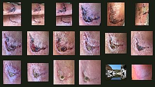
Fibrosis, also known as fibrotic scarring, is a pathological wound healing in which connective tissue replaces normal parenchymal tissue to the extent that it goes unchecked, leading to considerable tissue remodelling and the formation of permanent scar tissue.

Stromal cells, or mesenchymal stromal cells, are differentiating cells found in abundance within bone marrow but can also be seen all around the body. Stromal cells can become connective tissue cells of any organ, for example in the uterine mucosa (endometrium), prostate, bone marrow, lymph node and the ovary. They are cells that support the function of the parenchymal cells of that organ. The most common stromal cells include fibroblasts and pericytes. The term stromal comes from Latin stromat-, "bed covering", and Ancient Greek στρῶμα, strôma, "bed".

<span class="mw-page-title-main">Perlecan</span>

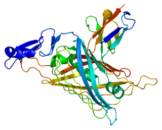
Perlecan (PLC) also known as basement membrane-specific heparan sulfate proteoglycan core protein (HSPG) or heparan sulfate proteoglycan 2 (HSPG2), is a protein that in humans is encoded by the HSPG2 gene. The HSPG2 gene codes for a 4,391 amino acid protein with a molecular weight of 468,829. It is one of the largest known proteins. The name perlecan comes from its appearance as a "string of pearls" in rotary shadowed images.

<span class="mw-page-title-main">Stroma (tissue)</span> Part of a tissue or organ with a structural or connective role

Stroma is the part of a tissue or organ with a structural or connective role. It is made up of all the parts without specific functions of the organ - for example, connective tissue, blood vessels, ducts, etc. The other part, the parenchyma, consists of the cells that perform the function of the tissue or organ.

<span class="mw-page-title-main">Mesenchyme</span> Type of animal embryonic connective tissue

Mesenchyme is a type of loosely organized animal embryonic connective tissue of undifferentiated cells that give rise to most tissues, such as skin, blood or bone. The interactions between mesenchyme and epithelium help to form nearly every organ in the developing embryo.

A fibrocyte is an inactive mesenchymal cell, that is, a cell showing minimal cytoplasm, limited amounts of rough endoplasmic reticulum and lacks biochemical evidence of protein synthesis.

<span class="mw-page-title-main">MMP2</span> Protein-coding gene in the species Homo sapiens

72 kDa type IV collagenase also known as matrix metalloproteinase-2 (MMP-2) and gelatinase A is an enzyme that in humans is encoded by the MMP2 gene. The MMP2 gene is located on chromosome 16 at position 12.2.

<span class="mw-page-title-main">Mucin short variant S1</span> Human protein

Mucin short variant S1, also called polymorphic epithelial mucin (PEM) or epithelial membrane antigen (EMA), is a mucin encoded by the MUC1 gene in humans. Mucin short variant S1 is a glycoprotein with extensive O-linked glycosylation of its extracellular domain. Mucins line the apical surface of epithelial cells in the lungs, stomach, intestines, eyes and several other organs. Mucins protect the body from infection by pathogen binding to oligosaccharides in the extracellular domain, preventing the pathogen from reaching the cell surface. Overexpression of MUC1 is often associated with colon, breast, ovarian, lung and pancreatic cancers. Joyce Taylor-Papadimitriou identified and characterised the antigen during her work with breast and ovarian tumors.

<span class="mw-page-title-main">Epithelial cell adhesion molecule</span> Transmembrane glycoprotein

Epithelial cell adhesion molecule (EpCAM), also known as CD326 among other names, is a transmembrane glycoprotein mediating Ca2+-independent homotypic cell–cell adhesion in epithelia. EpCAM is also involved in cell signaling, migration, proliferation, and differentiation. Additionally, EpCAM has oncogenic potential via its capacity to upregulate c-myc, e-fabp, and cyclins A & E. Since EpCAM is expressed exclusively in epithelia and epithelial-derived neoplasms, EpCAM can be used as diagnostic marker for various cancers. It appears to play a role in tumorigenesis and metastasis of carcinomas, so it can also act as a potential prognostic marker and as a potential target for immunotherapeutic strategies.

<span class="mw-page-title-main">Desmoplasia</span> Growth of fibrous or connective tissue

In medicine, desmoplasia is the growth of fibrous connective tissue. It is also called a desmoplastic reaction to emphasize that it is secondary to an insult. Desmoplasia may occur around a neoplasm, causing dense fibrosis around the tumor, or scar tissue (adhesions) within the abdomen after abdominal surgery.

Cenderitide is a natriuretic peptide developed by the Mayo Clinic as a potential treatment for heart failure. Cenderitide is created by the fusion of the 15 amino acid C-terminus of the snake venom dendroaspis natriuretic peptide (DNP) with the full C-type natriuretic peptide (CNP) structure. This peptide chimera is a dual activator of the natriuretic peptide receptors NPR-A and NPR-B and therefore exhibits the natriuretic and diuretic properties of DNP, as well as the antiproliferative and antifibrotic properties of CNP.

Dermal fibroblasts are cells within the dermis layer of skin which are responsible for generating connective tissue and allowing the skin to recover from injury. Using organelles, dermal fibroblasts generate and maintain the connective tissue which unites separate cell layers. Furthermore, these dermal fibroblasts produce the protein molecules including laminin and fibronectin which comprise the extracellular matrix. By creating the extracellular matrix between the dermis and epidermis, fibroblasts allow the epithelial cells of the epidermis to affix the matrix, thereby allowing the epidermal cells to effectively join together to form the top layer of the skin.

<span class="mw-page-title-main">Tumor microenvironment</span> Surroundings of tumors including nearby cells and blood vessels

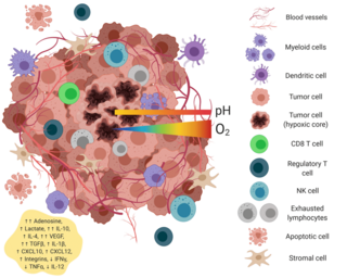
The tumor microenvironment is a complex ecosystem surrounding a tumor, composed of cancer cells, stromal tissue and the extracellular matrix. Mutual interaction between cancer cells and the different components of the tumor microenvironment support its growth and invasion in healthy tissues which correlates with tumor resistance to current treatments and poor prognosis. The tumor microenvironment is in constant change because of the tumor's ability to influence the microenvironment by releasing extracellular signals, promoting tumor angiogenesis and inducing peripheral immune tolerance, while the immune cells in the microenvironment can affect the growth and evolution of cancerous cells.

<span class="mw-page-title-main">Mucin-1</span> Protein of the mucin family

Mucin-1(MUC-1) is a heterodimer transmembrane protein of the mucin family encoded in humans by the MUC1 gene. It is cleaved into two chains: mucin-1 subunit alpha and mucin-1 subunit beta. These subunits differ in size due to proteolytic cleavage of the translated precursor protein in the endoplasmic reticulum. The larger subunit of MUC-1 is characterized by numerous O-glycosylated bonds and a terminal sialic acid, creating a net negative charge on MUC-1. The smaller subunit contains a juxtamembrane region of the extracellular area, a transmembrane domain, and the cytoplasmic tail. The extracellular domain of MUC-1 is composed of 20 identical amino acid tandem repeats (TR). Each tandem repeat contains two serine and three threonine amino acid residues, providing five sites for potential O-glycosylation. MUC-1 protein is estimated to weigh 120 to 225 kDA.

A cancer-associated fibroblast (CAF) is a cell type within the tumor microenvironment that promotes tumorigenic features by initiating the remodelling of the extracellular matrix or by secreting cytokines. CAFs are a complex and abundant cell type within the tumour microenvironment; the number cannot decrease, as they are unable to undergo apoptosis.

Physical oncology (PO) is defined as the study of the role of mechanical signals in a cancerous tumor. The mechanical signals can be forces, pressures. If we generalize we will speak of "stress field" and "stress tensor".

References

  1. Murray, Lynne A.; Knight, Darryl A.; Laurent, Geoffrey J. (2009). "Fibroblasts". Asthma and COPD: 193–200. doi:10.1016/B978-0-12-374001-4.00015-8.
  2. "Fibroblast". Genetics Home Reference. U.S. National Library of Medicine. 2014-05-05. Retrieved 2014-05-10.
  3. "Fibroblasts" . Retrieved 16 August 2018.
  4. Weissman-Shomer P, Fry M (1975). "Chick embryo fibroblasts senscence in vitro: pattern of cell division and life span as a function of cell density". Mechanisms of Ageing and Development. 4 (2): 159–166. doi:10.1016/0047-6374(75)90017-2. PMID   1152547. S2CID   9299977.
  5. Advances in Extracellular Space Research and Application. Scholarly Editions. 2013. p. 251. ISBN   9781481682626.
  6. Dave JM, Bayless KJ (May 2014). "Vimentin as an integral regulator of cell adhesion and endothelial sprouting". Microcirculation. 21 (4): 333–344. doi:10.1111/micc.12111. PMID   24387004. S2CID   26292524.
  7. Pilling, Darrell; Vakil, Varsha; Cox, Nehemiah; Gomer, Richard H. (2015-09-22). "TNF-α–stimulated fibroblasts secrete lumican to promote fibrocyte differentiation". Proceedings of the National Academy of Sciences. 112 (38): 11929–11934. doi:10.1073/pnas.1507387112. ISSN   0027-8424. PMC   4586854 . PMID   26351669.
  8. Smith RS, Smith TJ, Blieden TM, Phipps RP (August 1997). "Fibroblasts as sentinel cells. Synthesis of chemokines and regulation of inflammation". The American Journal of Pathology. 151 (2): 317–322. PMC   1858004 . PMID   9250144.
  9. 1 2 3 Silzle T, Randolph GJ, Kreutz M, Kunz-Schughart LA (January 2004). "The fibroblast: sentinel cell and local immune modulator in tumor tissue". International Journal of Cancer. 108 (2): 173–180. doi:10.1002/ijc.11542. PMID   14639599. S2CID   10936034.
  10. Bonnans C, Chou J, Werb Z (December 2014). "Remodelling the extracellular matrix in development and disease". Nature Reviews. Molecular Cell Biology. 15 (12): 786–801. doi:10.1038/nrm3904. PMC   4316204 . PMID   25415508.
  11. Llames, S.; García-Pérez, E.; Meana, A.; Larcher, F.; del Río, M. (2015). "Feeder Layer Cell Actions and Applications". Tissue Eng Part B Rev. 21 (4): 345–353. doi:10.1089/ten.teb.2014.0547. PMC   4533020 . PMID   25659081.
  12. Hynds, R.E.; Bonfanti, P.; Janes, S.M. (2018). "Regenerating human epithelia with cultured stem cells: feeder cells, organoids and beyond". EMBO Molecular Medicine. 10 (2): 139–150. doi:10.15252/emmm.201708213. PMC   5801505 . PMID   29288165.
  13. Hagbard, L.; Cameron, K.; August, P.; Penton, C.; Parmar, M.; Hay, D.C.; Kallur, T. (2018). "Developing defined substrates for stem cell culture and differentiation". Philosophical Transactions of the Royal Society B. 373 (1750). doi:10.1098/rstb.2017.0230. PMC   5974452 . PMID   29786564.
  14. Desai N, Rambhia P, Gishto A (February 2015). "Human embryonic stem cell cultivation: historical perspective and evolution of xeno-free culture systems". Reproductive Biology and Endocrinology. 13 (1): 9. doi: 10.1186/s12958-015-0005-4 . PMC   4351689 . PMID   25890180.
  15. Matsuda Y, Takahashi K, Kamioka H, Naruse K (September 2018). "Human gingival fibroblast feeder cells promote maturation of induced pluripotent stem cells into cardiomyocytes". Biochemical and Biophysical Research Communications. 503 (3): 1798–1804. doi: 10.1016/j.bbrc.2018.07.116 . PMID   30060947.
  16. 1 2 Krausgruber T, Fortelny N, Fife-Gernedl V, Senekowitsch M, Schuster LC, Lercher A, et al. (July 2020). "Structural cells are key regulators of organ-specific immune responses". Nature. 583 (7815): 296–302. Bibcode:2020Natur.583..296K. doi: 10.1038/s41586-020-2424-4 . PMC   7610345 . PMID   32612232. S2CID   220295181.
  17. Armingol E, Officer A, Harismendy O, Lewis NE (November 2020). "Deciphering cell-cell interactions and communication from gene expression". Nature Reviews. Genetics. 22 (2): 71–88. doi:10.1038/s41576-020-00292-x. PMC   7649713 . PMID   33168968.
  18. Minton K (September 2020). "A gene atlas of 'structural immunity'". Nature Reviews. Immunology. 20 (9): 518–519. doi:10.1038/s41577-020-0398-y. PMID   32661408. S2CID   220491226.