Flavobacteriia

Last updated

Flavobacteriia
Elizabethkingia meningoseptica Blood agar plate.JPG
Elizabethkingia meningoseptica on blood agar
Scientific classification OOjs UI icon edit-ltr.svg
Domain: Bacteria
Phylum: Bacteroidota
Class: Flavobacteriia
Bernardet 2012 [1]
Orders [2]
  • Flavobacteriales Bernardet 2012
  • Genera not assigned to an order or family
    • "Candidatus Walczuchella monophlebidarum" Rosas-Perez et al. 2014

The class Flavobacteriia is composed of a single class of environmental bacteria. [3] It contains the family Flavobacteriaceae, which is the largest family in the phylum Bacteroidota. [4] This class is widely distributed in soil, fresh, and seawater habitats. [5] The name is often spelt Flavobacteria, but was officially named Flavobacteriia in 2012. [6] [7]

Contents

Flavobacteriia are gram-negative aerobic rods, 25 μm long, 0.10.5 μm wide, with rounded or tapered ends. [6] They form circular cream to orange coloured colonies on agar, and are typically simple to successfully culture. [5] Flavobacteriia is a chemoorganotroph and are known for their ability to mineralize or degrade dissolved organic matter of high molecular weight and particulate plant material. [8]

Flavobacteriia have impacts on both the environment and human society, as they are able to cause diseases in many organisms. They are important in the decomposition of organic matter and pollutants, and are key members in the formation of marine biofilms. [9] They also have been known to cause diseases in some animal species, specifically bacterial cold water disease and columnaris disease. [10] [11]

Taxonomy

Flavobacteriia is the largest of the four classes of phylum Bacteroidota. It is a single-order class, and its largest family is Flavobacteriaceae. [4] Flavobacteriaceae is the largest family in the phylum Bacteroidota . The family has over 90 genera and hundreds of species. [4] The genus Flavobacterium is most commonly used in studies of Flavobacteriia. This genus has 100 classified species with many additional unclassified species. [12] Recent taxonomic updates have reclassified several Flavobacterium species to new genera such as Microbacterium, Salegentibacter, and Planococcus. [13]

History

Department disease laboratory researchers (Harold Wolf and Bill Schafer) investigate a trout disease threat in a commercial hatchery. California fish and game (19891740074).jpg
Department disease laboratory researchers (Harold Wolf and Bill Schafer) investigate a trout disease threat in a commercial hatchery.

The genus Flavobacterium was established in 1889. [13] It was first written about in 1923 in Bergey's manual of determinative bacteriology and contained one of the first of 46 discussed species. [14] The manual defines Flavobacteriia as gram-negative, non-spore-forming, aerobic, non-gliding rods. [14] In 1999, Flavobacteriia was discovered to have a yellow pigment in colonies. It was also identified that they move through gliding and only grow in the presence of oxygen. [15]

In 1922, Flavobacterium columnare , an agent of columnaris disease with significant effects on fish, was discovered in the Mississippi River, making it one of the earliest known diseases of its kind. [11] The disease was originally labelled as a Myxobacteria in 1944, but was renamed to Flavobacterium columnare in 1996 with 10 species. [16] [4] Flavobacteriia used to contain many non-related species of yellow, rod-shaped bacteria, but taxonomy has changed and stabilized due to the sequencing of rRNA to deduce phylogenetic relationships. [4]

Habitat

Flavobacteriia forming mudflat biofilms, as shown through confocal laser scanning microscopy in a 3D view. Growth took place over 24 hours on glass surfaces under dynamic conditions and stained with Syto 61 red. Scale bar: 67.3mm. Flavobacteriia forming Biofilms.png
Flavobacteriia forming mudflat biofilms, as shown through confocal laser scanning microscopy in a 3D view. Growth took place over 24 hours on glass surfaces under dynamic conditions and stained with Syto 61 red. Scale bar: 67.3μm.

Flavobacteriia are widely distributed with high abundances in aquatic systems. [5] They have been found in diseased fish, microbial mats, freshwater and river sediments, seawater and marine sediments, soil, glaciers, and Antarctic lakes. [18] [5] Increases in abundance are found in areas of high organic substrate inputs due to their role in the uptake, degradation, and decomposition of organic matter and can result in bacterial dominance. [13] Flavobacteriia is prominent in ocean sediments and decreases with increasing depth, and prefer sediments lacking vegetation. [19]

These bacteria also are highly abundant in melt ponds, solid ice cores, sea ice, and brine, as well as the photic zone. [18] More specifically, these photic zones show that Flavobacteriia are prominent in productive environments such as phytoplankton blooms and upwelling zones. [19] Flavobacteriia are prominent members of marine biofilms. [9] They have large impacts on the functioning of marine biofilms, however their abundance is believed to be heavily underestimated. [9]

Flavobacteriia can also be found in non-marine systems and are most common in Asian regions, specifically Korea and China, as well as Japan and India. [13]

Morphology

Colony morphology of Flavobacteriia species Bergeyella zoohelcum on blood agar. Photo credit: Stefan Walkowski Bergeyella zoohelcum 2.jpg
Colony morphology of Flavobacteriia species Bergeyella zoohelcum on blood agar. Photo credit: Stefan Walkowski

Flavobacteriia are a type of gram-negative rod-shaped bacteria with sizes typically ranging from 0.1μm to 0.5μm wide and 2μm to 5μm long. [6] [20] Depending on the species of Flavobacteriia, the genome size can range from 1.85x10⁹ daltons to 3.9x10⁹ daltons. [21] Flavobacteriia are also unable to form endospores. [20] They are classified as gram-negative due to the composition of their cell wall, which consists of a thin layer of peptidoglycan surrounded by an outer membrane composed of lipopolysaccharides. [22] The rod-shape of these bacteria typically have straight or slightly curved parallel sides with rounded or slightly tapered ends. [6] [8] The overall colony morphology of Flavobacteriia exhibit a circular shape that is either convex or slightly convex with a smooth appearance. [8] These colonies typically appear slightly translucent and can range in colour from pale yellow/cream to orange due to the presence of pigments such as carotenoids or flexirubin. [5] [8]

Flavobacteriia do not possess flagella and rely on either a gliding motion or are non-motile. [21] [23] The gliding motion allows them to move over wet surfaces such as a wet mount glass slide or agar plate. [23] [24] Flavobacteriia exhibit a predominant forward gliding motion, but can also reverse direction and show flipping movements . [24] Research suggests that the gliding motion is facilitated by the proton gradient across the cytoplasmic membrane. [23] [24]

Metabolism

Bacteria from the class Flavobacteriia have diverse metabolism. Flavobacteriia are chemoorganotrophic, meaning they use organic molecules as a source of energy. [8] Most species have obligately aerobic type of respiration, while some species can grow under weak microaerobic to anaerobic conditions. [8] Some species of Flavobacteriia have the ability to use a broad range of carbohydrates as energy sources, while others have a limited capacity or none at all, and instead prefer to utilize amino acids and proteins. [8] Approximately half of the species belonging to Flavobacteriia are capable of breaking down carbohydrates into acid and can degrade tyrosine and tween compounds. [8] Only a few species can degrade urea and DNA. [8] Many species also play a significant role in the mineralization of organic matter in both aquatic and soil environments due to their capability of breaking down various types of biomacromolecules. [8]

Diagram of bacterial cell with transporters. Genome analysis of proteorhodopsin in Flavobacteriia. MED152 Transporters.png
Diagram of bacterial cell with transporters. Genome analysis of proteorhodopsin in Flavobacteriia.

Flavobacteriia are not photosynthetic, but some marine species use proteorhodopsin for energy through the harvesting of light. [4] Proteorhodopsin (PR) is a proton pump that uses light, however species who use PR need to adapt to different environments to combat ultraviolet (UV) damage, and adopt the ability to mend DNA that has been damaged by UV. [25]

Proteorhodopsin is useful in the active transport of protons across the cell membrane. This is useful in the creation of ATP as energy in Flavobacteriia. The diagram to the right shows how proteorhodopsin is used in Flavobacteriia cells, and provides specifics about how it used in the bacteria. [25]

Culture

Typical culturing methods are used to isolate Flavobacteriia, such as simple dilutions. Techniques vary by species due to the high diversity of the class, however many are cultivated on simple media using yeast extract and a protein hydrolysate. [4] Sugars may need to be added or certain salts for marine species. Fish and bird pathogens may have additional requirements for culture methodology. [4]

Marine Flavobacteriia are cultured on marine agar or cytophaga agar. Non-marine Flavobacteriia are culture on rich media including nutrient agar, casitone-yeast extract agar, PYG agar, and TYES agar, or nutrient-poor media such as AO agar, PY2 agar, and R2A agar. [4] Flavobacteriia species that inhabit cold environments exhibit optimal growth at temperatures between 15 °C to 20 °C, while those that inhabit temperate environments exhibit optimal growth at temperatures between 20 °C to 30 °C. [8] Therefore, temperatures for culturing are between 20 °C and 30 °C, with an optimal temperature of 37˚C. [4]

Many psychrophilic and psychrotrophic species have been isolated through culture methods, mainly from polar regions. [4] Additional mesophilic species have been isolated as well as a few thermophiles, while extreme halophiles have not yet been identified. [4]

Environmental impact

Though the majority of Flavobacteriia are harmless, some infect opportunistically or cause severe diseases. This means that they can cause disease in many types organisms such as plants or fish. [13] They have proteins that discharge factors able to cause the development of a disease. [26] Fish pathogens are common on or in fish or the surrounding water. Bird pathogens cause outbreaks in domestic poultry or wild birds. [4]

Columnaris disease (Flavobacterium columnaris) in the gill of a chinook salmon. Columnaris disease.jpg
Columnaris disease ( Flavobacterium columnaris ) in the gill of a chinook salmon.

One possible disease is bacterial cold water disease caused by Flavobacterium psychrophilum in rainbow trout, which can cause tissue erosion, jaw ulcerations, inflammation, and behavioural issues. [10] It can also cause acute losses in young rainbow trout, known as rainbow trout fry syndrome. [27] In 2005, the National Center for Cool and Cold Water Aquaculture measured survival rate to be 29.3% from these diseases. [27]

Additionally, Flavobacterium columare causes columnaris disease in freshwater fish species. Columnaris disease causes skin lesions, fin erosion, and gill necrosis, leading to mortality. [11]

Marine biofilms are a biological element that significantly affects the productivity and operation of marine habitats by assisting in basic microbial processes like photosynthesis, the cycling of nitrogen, and the degradation of organic matter and pollutants. [9] In the early stage of marine biofilms formation, Flavobacteriia colonize and form microcolonies to serve as a foundation for establishing other microorganisms in a community. In the biofilms community, Flavobacteriia also engage in a variety of cooperated interactions with other microbes rather than competition, including quorum sensing, nutrient sharing and scavenging. In sum, these interactions are essential for establishing and maintaining complex microbial communities in marine biofilms. [9]

Human impact

Food

A culture of Cytophaga strains to study enzymes in relation to polysaccharides. Specifically, yeast is used industrial and laboratory processes and Cytophaga enzymes can destroy the yeast cell wall. The culture was obtained using a microbiological loop to streak on agar nutrient medium. Bakterii roda Cytophaga, vyrosshie na tviordoi pitatel'noi srede.jpg
A culture of Cytophaga strains to study enzymes in relation to polysaccharides. Specifically, yeast is used industrial and laboratory processes and Cytophaga enzymes can destroy the yeast cell wall. The culture was obtained using a microbiological loop to streak on agar nutrient medium.

Flavobacteriia have been linked to food and food product deterioration. The relative humidity of the shop where the product is located affects the growth of psychrophilic or psychrotrophic microorganisms. [13] Due to the formation of metabolic byproducts, spoilage of uncooked red flesh causes unpleasant smells, potential slime production, localised discolouration and unwanted flavours. Similarly, while Flavobacteriia are a continuous component of the initial flora in cold meats and fowl, they are unable to outcompete pseudomonads during preservation. [13] Poultry has a much greater prevalence of flavobacteriia than other fresh flesh. [13]

Flavobacteriia create pasteurisation-resistant extracellular enzymes, which causes the psychrotrophic deterioration of milk and dairy products. [13] Due to the creation of phospholipase C, they are also to blame for a decrease in cheddar cheese output and bitterness in milk. Given that phospholipases have the capacity to degrade the phospholipids that make up the milk fat globule membrane and thereby increase the vulnerability of the milk fat (triglycerides) to lipolytic assault, they may be significant in milk and milk products. [13]

Disease

Members of the Flavobacteriia also cause disease in humans. [13] However, as strains within the Flavobacterium were reclassified, many strains that cause human disease were transferred to new or different genera [28] such as Chryseobacterium, Myroides, Empedobacter and Sphingobacterium. [29] Their main infected populations are newborns and people with immunodeficiencies. Neonatal infections usually manifest as meningitis, and the mortality rate of neonatal meningitis is high. Meningitis can also cause bacteremia and pneumonia. In adults, infections can manifest in a variety of ways, including pneumonia, sepsis, meningitis, endocarditis, post-surgery, and post-burn. [29] To this point, existing pathogenic Flavobacteriia are currently very rare and difficult to detect, but remain a concern because they are resistant to many antimicrobial drugs. [28]

Industrial uses

The decomposition abilities of Flavobacteriia are also used to benefit humans industries. The bacteria commonly is found in sewage treatment facilities. They are used to treat wastewater because of their ability to digest chemicals and other molecules, including polycyclic aromatic hydrocarbons. [4]

Flavobacteriia is used to promote plant growth in the agricultural sector. The bacteria is able to solubilise inorganic phosphate and produce additional beneficial elements such as indole-3-acetic acid (the key plant hormone IAA) and 1-aminocyclopropane-1-carboxylatedeaminase (a hydrolase), which can be taken up and used by plants or alter their signalling. [30] It can act as a microbial agent to protect plants from other diseases, and even has benefits in the development of antimicrobial medicines. [4]

Related Research Articles

<span class="mw-page-title-main">Biofilm</span> Aggregation of bacteria or cells on a surface

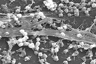
A biofilm comprises any syntrophic consortium of microorganisms in which cells stick to each other and often also to a surface. These adherent cells become embedded within a slimy extracellular matrix that is composed of extracellular polymeric substances (EPSs). The cells within the biofilm produce the EPS components, which are typically a polymeric conglomeration of extracellular polysaccharides, proteins, lipids and DNA. Because they have three-dimensional structure and represent a community lifestyle for microorganisms, they have been metaphorically described as "cities for microbes".

<span class="mw-page-title-main">Bacteroidota</span> Phylum of Gram-negative bacteria

The phylum Bacteroidota is composed of three large classes of Gram-negative, nonsporeforming, anaerobic or aerobic, and rod-shaped bacteria that are widely distributed in the environment, including in soil, sediments, and sea water, as well as in the guts and on the skin of animals.

<i>Chlorobium</i> Genus of bacteria

Chlorobium is a genus of green sulfur bacteria. They are photolithotrophic oxidizers of sulfur and most notably utilise a noncyclic electron transport chain to reduce NAD+. Photosynthesis is achieved using a Type 1 Reaction Centre using bacteriochlorophyll (BChl) a. Two photosynthetic antenna complexes aid in light absorption: the Fenna-Matthews-Olson complex, and the chlorosomes which employ mostly BChl c, d, or e. Hydrogen sulfide is used as an electron source and carbon dioxide its carbon source.

<i>Beggiatoa</i> Genus of bacteria

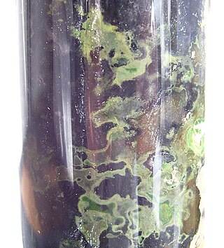
Beggiatoa is a genus of Gammaproteobacteria belonging to the order Thiotrichales, in the Pseudomonadota phylum. This genus was one of the first bacteria discovered by Ukrainian botanist Sergei Winogradsky. During his research in Anton de Bary's laboratory of botany in 1887, he found that Beggiatoa oxidized hydrogen sulfide (H2S) as an energy source, forming intracellular sulfur droplets, with oxygen as the terminal electron acceptor and CO2 used as a carbon source. Winogradsky named it in honor of the Italian doctor and botanist Francesco Secondo Beggiato (1806 - 1883), from Venice. Winogradsky referred to this form of metabolism as "inorgoxidation" (oxidation of inorganic compounds), today called chemolithotrophy. These organisms live in sulfur-rich environments such as soil, both marine and freshwater, in the deep sea hydrothermal vents and in polluted marine environments. The finding represented the first discovery of lithotrophy. Two species of Beggiatoa have been formally described: the type species Beggiatoa alba and Beggiatoa leptomitoformis, the latter of which was only published in 2017. This colorless and filamentous bacterium, sometimes in association with other sulfur bacteria (for example the genus Thiothrix), can be arranged in biofilm visible to the naked eye formed by a very long white filamentous mat, the white color is due to the stored sulfur. Species of Beggiatoa have cells up to 200 µm in diameter and they are one of the largest prokaryotes on Earth.

<i>Elizabethkingia meningoseptica</i> Species of bacterium

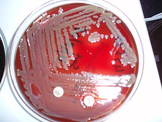
Elizabethkingia meningoseptica is a Gram-negative, rod-shaped bacterium widely distributed in nature. It may be normally present in fish and frogs; it may be isolated from chronic infectious states, as in the sputum of cystic fibrosis patients. In 1959, American bacteriologist Elizabeth O. King was studying unclassified bacteria associated with pediatric meningitis at the Centers for Disease Control and Prevention in Atlanta, when she isolated an organism that she named Flavobacterium meningosepticum. In 1994, it was reclassified in the genus Chryseobacterium and renamed Chryseobacterium meningosepticum(chryseos = "golden" in Greek, so Chryseobacterium means a golden/yellow rod similar to Flavobacterium). In 2005, a 16S rRNA phylogenetic tree of Chryseobacteria showed that C. meningosepticum along with C. miricola were close to each other but outside the tree of the rest of the Chryseobacteria and were then placed in a new genus Elizabethkingia named after the original discoverer of F. meningosepticum.

<span class="mw-page-title-main">Gammaproteobacteria</span> Class of bacteria

Gammaproteobacteria is a class of bacteria in the phylum Pseudomonadota. It contains about 250 genera, which makes it the most genus-rich taxon of the Prokaryotes. Several medically, ecologically, and scientifically important groups of bacteria belong to this class. It is composed by all Gram-negative microbes and is the most phylogenetically and physiologically diverse class of Proteobacteria.

<span class="mw-page-title-main">Gliding motility</span>

Gliding motility is a type of translocation used by microorganisms that is independent of propulsive structures such as flagella, pili, and fimbriae. Gliding allows microorganisms to travel along the surface of low aqueous films. The mechanisms of this motility are only partially known.

<i>Chryseobacterium</i> Genus of bacteria

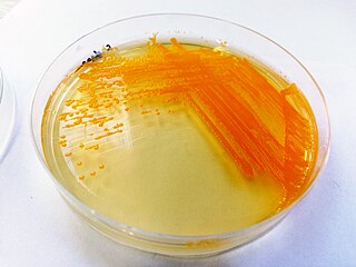
Chryseobacterium is a genus of Gram-negative bacteria. Chryseobacterium species are chemoorganotrophic, rod shape gram-negative bacteria. Chryseobacterium form typical yellow-orange color colonies due to flexirubin-type pigment. The genus contains more than 100 described species from diverse habitats, including freshwater sources, soil, marine fish, and human hosts.

<i>Cytophaga</i> Genus of bacteria

Cytophaga is a genus of Gram-negative, gliding, rod-shaped bacteria. This bacterium is commonly found in soil, rapidly digests crystalline cellulose C. hutchinsonii is able to use its gliding motility to move quickly over surfaces. Although the mechanism for this is not known, there is a belief that the flagellum is not used

Actibacter is a genus in the phylum Bacteroidota (Bacteria). The genus contains a single species, namely A. sediminis.

Tamlana is a genus in the phylum Bacteroidota (Bacteria). Two species have been described so far: T. agarivorans and T. crocina.

Bacterial cold water disease (BCWD) is a bacterial disease of freshwater fish, specifically salmonid fish. It is caused by the bacterium Flavobacterium psychrophilum, a psychrophilic, gram-negative rod-shaped bacterium of the family Flavobacteriaceae. This bacterium is found in fresh waters with the optimal growth temperature below 13 °C, and it can be seen in any area with water temperatures consistently below 15 °C. Salmon are the most commonly affected species. This disease is not zoonotic.

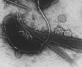
Flavobacterium psychrophilum is a psychrophilic, gram-negative bacterial rod, belonging to the Bacteroidota. It is the causative agent of bacterial coldwater disease (BCWD) and was first isolated in 1948 during a die-off in the salmonid Oncorhynchus kisutch.

Tenacibaculum is a Gram-negative and motile bacterial genus from the family of Flavobacteriaceae.

Dokdonia donghaensis is a strictly aerobic, gram-negative, phototrophic bacterium that thrives in marine environments. The organism can grow at a broad range of temperatures on seawater media. It has the ability to form biofilms, which increases the organism's resistance to antimicrobial agents, such as tetracycline.

Polaribacter is a genus in the family Flavobacteriaceae. They are gram-negative, aerobic bacteria that can be heterotrophic, psychrophilic or mesophilic. Most species are non-motile and species range from ovoid to rod-shaped. Polaribacter forms yellow- to orange-pigmented colonies. They have been mostly adapted to cool marine ecosystems, and their optimal growth range is at a temperature between 10 and 32 °C and at a pH of 7.0 to 8.0. They are oxidase and catalase-positive and are able to grow using carbohydrates, amino acids, and organic acids.

Flavobacterium akiainvivens, or koʻohonua ʻili akia, is a species of gram-negative bacteria in the Flavobacteriaceae family. The specific epithet akiainvivens is Latin and literally means "living on or in ʻākia." It was isolated originally from decaying wood of the endemic Hawai'ian shrub ʻākia.

Dokdonia is a genus of bacteria in the family Flavobacteriaceae and phylum Bacteroidota.

Mariniflexile is a genus in the phylum Bacteroidota (Bacteria). The various species have been recovered from sea water, sea urchins, springs, brackish water, and an oyster.

Cytophagales is an order of non-spore forming, rod-shaped, Gram-negative bacteria that move through a gliding or flexing motion. These chemoorganotrophs are important remineralizers of organic materials into micronutrients. They are widely dispersed in the environment, found in ecosystems including soil, freshwater, seawater and sea ice. Cytophagales is included in the Bacteroidota phylum.

References

  1. Bernardet JF (2010). "Class II. Flavobacteriia class. nov.". In Krieg NR, Staley JT, Brown DR, Hedlund BP, Paster BJ, Ward NL, Ludwig W, Whitman WB (eds.). Bergey's Manual of Systematic Bacteriology. Vol. 4 (2nd ed.). New York, NY: Springer. p. 105.
  2. Euzéby JP, Parte AC. "Flavobacteriia". List of Prokaryotic names with Standing in Nomenclature (LPSN). Retrieved June 29, 2021.
  3. Boone DR, Castenholz RW, eds. (2001). Bergey's Manual of Systematic Bacteriology. Vol. 1 (The Archaea and the deeply branching and phototrophic Bacteria) (2nd ed.). New York: Springer-Verlag. pp. 465–466. ISBN   978-0-443-05615-4.
  4. 1 2 3 4 5 6 7 8 9 10 11 12 13 14 15 McBride M (2014). "The Family Flavobacteriaceae". The Prokaryotes. pp. 643–676. doi:10.1007/978-3-642-38954-2_130. ISBN   978-3-642-38953-5 via SpringerLink.
  5. 1 2 3 4 5 Buchan A, LeCleir GR, Gulvik CA, González JM (October 2014). "Master recyclers: features and functions of bacteria associated with phytoplankton blooms". Nature Reviews. Microbiology. 12 (10): 686–698. doi:10.1038/nrmicro3326. PMID   25134618. S2CID   26684717.
  6. 1 2 3 4 Bernardet JF, Bowman JP (July 2013). "International Committee on Systematics of Prokaryotes Subcommittee on the taxonomy of Flavobacterium and Cytophaga-like bacteria: Minutes of the meetings, 7 September 2011, Sapporo, Japan". International Journal of Systematic and Evolutionary Microbiology. 63 (Pt 7): 2752–2754. doi: 10.1099/ijs.0.053926-0 . PMID   23825377.
  7. "Flavobacteriia class". allmicrobes.com. Archived from the original on 1 November 2014. Retrieved 1 November 2014.
  8. 1 2 3 4 5 6 7 8 9 10 11 Whitman WB, Rainey F, Kämpfer P, Trujillo M, Chun J, DeVos P, et al., eds. (2015-04-17). Bergey's Manual of Systematics of Archaea and Bacteria (1st ed.). Wiley. doi:10.1002/9781118960608.gbm00312. ISBN   978-1-118-96060-8. S2CID   242820836.
  9. 1 2 3 4 5 Pollet T, Berdjeb L, Garnier C, Durrieu G, Le Poupon C, Misson B, Jean-François B (June 2018). "Prokaryotic community successions and interactions in marine biofilms: the key role of Flavobacteriia". FEMS Microbiology Ecology. 94 (6). doi: 10.1093/femsec/fiy083 . PMID   29733333.
  10. 1 2 Barnes ME (2011). "A Review of Flavobacterium Psychrophilum Biology, Clinical Signs, and Bacterial Cold Water Disease Prevention and Treat" (PDF). The Open Fish Science Journal. 4 (1): 40–48. doi: 10.2174/1874401X01104010040 .
  11. 1 2 3 Declercq AM, Haesebrouck F, Van den Broeck W, Bossier P, Decostere A (April 2013). "Columnaris disease in fish: a review with emphasis on bacterium-host interactions". Veterinary Research. 44 (1): 27. doi: 10.1186/1297-9716-44-27 . PMC   3648355 . PMID   23617544.
  12. 1 2 3 4 5 6 7 8 9 10 11 12 13 14 15 "Flavobacteriia". NCBI taxonomy database. National Center for Biotechnology Information (NCBI), U.S. National Library of Medicine. Retrieved 2023-03-07.
  13. 1 2 3 4 5 6 7 8 9 10 11 12 Waśkiewicz A, Irzykowska L (2014). "Flavobacterium spp. – Characteristics, Occurrence, and Toxicity". Encyclopedia of Food Microbiology: 938–942. doi:10.1016/B978-0-12-384730-0.00126-9. ISBN   978-0-12-384733-1.
  14. 1 2 Loch TP, Faisal M (May 2015). "Emerging flavobacterial infections in fish: A review". Journal of Advanced Research. 6 (3): 283–300. doi:10.1016/j.jare.2014.10.009. PMC   4522593 . PMID   26257926.
  15. "Species Flavobacterium aquatile". LPSN. Retrieved March 25, 2023.
  16. Kaiser D (2009). "Myxococcus". Encyclopedia of Microbiology (Third ed.): 220–244. doi:10.1016/B978-012373944-5.00018-3. ISBN   978-0-12-373944-5.
  17. Doghri I, Rodrigues S, Bazire A, Dufour A, Akbar D, Sopena V, Sablé S, Lanneluc I (October 2015). "Marine bacteria from the French Atlantic coast displaying high forming-biofilm abilities and different biofilm 3D architectures". BMC Microbiology. 15: 231. doi: 10.1186/s12866-015-0568-4 . PMC   4619314 . PMID   26498445.
  18. 1 2 Fernández-Gómez B, Díez B, Polz MF, Arroyo JI, Alfaro FD, Marchandon G, et al. (February 2019). "Bacterial community structure in a sympagic habitat expanding with global warming: brackish ice brine at 85-90 °N". The ISME Journal. 13 (2): 316–333. Bibcode:2019ISMEJ..13..316F. doi:10.1038/s41396-018-0268-9. PMC   6331608 . PMID   30228379.
  19. 1 2 Cleary DF, Coelho FJ, Oliveira V, Gomes NC, Polónia AR (2017). "Sediment depth and habitat as predictors of the diversity and composition of sediment bacterial communities in an inter-tidal estuarine environment". Marine Ecology. 38 (2): e12411. Bibcode:2017MarEc..38E2411C. doi:10.1111/maec.12411.
  20. 1 2 Zamora L, Vela AI, Sánchez-Porro C, Palacios MA, Domínguez L, Moore ER, Ventosa A, Fernández-Garayzábal JF (December 2013). "Characterization of flavobacteria possibly associated with fish and fish farm environment. Description of three novel Flavobacterium species: Flavobacterium collinsii sp. nov., Flavobacterium branchiarum sp. nov., and Flavobacterium branchiicola sp. nov". Aquaculture. 416–417: 346–353. Bibcode:2013Aquac.416..346Z. doi:10.1016/j.aquaculture.2013.09.019.
  21. 1 2 Shewan JM, McMeekin TA (October 1983). "Taxonomy (and ecology) of Flavobacterium and related genera". Annual Review of Microbiology. 37 (1): 233–252. doi:10.1146/annurev.mi.37.100183.001313. PMID   6357052.
  22. Lüderitz O, Freudenberg MA, Galanos C, Lehmann V, Rietschel ET, Shaw DH (January 1982). "Lipopolysaccharides of Gram-Negative Bacteria". Current Topics in Membranes and Transport. Vol. 17. Elsevier. pp. 79–151. doi:10.1016/s0070-2161(08)60309-3. ISBN   978-0-12-153317-5.
  23. 1 2 3 Shrivastava A, Berg HC (December 2015). "Towards a model for Flavobacterium gliding". Current Opinion in Microbiology. 28: 93–97. doi:10.1016/j.mib.2015.07.018. PMC   4688146 . PMID   26476806.
  24. 1 2 3 McBride MJ, Zhu Y (January 2013). "Gliding motility and Por secretion system genes are widespread among members of the phylum bacteroidetes". Journal of Bacteriology. 195 (2): 270–278. doi:10.1128/JB.01962-12. PMC   3553832 . PMID   23123910.
  25. 1 2 Olson DK, Yoshizawa S, Boeuf D, Iwasaki W, DeLong EF (April 2018). "Proteorhodopsin variability and distribution in the North Pacific Subtropical Gyre". The ISME Journal. 12 (4): 1047–1060. Bibcode:2018ISMEJ..12.1047O. doi:10.1038/s41396-018-0074-4. PMC   5864233 . PMID   29476140.
  26. "Recent Advancements in Flavobacteria: Pathogenesis Mechanisms, Novel Genetic and Physiological Features, and Interaction with Hosts". Microbial Physiology and Metabolism via Frontiers.
  27. 1 2 Silverstein JT, Vallejo RL, Palti Y, Leeds TD, Rexroad CE, Welch TJ, et al. (March 2009). "Rainbow trout resistance to bacterial cold-water disease is moderately heritable and is not adversely correlated with growth" (PDF). Journal of Animal Science. 87 (3): 860–867. doi:10.2527/jas.2008-1157. PMID   19028851.
  28. 1 2 García-López M, Santos J, Otero A (January 1999), "FLAVOBACTERIUM", in Robinson RK (ed.), Encyclopedia of Food Microbiology, Oxford: Elsevier, pp. 820–826, doi:10.1006/rwfm.1999.0660, ISBN   978-0-12-227070-3
  29. 1 2 Booth J (January 2007), Enna SJ, Bylund DB (eds.), "Chryseobacterium and Related Genera Infections", xPharm: The Comprehensive Pharmacology Reference, New York: Elsevier, pp. 1–4, doi:10.1016/b978-008055232-3.62958-7, ISBN   978-0-08-055232-3
  30. Nishioka T, Elsharkawy MM, Suga H, Kageyama K, Hyakumachi M, Shimizu M (June 2016). "Development of Culture Medium for the Isolation of Flavobacterium and Chryseobacterium from Rhizosphere Soil". Microbes and Environments. 31 (2): 104–10. doi:10.1264/jsme2.ME15144. PMC   4912144 . PMID   27098502.