Formins

Last updated
formin 1
Identifiers
SymbolFMN1
Alt. symbolsLD, FMN
NCBI gene 342184
HGNC 3768
OMIM 136535
RefSeq NM_001103184
UniProt Q68DA7
Other data
Locus Chr. 15 q13-q14
Search for
Structures Swiss-model
Domains InterPro
Formin Homology Region 1
Identifiers
SymbolDrf_FH1
Pfam PF06346
InterPro IPR009408
Available protein structures:
Pfam   structures / ECOD  
PDB RCSB PDB; PDBe; PDBj
PDBsum structure summary
Domain structure of formin proteins across phyla. Formin proteins.png
Domain structure of formin proteins across phyla.
Formin Homology 2 Domain
PDB 1ux4 EBI.jpg
crystal structures of a formin homology-2 domain reveal a tethered-dimer architecture
Identifiers
SymbolFH2
Pfam PF02181
InterPro IPR015425
SMART FH2
SCOP2 1ux5 / SCOPe / SUPFAM
Available protein structures:
Pfam   structures / ECOD  
PDB RCSB PDB; PDBe; PDBj
PDBsum structure summary
Diaphanous FH3 Domain
PDB 1z2c EBI.jpg
crystal structure of mdia1 gbd-fh3 in complex with rhoc-gmppnp
Identifiers
SymbolDrf_FH3
Pfam PF06367
Pfam clan CL0020
InterPro IPR010472
Available protein structures:
Pfam   structures / ECOD  
PDB RCSB PDB; PDBe; PDBj
PDBsum structure summary
DRF Autoregulatory Domain
PDB 2bap EBI.jpg
crystal structure of the n-terminal mdia1 armadillo repeat region and dimerisation domain in complex with the mdia1 autoregulatory domain (dad)
Identifiers
SymbolDrf_DAD
Pfam PF06345
InterPro IPR010465
Available protein structures:
Pfam   structures / ECOD  
PDB RCSB PDB; PDBe; PDBj
PDBsum structure summary
Diaphanous GTPase-binding Domain
PDB 1z2c EBI.jpg
crystal structure of mdia1 gbd-fh3 in complex with rhoc-gmppnp
Identifiers
SymbolDrf_GBD
Pfam PF06371
Pfam clan CL0020
InterPro IPR010473
Available protein structures:
Pfam   structures / ECOD  
PDB RCSB PDB; PDBe; PDBj
PDBsum structure summary

Formins (formin homology proteins) are a group of proteins that are involved in the polymerization of actin and associate with the fast-growing end (barbed end) of actin filaments. [2] Most formins are Rho-GTPase effector proteins. Formins regulate the actin and microtubule cytoskeleton [3] [4] and are involved in various cellular functions such as cell polarity, cytokinesis, cell migration and SRF transcriptional activity. [5] Formins are multidomain proteins that interact with diverse signalling molecules and cytoskeletal proteins, although some formins have been assigned functions within the nucleus.

Contents

Diversity

Formins have been found in all eukaryotes studied. [1] In humans, 15 different formin proteins are present that have been classified in 7 subgroups. [6] By contrast, yeasts contain only 2-3 formins. [7]

Structure and interactions

Formins are characterized by the presence of three formin homology (FH) domains (FH1, FH2 and FH3), although members of the formin family do not necessarily contain all three domains. [8] [9] In addition, other domains are usually present, such as PDZ, DAD, WH2, or FHA domains.

The proline-rich FH1 domain mediates interactions with a variety of proteins, including the actin-binding protein profilin, [10] SH3 (Src homology 3) domain proteins, [11] and WW domain proteins. The actin nucleation-promoting activity of S. cerevisiae formins has been localized to the FH2 domain. [4] The FH2 domain is required for the self-association of formin proteins through the ability of FH2 domains to directly bind each other, and may also act to inhibit actin polymerization. [12] [13] The FH3 domain is less well conserved and is required for directing formins to the correct intracellular location, such as the mitotic spindle, or the projection tip during conjugation. [14] [15] In addition, some formins can contain a GTPase-binding domain (GBD) required for binding to Rho small GTPases, and a C-terminal conserved Dia-autoregulatory domain (DAD). The GBD is a bifunctional autoinhibitory domain that interacts with and is regulated by activated Rho family members. Mammalian Drf3 contains a CRIB-like motif within its GBD for binding to Cdc42, which is required for Cdc42 to activate and guide Drf3 towards the cell cortex where it remodels the actin skeleton. [16] The DAD binds the N-terminal GBD; this link is broken when GTP-bound Rho binds to the GBD and activates the protein. The addition of the DAD to mammalian cells induces actin filament formation, stabilizes microtubules, and activates SRF mediated transcription. [16] Another commonly found domain is an armadillo repeat region (ARR) located in the FH3 domain.

The FH2 domain, has been shown by X-ray crystallography to have an elongated, crescent shape containing three helical subdomains. [17] [18]

Formins also directly bind to microtubules via their FH2 domain. This interaction is important in promoting the capture and stabilization of a subset of microtubules oriented towards the leading edge of migrating cells. Formins also promote the capture of microtubules by the kinetochore during mitosis and for aligning microtubules along actin filaments. [19] [20]

See also

Related Research Articles

<span class="mw-page-title-main">Microfilament</span> Filament in the cytoplasm of eukaryotic cells

Microfilaments, also called actin filaments, are protein filaments in the cytoplasm of eukaryotic cells that form part of the cytoskeleton. They are primarily composed of polymers of actin, but are modified by and interact with numerous other proteins in the cell. Microfilaments are usually about 7 nm in diameter and made up of two strands of actin. Microfilament functions include cytokinesis, amoeboid movement, cell motility, changes in cell shape, endocytosis and exocytosis, cell contractility, and mechanical stability. Microfilaments are flexible and relatively strong, resisting buckling by multi-piconewton compressive forces and filament fracture by nanonewton tensile forces. In inducing cell motility, one end of the actin filament elongates while the other end contracts, presumably by myosin II molecular motors. Additionally, they function as part of actomyosin-driven contractile molecular motors, wherein the thin filaments serve as tensile platforms for myosin's ATP-dependent pulling action in muscle contraction and pseudopod advancement. Microfilaments have a tough, flexible framework which helps the cell in movement.

<span class="mw-page-title-main">Wiskott–Aldrich syndrome protein</span> Mammalian protein found in humans

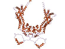
The Wiskott–Aldrich Syndrome protein (WASp) is a 502-amino acid protein expressed in cells of the hematopoietic system that in humans is encoded by the WAS gene. In the inactive state, WASp exists in an autoinhibited conformation with sequences near its C-terminus binding to a region near its N-terminus. Its activation is dependent upon CDC42 and PIP2 acting to disrupt this interaction, causing the WASp protein to 'open'. This exposes a domain near the WASp C-terminus that binds to and activates the Arp2/3 complex. Activated Arp2/3 nucleates new F-actin.

<span class="mw-page-title-main">Cell cortex</span> Layer on the inner face of a cell membrane

The cell cortex, also known as the actin cortex, cortical cytoskeleton or actomyosin cortex, is a specialized layer of cytoplasmic proteins on the inner face of the cell membrane. It functions as a modulator of membrane behavior and cell surface properties. In most eukaryotic cells lacking a cell wall, the cortex is an actin-rich network consisting of F-actin filaments, myosin motors, and actin-binding proteins. The actomyosin cortex is attached to the cell membrane via membrane-anchoring proteins called ERM proteins that plays a central role in cell shape control. The protein constituents of the cortex undergo rapid turnover, making the cortex both mechanically rigid and highly plastic, two properties essential to its function. In most cases, the cortex is in the range of 100 to 1000 nanometers thick.

<span class="mw-page-title-main">CDC42</span> Protein-coding gene in the species Homo sapiens

Cell division control protein 42 homolog is a protein that in humans is encoded by the CDC42 gene. Cdc42 is involved in regulation of the cell cycle. It was originally identified in S. cerevisiae (yeast) as a mediator of cell division, and is now known to influence a variety of signaling events and cellular processes in a variety of organisms from yeast to mammals.

<span class="mw-page-title-main">FGD1</span> Protein-coding gene in the species Homo sapiens

FYVE, RhoGEF and PH domain-containing protein 1 (FGD1) also known as faciogenital dysplasia 1 protein (FGDY), zinc finger FYVE domain-containing protein 3 (ZFYVE3), or Rho/Rac guanine nucleotide exchange factor FGD1 is a protein that in humans is encoded by the FGD1 gene that lies on the X chromosome. Orthologs of the FGD1 gene are found in dog, cow, mouse, rat, and zebrafish, and also budding yeast and C. elegans. It is a member of the FYVE, RhoGEF and PH domain containing family.

<span class="mw-page-title-main">Transforming protein RhoA</span> Protein and coding gene in humans

Transforming protein RhoA, also known as Ras homolog family member A (RhoA), is a small GTPase protein in the Rho family of GTPases that in humans is encoded by the RHOA gene. While the effects of RhoA activity are not all well known, it is primarily associated with cytoskeleton regulation, mostly actin stress fibers formation and actomyosin contractility. It acts upon several effectors. Among them, ROCK1 and DIAPH1 are the best described. RhoA, and the other Rho GTPases, are part of a larger family of related proteins known as the Ras superfamily, a family of proteins involved in the regulation and timing of cell division. RhoA is one of the oldest Rho GTPases, with homologues present in the genomes since 1.5 billion years. As a consequence, RhoA is somehow involved in many cellular processes which emerged throughout evolution. RhoA specifically is regarded as a prominent regulatory factor in other functions such as the regulation of cytoskeletal dynamics, transcription, cell cycle progression and cell transformation.

<span class="mw-page-title-main">IQGAP1</span>

Ras GTPase-activating-like protein IQGAP1 (IQGAP1) also known as p195 is a ubiquitously expressed protein that in humans is encoded by the IQGAP1 gene. IQGAP1 is a scaffold protein involved in regulating various cellular processes ranging from organization of the actin cytoskeleton, transcription, and cellular adhesion to regulating the cell cycle.

<span class="mw-page-title-main">DIAPH1</span> Protein and coding gene in humans

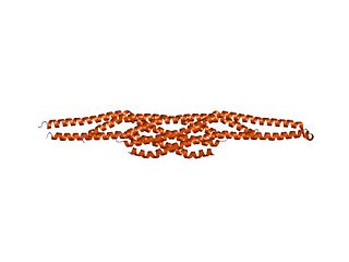
Protein diaphanous homolog 1 is a protein that in humans is encoded by the DIAPH1 gene.

<span class="mw-page-title-main">RhoG</span> Protein-coding gene in the species Homo sapiens

RhoG is a small monomeric GTP-binding protein, and is an important component of many intracellular signalling pathways. It is a member of the Rac subfamily of the Rho family of small G proteins and is encoded by the gene RHOG.

<span class="mw-page-title-main">FHOD1</span> Protein-coding gene in the species Homo sapiens

FH1/FH2 domain-containing protein 1 is a protein that in humans is encoded by the FHOD1 gene.

<span class="mw-page-title-main">Citron kinase</span> Enzyme found in humans

Citron Rho-interacting kinase is an enzyme that in humans is encoded by the CIT gene.

<span class="mw-page-title-main">FNBP1L</span> Protein-coding gene in the species Homo sapiens

Formin-binding protein 1-like is a protein that in humans is encoded by the FNBP1L gene.

<span class="mw-page-title-main">Stress fiber</span> Contractile actin bundles found in non-muscle cells

Stress fibers are contractile actin bundles found in non-muscle cells. They are composed of actin (microfilaments) and non-muscle myosin II (NMMII), and also contain various crosslinking proteins, such as α-actinin, to form a highly regulated actomyosin structure within non-muscle cells. Stress fibers have been shown to play an important role in cellular contractility, providing force for a number of functions such as cell adhesion, migration and morphogenesis.

<span class="mw-page-title-main">DAAM1</span> Protein-coding gene in the species Homo sapiens

Dishevelled-associated activator of morphogenesis 1 is a protein that in humans is encoded by the DAAM1 gene. Evidence of alternative splicing has been observed for this gene but the full-length nature of these variants has not been determined.

<span class="mw-page-title-main">Cordon-bleu protein</span> Protein found in humans

Protein cordon-bleu is a protein that in humans is encoded by the COBL gene.

<span class="mw-page-title-main">Actin assembly-inducing protein</span>

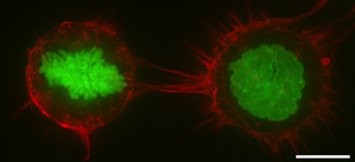
The Actin assembly-inducing protein (ActA) is a protein encoded and used by Listeria monocytogenes to propel itself through a mammalian host cell. ActA is a bacterial surface protein comprising a membrane-spanning region. In a mammalian cell the bacterial ActA interacts with the Arp2/3 complex and actin monomers to induce actin polymerization on the bacterial surface generating an actin comet tail. The gene encoding ActA is named actA or prtB.

<span class="mw-page-title-main">Rho-associated protein kinase</span>

Rho-associated protein kinase (ROCK) is a kinase belonging to the AGC family of serine-threonine specific protein kinases. It is involved mainly in regulating the shape and movement of cells by acting on the cytoskeleton.

<span class="mw-page-title-main">MDia1</span> Protein

mDia1 is a member of the protein family called the formins and is a Rho effector. It is the mouse version of the diaphanous homolog 1 of Drosophila. mDia1 localizes to cells' mitotic spindle and midbody, plays a role in stress fiber and filopodia formation, phagocytosis, activation of serum response factor, formation of adherens junctions, and it can act as a transcription factor. mDia1 accelerates actin nucleation and elongation by interacting with barbed ends of actin filaments. The gene encoding mDia1 is located on Chromosome 18 of Mus musculus and named Diap1.

<span class="mw-page-title-main">Cyclase-associated protein family</span>

In molecular biology, the cyclase-associated protein family (CAP) is a family of highly conserved actin-binding proteins present in a wide range of organisms including yeast, flies, plants, and mammals. CAPs are multifunctional proteins that contain several structural domains. CAP is involved in species-specific signalling pathways. In Drosophila, CAP functions in Hedgehog-mediated eye development and in establishing oocyte polarity. In Dictyostelium discoideum, CAP is involved in microfilament reorganisation near the plasma membrane in a PIP2-regulated manner and is required to perpetuate the cAMP relay signal to organise fruitbody formation. In plants, CAP is involved in plant signalling pathways required for co-ordinated organ expansion. In yeast, CAP is involved in adenylate cyclase activation, as well as in vesicle trafficking and endocytosis. In both yeast and mammals, CAPs appear to be involved in recycling G-actin monomers from ADF/cofilins for subsequent rounds of filament assembly. In mammals, there are two different CAPs that share 64% amino acid identity.

<span class="mw-page-title-main">IMD domain</span>

In molecular biology, the IMD domain is a BAR-like domain of approximately 250 amino acids found at the N-terminus in the insulin receptor tyrosine kinase substrate p53 (IRSp53/BAIAP2) and in the evolutionarily related IRSp53/MIM (MTSS1) family. In IRSp53, a ubiquitous regulator of the actin cytoskeleton, the IMD domain acts as conserved F-actin bundling domain involved in filopodium formation. Filopodium-inducing IMD activity is regulated by Cdc42 and Rac1 and is SH3-independent. The IRSp53/MIM family is a novel F-actin bundling protein family that includes invertebrate relatives:

References

  1. 1 2 Chalkia D, Nikolaidis N, Makalowski W, Klein J, Nei M (December 2008). "Origins and evolution of the formin multigene family that is involved in the formation of actin filaments". Molecular Biology and Evolution. 25 (12): 2717–33. doi:10.1093/molbev/msn215. PMC   2721555 . PMID   18840602.
  2. Evangelista M, Zigmond S, Boone C (July 2003). "Formins: signaling effectors for assembly and polarization of actin filaments". Journal of Cell Science. 116 (Pt 13): 2603–11. doi: 10.1242/jcs.00611 . PMID   12775772.
  3. Gunning PW, Ghoshdastider U, Whitaker S, Popp D, Robinson RC (June 2015). "The evolution of compositionally and functionally distinct actin filaments". Journal of Cell Science. 128 (11): 2009–19. doi: 10.1242/jcs.165563 . PMID   25788699.
  4. 1 2 Goode BL, Eck MJ (2007). "Mechanism and function of formins in the control of actin assembly". Annual Review of Biochemistry. 76: 593–627. doi:10.1146/annurev.biochem.75.103004.142647. PMID   17373907.
  5. Faix J, Grosse R (June 2006). "Staying in shape with formins". Developmental Cell. 10 (6): 693–706. doi: 10.1016/j.devcel.2006.05.001 . PMID   16740473.
  6. Higgs HN, Peterson KJ (January 2005). "Phylogenetic analysis of the formin homology 2 domain". Molecular Biology of the Cell. 16 (1): 1–13. doi:10.1091/mbc.E04-07-0565. PMC   539145 . PMID   15509653.
  7. Kitayama C, Uyeda TQ (February 2003). "ForC, a novel type of formin family protein lacking an FH1 domain, is involved in multicellular development in Dictyostelium discoideum". Journal of Cell Science. 116 (Pt 4): 711–23. doi: 10.1242/jcs.00265 . PMID   12538772.
  8. Wallar BJ, Alberts AS (August 2003). "The formins: active scaffolds that remodel the cytoskeleton". Trends in Cell Biology. 13 (8): 435–46. doi:10.1016/S0962-8924(03)00153-3. PMID   12888296.
  9. Uetz, Peter (1997). Biochemische Studien am limb deformity-Protein der Vertebraten: Inaugural-Dissertation zur Erlangung der Doktorwürde der Naturwissenschaftlich-Mathematischen Gesamtfakultät der Ruprecht-Karls-Universität Heidelberg. Developmental Biology, European Molecular Biology Laboratory. Heidelberg: European Molecular Biology Laboratory.
  10. Uetz P, Fumagalli S, James D, Zeller R (December 1996). "Molecular interaction between limb deformity proteins (formins) and Src family kinases". The Journal of Biological Chemistry. 271 (52): 33525–30. doi: 10.1074/jbc.271.52.33525 . PMID   8969217.
  11. Takeya R, Sumimoto H (November 2003). "Fhos, a mammalian formin, directly binds to F-actin via a region N-terminal to the FH1 domain and forms a homotypic complex via the FH2 domain to promote actin fiber formation". Journal of Cell Science. 116 (Pt 22): 4567–75. doi: 10.1242/jcs.00769 . PMID   14576350.
  12. Shimada A, Nyitrai M, Vetter IR, Kühlmann D, Bugyi B, Narumiya S, Geeves MA, Wittinghofer A (February 2004). "The core FH2 domain of diaphanous-related formins is an elongated actin binding protein that inhibits polymerization". Molecular Cell. 13 (4): 511–22. doi: 10.1016/S1097-2765(04)00059-0 . PMID   14992721.
  13. Kato T, Watanabe N, Morishima Y, Fujita A, Ishizaki T, Narumiya S (February 2001). "Localization of a mammalian homolog of diaphanous, mDia1, to the mitotic spindle in HeLa cells". Journal of Cell Science. 114 (Pt 4): 775–84. doi:10.1242/jcs.114.4.775. hdl: 2433/150544 . PMID   11171383.
  14. Petersen J, Nielsen O, Egel R, Hagan IM (June 1998). "FH3, a domain found in formins, targets the fission yeast formin Fus1 to the projection tip during conjugation". The Journal of Cell Biology. 141 (5): 1217–28. doi:10.1083/jcb.141.5.1217. PMC   2137179 . PMID   9606213.
  15. 1 2 Peng J, Wallar BJ, Flanders A, Swiatek PJ, Alberts AS (April 2003). "Disruption of the Diaphanous-related formin Drf1 gene encoding mDia1 reveals a role for Drf3 as an effector for Cdc42". Current Biology. 13 (7): 534–45. doi: 10.1016/S0960-9822(03)00170-2 . PMID   12676083. S2CID   13902104.
  16. Xu Y, Moseley JB, Sagot I, Poy F, Pellman D, Goode BL, Eck MJ (March 2004). "Crystal structures of a Formin Homology-2 domain reveal a tethered dimer architecture". Cell. 116 (5): 711–23. doi: 10.1016/S0092-8674(04)00210-7 . PMID   15006353. S2CID   15855545.
  17. Thompson ME, Heimsath EG, Gauvin TJ, Higgs HN, Kull FJ (January 2013). "FMNL3 FH2-actin structure gives insight into formin-mediated actin nucleation and elongation". Nature Structural & Molecular Biology. 20 (1): 111–8. doi:10.1038/nsmb.2462. PMC   3876896 . PMID   23222643.
  18. Palazzo AF, Cook TA, Alberts AS, Gundersen GG (August 2001). "mDia mediates Rho-regulated formation and orientation of stable microtubules". Nature Cell Biology. 3 (8): 723–9. doi:10.1038/35087035. PMID   11483957. S2CID   7374170.
  19. Bartolini F, Gundersen GG (February 2010). "Formins and microtubules". Biochimica et Biophysica Acta (BBA) - Molecular Cell Research. 1803 (2): 164–73. doi:10.1016/j.bbamcr.2009.07.006. PMC   2856479 . PMID   19631698.
This article incorporates text from the public domain Pfam and InterPro: IPR010472
This article incorporates text from the public domain Pfam and InterPro: IPR015425
This article incorporates text from the public domain Pfam and InterPro: IPR010465
This article incorporates text from the public domain Pfam and InterPro: IPR010473