
Gas vesicles, also known as gas vacuoles, are nanocompartments in certain prokaryotic organisms, which help in buoyancy. [1] Gas vesicles are composed entirely of protein; no lipids or carbohydrates have been detected.
Gas vesicles, also known as gas vacuoles, are nanocompartments in certain prokaryotic organisms, which help in buoyancy. [1] Gas vesicles are composed entirely of protein; no lipids or carbohydrates have been detected.
Gas vesicles occur primarily in aquatic organisms as they are used to modulate the cell's buoyancy and modify the cell's position in the water column so it can be optimally located for photosynthesis or move to locations with more or less oxygen. [1] Organisms that could float to the air–liquid interface out competes other aerobes that cannot rise in a water column, through using up oxygen in the top layer.
In addition, gas vesicles can be used to maintain optimum salinity by positioning the organism in specific locations in a stratified body of water to prevent osmotic shock. [2] High concentrations of solute will cause water to be drawn out of the cell by osmosis, causing cell lysis. The ability to synthesize gas vesicles is one of many strategies that allow halophilic organisms to tolerate environments with high salt content.
Gas vesicles are likely one of the most early mechanisms of motility among microscopic organisms due to the fact that it is the most widespread form of motility conserved within the genome of prokaryotes, some of which have evolved about 3 billion years ago. [3] [4] Modes of active motility such as flagella movement require a mechanism that could convert chemical energy into mechanical energy, and thus is much more complex and would have evolved later. Functions of the gas vesicles are also largely conserved among species, although the mode of regulation might differ, suggesting the importance of gas vesicles as a form of motility. In certain organism such as enterobacterium Serratia sp. flagella-based motility and gas vesicle production are regulated oppositely by a single RNA binding protein, RsmA, suggesting alternate modes of environmental adaptation which would have developed into different taxons through regulation of the development between motility and flotation. [5]
Although there is evidence suggesting the early evolution of gas vesicles, plasmid transfer serves as an alternate explanation of the widespread and conserved nature of the organelle. [4] Cleavage of a plasmid in Halobacterium halobium resulted in the loss of the ability to biosynthesize gas vesicles, indicating the possibility of horizontal gene transfer, which could result in a transfer of the ability to produce gas vesicles among different strains of bacteria. [6]

Gas vesicles are generally lemon-shaped or cylindrical, hollow tubes of protein with conical caps on both ends. The vesicles vary most in their diameter. Larger vesicles can hold more air and use less protein making them the most economic in terms of resource use, however, the larger a vesicle is the structurally weaker it is under pressure and the less pressure required before the vesicle would collapse. Organisms have evolved to be the most efficient with protein use and use the largest maximum vesicle diameter that will withstand the pressure the organism could be exposed to. In order for natural selection to have affected gas vesicles, the vesicles' diameter must be controlled by genetics. Although genes encoding gas vesicles are found in many species of haloarchaea, only a few species produce them. The first Haloarchaeal gas vesicle gene, GvpA was cloned from Halobacterium sp. NRC-1. [7] 14 genes are involved in forming gas vesicles in haloarchaea. [8]
The first gas vesicle gene, GvpA was identified in Calothrix. [9] There are at least two proteins that compose a cyanobacterium's gas vesicle: GvpA, and GvpC. GvpA forms ribs and much of the mass (up to 90%) of the main structure. GvpA is strongly hydrophobic and may be one of the most hydrophobic proteins known. GvpC is hydrophilic and helps to stabilize the structure by periodic inclusions into the GvpA ribs. GvpC is capable of being washed out of the vesicle and a consequential decreases in the vesicle's strength. The thickness of the vesicle's wall may range from 1.8 to 2.8 nm. The ribbed structure of the vesicle is evident on both inner and outer surfaces with a spacing of 4–5 nm between ribs. Vesicles may be 100–1400 nm long and 45–120 nm in diameter.
Within a species gas vesicle sizes are relatively uniform with a standard deviation of ±4%.

It appears that gas vesicles begin their existence as small biconical (two cones with the flat bases joined together) structures which enlarge to the specific diameter than grow and expand their length. It is unknown exactly what controls the diameter but it may be a molecule that interferes with GvpA or the shape of GvpA may change.
Formation of gas vesicles are regulated by two Gvp proteins: GvpD, which represses the expression of GvpA and GvpC proteins, and GvpE, which induces expression. [10] Extracellular environmental factors also affect vesicle formation, either by regulating Gvp protein production or by directly disturbing the vesicle structure. [8] [11]
Light intensity has been found to affect gas vesicles production and maintenance differently between different bacteria and archaea. For Anabaena flos-aquae, higher light intensities leads to vesicle collapse from an increase in turgor pressure and greater accumulation of photosynthetic products. In cyanobacteria, vesicle production decreases at high light intensity due to exposure of the bacterial surface to UV radiation, which can damage the bacterial genome. [11]
Accumulation of glucose, maltose, or sucrose in Haloferax mediterranei and Haloferax volcanii were found to inhibit the expression of GvpA proteins and, therefore, a decrease of gas vesicle production. However, this only occurred at the cell's early exponential growth phase. Vesicle formation could also be induced in decreasing extracellular glucose concentrations. [12]
A lack of oxygen was found to negatively affect gas vesicle formation in halophilic archaea. Halobacterium salinarum produce little or no vesicles under anaerobic conditions due to reduced synthesis of mRNA transcripts encoding for Gvp proteins. H. mediterranei and H. volcanii do not produce any vesicles under anoxic conditions due to a decrease in synthesized transcripts encoding for GvpA and truncated transcripts expressing GvpD. [12]
Increased extracellular pH levels have been found to increase vesicle formation in Microcytis species. Under increased pH, levels of gvpA and gvpC transcripts increase, allowing more exposure to ribosomes for expression and leading to upregulation of Gvp proteins. It may be attributed to greater transcription of these genes, decreased decay of the synthesized transcripts or the higher stability of the mRNA. [13]
Ultrasonic irradiation, at certain frequencies, was found to collapse gas vesicles in cyanobacteria Spirulina platensis, preventing them from blooming. [14]
In enterobacterium; Serratia sp. strain ATCC39006, gas vesicle is produced only when there is sufficient concentration of a signalling molecule, N-acyl homoserine lactone. In this case, the quorum sensing molecule, N-acyl homoserine lactone acts as a morphogen initiating organelle development. [5] This is advantageous to the organism as resources for gas vesicle production are utilized only when there is oxygen limitation caused by an increase in bacterial population.
Gas vesicle gene gvpC from Halobacterium sp. is used as delivery system for vaccine studies.
Several characteristics of the protein encoded by the gas vesicle gene gvpC allow it to be used as carrier and adjuvant for antigens: it is stable, resistant to biological degradation, tolerates relatively high temperatures (up to 50 °C), and non-pathogenic to humans. [15] Several antigens from various human pathogens have been recombined into the gvpC gene to create subunit vaccines with long-lasting immunologic responses. [16]
Different genomic segments encoding for several Chlamydia trachomatis pathogen's proteins, including MOMP, OmcB, and PompD, are joined to the gvpC gene of Halobacteria. In vitro assessments of cells show expression of the Chlamydia genes on cell surfaces through imaging techniques and show characteristic immunologic responses such as TLRs activities and pro-inflammatory cytokines production. [17] Gas vesicle gene can be exploited as a delivery vehicle to generate a potential vaccine for Chlamydia. Limitations of this method include the need to minimize the damage of the GvpC protein itself while including as much of the vaccine target gene into the gvpC gene segment. [17]
A similar experiment uses the same gas vesicle gene and Salmonella enterica pathogen's secreted inosine phosphate effector protein SopB4 and SopB5 to generate a potential vaccine vector. Immunized mice secrete pro-inflammatory cytokines IFN-γ, IL-2, and IL-9. Antibody IgG is also detected. After an infection challenge, none or significantly less amount of bacteria were found in the harvested organs such as the spleen and the liver. Potential vaccines using gas vesicle as an antigen display can be given via the mucosal route as an alternative administration pathway, increasing its accessibility to more people and eliciting a wider range of immune responses within the body. [15]

Gas vesicles have several physical properties that make them visible on various medical imaging modalities. [18] The ability of gas vesicle to scatter light has been used for decades for estimating their concentration and measuring their collapse pressure . The optical contrast of gas vesicles also enables them to serve as contrast agents in optical coherence tomography, with applications in ophthalmology. [19] The difference in acoustic impedance between the gas in their cores and the surrounding fluid gives gas vesicles robust acoustic contrast. [20] Moreover, the ability of some gas vesicle shells to buckle generates harmonic ultrasound echoes that improves the contrast to tissue ratio. [21] Finally, gas vesicles can be used as contrast agents for magnetic resonance imaging (MRI), relying on the difference between the magnetic susceptibility of air and water. [22] The ability to non-invasively collapse gas vesicles using pressure waves provides a mechanism for erasing their signal and improving their contrast. Subtracting the images before and after acoustic collapse can eliminate background signals enhancing the detection of gas vesicles.
Heterologous expression of gas vesicles in bacterial [23] and mammalian [24] cells enabled their use as the first family of acoustic reporter genes. [25] While fluorescent reporter genes like green fluorescent protein (GFP) had widespread use in biology, their in vivo applications are limited by the penetration depth of light in tissue, typically a few mm. Luminescence can be detected deeper within the tissue, but have a low spatial resolution. Acoustic reporter genes provide sub-millimeter spatial resolution and a penetration depth of several centimeters, enabling the in vivo study of biological processes deep within the tissue.

A pilus is a hair-like appendage found on the surface of many bacteria and archaea. The terms pilus and fimbria can be used interchangeably, although some researchers reserve the term pilus for the appendage required for bacterial conjugation. All conjugative pili are primarily composed of pilin – fibrous proteins, which are oligomeric.

A vacuole is a membrane-bound organelle which is present in plant and fungal cells and some protist, animal, and bacterial cells. Vacuoles are essentially enclosed compartments which are filled with water containing inorganic and organic molecules including enzymes in solution, though in certain cases they may contain solids which have been engulfed. Vacuoles are formed by the fusion of multiple membrane vesicles and are effectively just larger forms of these. The organelle has no basic shape or size; its structure varies according to the requirements of the cell.

A DNA vaccine is a type of vaccine that transfects a specific antigen-coding DNA sequence into the cells of an organism as a mechanism to induce an immune response.
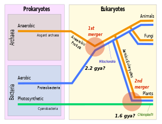
Symbiogenesis is the leading evolutionary theory of the origin of eukaryotic cells from prokaryotic organisms. The theory holds that mitochondria, plastids such as chloroplasts, and possibly other organelles of eukaryotic cells are descended from formerly free-living prokaryotes taken one inside the other in endosymbiosis. Mitochondria appear to be phylogenetically related to Rickettsiales bacteria, while chloroplasts are thought to be related to cyanobacteria.

Chlamydia trachomatis, commonly known as chlamydia, is a bacterium that causes chlamydia, which can manifest in various ways, including: trachoma, lymphogranuloma venereum, nongonococcal urethritis, cervicitis, salpingitis, pelvic inflammatory disease. C. trachomatis is the most common infectious cause of blindness and the most common sexually transmitted bacterium.
Mycoplasma pneumoniae is a very small bacterium in the class Mollicutes. It is a human pathogen that causes the disease mycoplasma pneumonia, a form of atypical bacterial pneumonia related to cold agglutinin disease. M. pneumoniae is characterized by the absence of a peptidoglycan cell wall and resulting resistance to many antibacterial agents. The persistence of M. pneumoniae infections even after treatment is associated with its ability to mimic host cell surface composition.

Motility is the ability of an organism to move independently, using metabolic energy.
Halobacterium is a genus in the family Halobacteriaceae.
Extrachromosomal DNA is any DNA that is found off the chromosomes, either inside or outside the nucleus of a cell. Most DNA in an individual genome is found in chromosomes contained in the nucleus. Multiple forms of extrachromosomal DNA exist, and, while some of these serve important biological functions, they can also play a role in diseases such as cancer.
The bacterium, despite its simplicity, contains a well-developed cell structure which is responsible for some of its unique biological structures and pathogenicity. Many structural features are unique to bacteria and are not found among archaea or eukaryotes. Because of the simplicity of bacteria relative to larger organisms and the ease with which they can be manipulated experimentally, the cell structure of bacteria has been well studied, revealing many biochemical principles that have been subsequently applied to other organisms.
Chlamydia felis is a Gram-negative, obligate intracellular bacterial pathogen that infects cats. It is endemic among domestic cats worldwide, primarily causing inflammation of feline conjunctiva, rhinitis and respiratory problems. C. felis can be recovered from the stomach and reproductive tract. Zoonotic infection of humans with C. felis has been reported. Strains FP Pring and FP Cello have an extrachromosomal plasmid, whereas the FP Baker strain does not. FP Cello produces lethal disease in mice, whereas the FP Baker does not. An attenuated FP Baker strain, and an attenuated 905 strain, are used as live vaccines for cats.
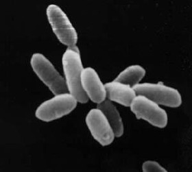
Halobacterium salinarum, formerly known as Halobacterium cutirubrum or Halobacterium halobium, is an extremely halophilic marine obligate aerobic archaeon. Despite its name, this is not a bacterium, but a member of the domain Archaea. It is found in salted fish, hides, hypersaline lakes, and salterns. As these salterns reach the minimum salinity limits for extreme halophiles, their waters become purple or reddish color due to the high densities of halophilic Archaea. H. salinarum has also been found in high-salt food such as salt pork, marine fish, and sausages. The ability of H. salinarum to survive at such high salt concentrations has led to its classification as an extremophile.
GvpA is a gas vesicle structural protein found in different phyla of bacteria and archaea for example in Halobacterium salinarum or Haloferax mediterranei. Gas vesicles are small, hollow, gas filled protein structures found in several cyanobacterial and archaebacterial microorganisms. They allow the positioning of the bacteria at a favourable depth for growth.
The archaellum is a unique structure on the cell surface of many archaea, that allows for swimming motility. The archaellum consists of a rigid helical filament that is attached to the cell membrane by a molecular motor. This molecular motor – composed of cytosolic, membrane, and pseudo-periplasmic proteins – is responsible for the assembly of the filament and, once assembled, for its rotation. The rotation of the filament propels archaeal cells in liquid medium, in a manner similar to the propeller of a boat. The bacterial analog of the archaellum is the flagellum, which is also responsible for their swimming motility and can also be compared to a rotating corkscrew. Although the movement of archaella and flagella is sometimes described as "whip-like", this is incorrect, as only cilia from Eukaryotes move in this manner. Indeed, even "flagellum" is a misnomer, as bacterial flagella also work as propeller-like structures.

Reverse genetics is a method in molecular genetics that is used to help understand the function(s) of a gene by analysing the phenotypic effects caused by genetically engineering specific nucleic acid sequences within the gene. The process proceeds in the opposite direction to forward genetic screens of classical genetics. While forward genetics seeks to find the genetic basis of a phenotype or trait, reverse genetics seeks to find what phenotypes are controlled by particular genetic sequences.
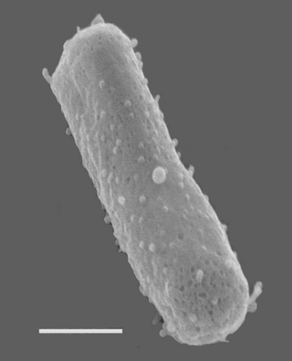
Halobacterium noricense is a halophilic, rod-shaped microorganism that thrives in environments with salt levels near saturation. Despite the implication of the name, Halobacterium is actually a genus of archaea, not bacteria. H. noricense can be isolated from environments with high salinity such as the Dead Sea and the Great Salt Lake in Utah. Members of the Halobacterium genus are excellent model organisms for DNA replication and transcription due to the stability of their proteins and polymerases when exposed to high temperatures. To be classified in the genus Halobacterium, a microorganism must exhibit a membrane composition consisting of ether-linked phosphoglycerides and glycolipids.

Shiladitya DasSarma is a molecular biologist well-known for contributions to the biology of halophilic and extremophilic microorganisms. He is a Professor in the University of Maryland Baltimore. He earned a PhD degree in Biochemistry from the Massachusetts Institute of Technology and a BS degree in Chemistry from Indiana University Bloomington. Prior to taking a faculty position, he conducted research at the Massachusetts General Hospital, Harvard Medical School, and Pasteur Institute, Paris.
Haloferax mediterranei is a species of archaea in the family Haloferacaceae.

Halorubrum lacusprofundi is a rod-shaped, halophilic Archaeon in the family of Halorubraceae. It was first isolated from Deep Lake in Antarctica in the 1980s.
Archaerhodopsin proteins are a family of retinal-containing photoreceptors found in the archaea genera Halobacterium and Halorubrum. Like the homologous bacteriorhodopsin (bR) protein, archaerhodopsins harvest energy from sunlight to pump H+ ions out of the cell, establishing a proton motive force that is used for ATP synthesis. They have some structural similarities to the mammalian GPCR protein rhodopsin, but are not true homologs.