Gliogenesis

Last updated

Gliogenesis is the generation of non-neuronal glia populations derived from multipotent neural stem cells.

Contents

Overview

Gliogenesis results in the formation of non-neuronal glia populations from neuronal cells. In this capacity, glial cells provide multiple functions to both the central nervous system (CNS) and the peripheral nervous system (PNS). Subsequent differentiation of glial cell populations results in function-specialized glial lineages. Glial cell-derived astrocytes are specialized lineages responsible for modulating the chemical environment by altering ion gradients and neurotransmitter transduction. Similarly derived, oligodendrocytes produce myelin, which insulates axons to facilitate electric signal transduction. Finally, microglial cells are derived from glial precursors and carry out macrophage-like properties to remove cellular and foreign debris within the central nervous system ref. Functions of glial-derived cell lineages are reviewed by Baumann and Hauw. [1] Gliogenesis itself, and differentiation of glial-derived lineages are activated upon stimulation of specific signaling cascades. Similarly, inhibition of these pathways is controlled by distinct signaling cascades that control proliferation and differentiation. Thus, elaborate intracellular-mechanisms based on environmental signals are present to regulate the formation of these cells. As regulation is much more known in the CNS, its mechanisms and components will be focused on here. Understanding the mechanisms in which gliogenesis is regulated provides the potential to harness the ability to control the fate of glial cells and, consequently, the ability to reverse neurodegenerative diseases.

Gliogenesis induction

Following the generation of neural stem cells, an option is presented to proceed to enter neurogenesis and form new neurons within the CNS, shift into gliogenesis, or remain in a pluripotent cell state. The mechanisms determining the ultimate fate of neural stem cells are conserved among both invertebrate and vertebrate species and are determined from extracellular cues generated from neighboring cells. [2] Most work to derive such mechanisms, however, began with invertebrate models. Conclusions reached from these studies have directed attention to specific signaling molecules and effector pathways that are responsible for mediating the cellular events required for maintaining or changing the neural stem cell fate.

Signaling effectors

Notch signaling is known to mediate prominent cellular events that result in gliogenesis. The Notch family proteins are transmembrane receptors that are ligand activated. In the presence of ligand effectors, the intracellular domain of the receptor is cleaved and sequestered to the nucleus where it acts to influence expression of transcription factors required for gliogenesis. Transcription factors synthesized as a result of the Notch signaling cascade bind to promoters of genes responsible for glial determination. [3] Additionally, Notch signaling also acts to downregulate many genes responsible for neuronal development, thus inhibiting a neuron phenotype from arising. [4] Both actions collectively function to promote glial fate.

In certain CNS tissue, JAK/STAT signaling is also known to promote gliogenesis [5] [6] Significant levels of the ciliary neurotrophic factor (CNTF) are expressed immediately preceding gliogensis in response to environmental cues allowing the activation of the JAK-STAT signaling pathway. Kinase activity phosphorylates STAT proteins which then are recruited by transcription factors. The STAT complex is targeted to promoters of genes responsible for gliogenesis activation. It is important to recognize that when isolated, receptor-mediated signaling cascades can produce distinct actions, however, when in vivo coopertivity often exists among receptor pathways and results in much more complicated cellular actions.

Signaling molecules

The receptor-proteins responsible for gliogenic pathways are often ligand activated. Upon binding of Delta or Jagged, the notch-mediated signaling cascades are activated leading to gliogenic transcription factor production as discussed above. [7] As noted for receptor-proteins, in vivo interactions among different growth factor responsible for gliogenesis and other cell fates produce very different roles than when isolated.

Gliogenesis regulation

To ensure proper temporal differentiation as well as correct quantities of glial cell formation, gliogenesis is subjected to stringent regulatory mechanisms. Proneural factors are expressed in high concentrations during times in which glial cells are not to form or neuron development is needed. These protein signals function to inhibit many of the signals utilized during the induction of gliogenesis. Additionally, the properties and abundance of receptor molecules that mediate gliogenesis are altered, consequently disrupting propagation of induction signals.

Signaling inhibition

Stem cell differentiation and Notch-Delta lateral inhibition in neural stem cells, resulting in the generation of neuronal and glia progenitors. Neuro-Gliogenesis via Lateral Inhibition.png
Stem cell differentiation and Notch-Delta lateral inhibition in neural stem cells, resulting in the generation of neuronal and glia progenitors.

During periods in which glial cell formation is discouraged, neural stem cells have the option to remain pluripotent or switch pathway lineages and begin forming neurons during neurogenesis. If neuron development is instructed, neurogenic factors, i.e. BMPs, [8] are present to induce expression of proneural transcription factors like Neurogenin and ASCL1. These transcription factors function to interact with transcription factors generated from Notch signaling. Consequently, this complex is sequestered away from promoters activating gliogenesis and now directed to promoters that influence activity directed for neuron development. [9] Neurogenin proteins regulate JAK/STAT signaling by similar mechanisms. [10]

Receptor insensitivity

Recently, an alternative mechanism to regulate differentiation has been proposed in addition to inhibition through growth factors. Changes in local sensitivity of neural stem cells have been shown to modulate the differentiation capacity of growth factors. Over developmental time, neural stem cells lose the ability to respond to growth factors that influence differentiation as intrinsic changes occur to receptor structure and function of these cells. [11] It has been shown Notch receptors require 50-fold higher concentrations of ligand effectors to initiate differentiation responses similar to that of developmentally earlier neural stem cells. [12] Decrease in sensitivity of Notch receptors reduces the activity of Notch-signaling required for gliogenesis to occur. Consequently, neural stem cells have developed a general mechanism limiting further differentiation after intense specialization during the early developmental periods.

Receptor internalization

The internalization, or endocytosis, of receptor proteins from the cell’s plasma membrane contributes to yet another mode of regulation of cellular function. [13] While receptor internalization has the potential to regulate cellular functions in both a positive and negative fashion, internalization of the Notch receptor is shown to down-regulate the events leading to gliogenesis as this process is Notch-signaling dependent [14]

During repression of gliogenesis, expression of the Notch-binding protein, Numb, is elevated. [15] Numb is suggested to function in two manners: 1) When expressed, Numb will interact with specific endocytic proteins and create a link between the notch receptor and the endocytic vesicles. The vesicle-receptor complex generated will be targeted back to the cell membrane and the membrane receptor will be recycled to the cell surface never reaching the nucleus. Alternatively, 2) Numb is suggested to recruit additional molecules other than endocytic proteins. In particular, ubiquitin ligases are shown to be recruited by Numb in mammals. The ubiquitin ligases ubiquitinates Notch and targets it for degradation [16] Whatever the mechanism of Numb, the Notch receptor does not reach the nucleus and the transcription factors required for gliogenesis are not generated.

Gliogenic-associated pathology

Recent work has demonstrated abnormalities in the signaling pathways responsible for gliogenesis and neurogenesis could contribute to the pathogenesis of neurodegenerative diseases and tumor development within the nervous system. [17] [18] Recognizing the distinct pathways controlling neural stem fate, as discussed above, provides one the opportunity to intervene in the pathogenesis of these diseases.

Gliogenesis and neurodegenerative disease

The pathology of neurodegenerative diseases is associated with the disruption of gliogenic pathways and has been recently reviewed. [19] The subventricular zone (SVZ) of the forebrain is of special interest when evaluating errant gliogenic pathways as it is the largest store of neural stem cells in the brain. [20] In multiple sclerosis (MS) patients, lesions in this area are frequently observed and often extend outward toward the lateral ventricles of the brain. [21] Immune cells infiltrate the gliogenic regions within the SVZ adjacent to the lesions and initiate inflammatory response mechanisms in response to damage in this region. [22] It is suggested that cytokine release during the inflammatory response reduces, foremost, the inherent neural stem cell populations, and jointly the potential of the remaining neural stem cell to differentiate to glial-fates. [23] Consequently, a reduction of glial-derived oligodendrocytes, among others, compromise maintenance of myelin production for axon insulation, a hallmark phenotype among MS patients.

Consequences of gliogenesis disruption among other neurodegenerative diseases, such as Huntington's, [24] Parkinson's, [25] and Alzheimer's Diseases [26] are currently being investigated and strong mechanistic evidence is shown for pathogenesis similar to MS.

Gliogenesis and glial tumors

Disruption of controlled glial generation subsequently results in tumorigenesis and glioma formation within the central nervous system. Loss of contact inhibition, cellular migration, and unregulated proliferation are characteristic of gliomas. Consistent with other tissues, these malignant phenotypes result most commonly from chromosome deletions, translocations, and point mutations. Linskey reviews both the genetic contributions and phenotypic observations of glioma [27]

In non-carcinogenic neural stem cells, key regulatory mechanisms prevent uncontrolled gliogenic proliferation. However, such mechanisms are disrupted upon genetic damage. Studies now suggest glioma formation may result from cellular insensitivity to regulatory growth factors and cell signals, like neurogenin, that would normally inhibit further proliferation of glial cells. [28] Conformational changes in receptor proteins are thought to occur, leaving the cell constitutively proliferating. [29]

Therapeutic intervention of gliogenic-derived pathogenesis

Understanding the pathology of these neurodegenerative diseases and establishment of therapeutic interventions require recognition of the processes of induction and inhibition of gliogenesis and the regulating mechanisms coordinating the intricate system established from both actions. Cell replacement strategies are now intensely studied as a possible therapeutic intervention of glial associated neurodegenerative disorders and glial tumors. Similar to any novel strategy, however, set-backs and liabilities accompany the promises this technique withholds. For cell replacement to function efficiently and demonstrate robust results, introduced cells must be 1) generated in sufficient yield and 2) immunocompatible with the host and 3) able to sustain self-growth. [30] New perspectives within stem cell biology and gliogenesis regulation have provided new insights within the past decade to begin addressing these challenges. Reprogramming terminally differentiated neural lineages back to neural stem cells permits regeneration of a multipotent self-lineage that can be redirected to cellular-fates affected during neurogenerative diseases, oligodendrocytes with MS patients or astrocytes in those affected with Alzheimer's, in the presence of proper environmental signals. [31]

It can be expected that as the signaling pathways discussed are shown as prominent regulators during glial cell generation, these same pathways will become therapeutic targets for glial-derived and other CNS cancers. In medulloblastomas, in vivo studies have begun targeting notch pathways by blocking Notch receptors with specific inhibitors preventing further differentiation. [32] When used, pathway inhibitors provided 10-fold greater sensitivity to apoptotic induction in medulloblastoma cells [33] Recognition of the regulatory mechanisms of gliogenesis provide new direction for intervention of neurogenic disorders.

Related Research Articles

The development of the nervous system, or neural development (neurodevelopment), refers to the processes that generate, shape, and reshape the nervous system of animals, from the earliest stages of embryonic development to adulthood. The field of neural development draws on both neuroscience and developmental biology to describe and provide insight into the cellular and molecular mechanisms by which complex nervous systems develop, from nematodes and fruit flies to mammals.

<span class="mw-page-title-main">Notch signaling pathway</span> Series of molecular signals

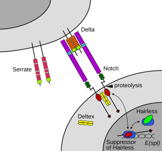
The Notch signaling pathway is a highly conserved cell signaling system present in most animals. Mammals possess four different notch receptors, referred to as NOTCH1, NOTCH2, NOTCH3, and NOTCH4. The notch receptor is a single-pass transmembrane receptor protein. It is a hetero-oligomer composed of a large extracellular portion, which associates in a calcium-dependent, non-covalent interaction with a smaller piece of the notch protein composed of a short extracellular region, a single transmembrane-pass, and a small intracellular region.

<span class="mw-page-title-main">Rostral migratory stream</span> One path neural stem cells take to reach the olfactory bulb


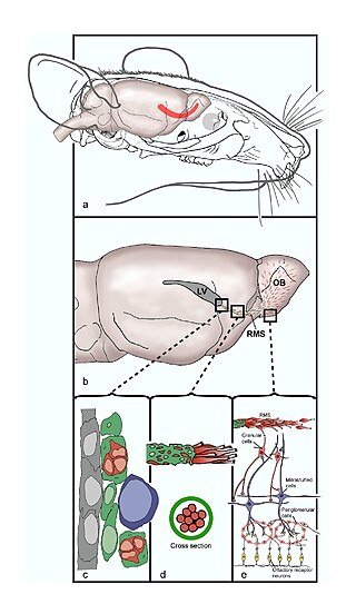
The rostral migratory stream (RMS) is a specialized migratory route found in the brain of some animals along which neuronal precursors that originated in the subventricular zone (SVZ) of the brain migrate to reach the main olfactory bulb (OB). The importance of the RMS lies in its ability to refine and even change an animal's sensitivity to smells, which explains its importance and larger size in the rodent brain as compared to the human brain, as our olfactory sense is not as developed. This pathway has been studied in the rodent, rabbit, and both the squirrel monkey and rhesus monkey. When the neurons reach the OB they differentiate into GABAergic interneurons as they are integrated into either the granule cell layer or periglomerular layer.

Neuroepithelial cells, or neuroectodermal cells, form the wall of the closed neural tube in early embryonic development. The neuroepithelial cells span the thickness of the tube's wall, connecting with the pial surface and with the ventricular or lumenal surface. They are joined at the lumen of the tube by junctional complexes, where they form a pseudostratified layer of epithelium called neuroepithelium.

Neural stem cells (NSCs) are self-renewing, multipotent cells that firstly generate the radial glial progenitor cells that generate the neurons and glia of the nervous system of all animals during embryonic development. Some neural progenitor stem cells persist in highly restricted regions in the adult vertebrate brain and continue to produce neurons throughout life. Differences in the size of the central nervous system are among the most important distinctions between the species and thus mutations in the genes that regulate the size of the neural stem cell compartment are among the most important drivers of vertebrate evolution.

<span class="mw-page-title-main">Floor plate</span> Embryonic structure

The floor plate is a structure integral to the developing nervous system of vertebrate organisms. Located on the ventral midline of the embryonic neural tube, the floor plate is a specialized glial structure that spans the anteroposterior axis from the midbrain to the tail regions. It has been shown that the floor plate is conserved among vertebrates, such as zebrafish and mice, with homologous structures in invertebrates such as the fruit fly Drosophila and the nematode C. elegans. Functionally, the structure serves as an organizer to ventralize tissues in the embryo as well as to guide neuronal positioning and differentiation along the dorsoventral axis of the neural tube.

<span class="mw-page-title-main">Radial glial cell</span> Bipolar-shaped progenitor cells of all neurons in the cerebral cortex and some glia

Radial glial cells, or radial glial progenitor cells (RGPs), are bipolar-shaped progenitor cells that are responsible for producing all of the neurons in the cerebral cortex. RGPs also produce certain lineages of glia, including astrocytes and oligodendrocytes. Their cell bodies (somata) reside in the embryonic ventricular zone, which lies next to the developing ventricular system.

Neuropoiesis is the process by which neural stem cells differentiate to form mature neurons, astrocytes, and oligodendrocytes in the adult mammal. This process is also referred to as adult neurogenesis.

<span class="mw-page-title-main">Subventricular zone</span> Region outside each lateral ventricle of the brain

The subventricular zone (SVZ) is a region situated on the outside wall of each lateral ventricle of the vertebrate brain. It is present in both the embryonic and adult brain. In embryonic life, the SVZ refers to a secondary proliferative zone containing neural progenitor cells, which divide to produce neurons in the process of neurogenesis. The primary neural stem cells of the brain and spinal cord, termed radial glial cells, instead reside in the ventricular zone (VZ).

<span class="mw-page-title-main">Subgranular zone</span>

The subgranular zone (SGZ) is a brain region in the hippocampus where adult neurogenesis occurs. The other major site of adult neurogenesis is the subventricular zone (SVZ) in the brain.

<span class="mw-page-title-main">NUMB (gene)</span> Protein-coding gene in the species Homo sapiens

Protein numb homolog is a protein that in humans is encoded by the NUMB gene. The protein encoded by this gene plays a role in the determination of cell fates during development. The encoded protein, whose degradation is induced in a proteasome-dependent manner by MDM2, is a membrane-bound protein that has been shown to associate with EPS15, LNX1, and NOTCH1. Four transcript variants encoding different isoforms have been found for this gene.

<span class="mw-page-title-main">HES1</span> Protein-coding gene in the species Homo sapiens

Transcription factor HES1 is a protein that is encoded by the Hes1 gene, and is the mammalian homolog of the hairy gene in Drosophila. HES1 is one of the seven members of the Hes gene family (HES1-7). Hes genes code nuclear proteins that suppress transcription.

Neurogenins are a family of bHLH transcription factors involved in specifying neuronal differentiation. It is one of many gene families related to the atonal gene in Drosophila. Other positive regulators of neuronal differentiation also expressed during early neural development include NeuroD and ASCL1.

<span class="mw-page-title-main">Ganglion mother cell</span>

Ganglion mother cells (GMCs) are cells involved in neurogenesis, in non-mammals, that divide only once to give rise to two neurons, or one neuron and one glial cell or two glial cells, and are present only in the central nervous system. They are also responsible for transcription factor expression. While each ganglion mother cell necessarily gives rise to two neurons, a neuroblast can asymmetrically divide multiple times. GMCs are the progeny of type I neuroblasts. Neuroblasts asymmetrically divide during embryogenesis to create GMCs. GMCs are only present in certain species and only during the embryonic and larval stages of life. Recent research has shown that there is an intermediate stage between a GMC and two neurons. The GMC forms two ganglion cells which then develop into neurons or glial cells. Embryonic neurogenesis has been extensively studied in Drosophila melanogaster embryos and larvae.

<span class="mw-page-title-main">P19 cell</span>

P19 cells is an embryonic carcinoma cell line derived from an embryo-derived teratocarcinoma in mice. The cell line is pluripotent and can differentiate into cell types of all three germ layers. Also, it is the most characterized embryonic carcinoma (EC) cell line that can be induced into cardiac muscle cells and neuronal cells by different specific treatments. Indeed, exposing aggregated P19 cells to dimethyl sulfoxide (DMSO) induces differentiation into cardiac and skeletal muscle. Also, exposing P19 cells to retinoic acid (RA) can differentiate them into neuronal cells.

Endogenous regeneration in the brain is the ability of cells to engage in the repair and regeneration process. While the brain has a limited capacity for regeneration, endogenous neural stem cells, as well as numerous pro-regenerative molecules, can participate in replacing and repairing damaged or diseased neurons and glial cells. Another benefit that can be achieved by using endogenous regeneration could be avoiding an immune response from the host.

Corticogenesis is the process during which the cerebral cortex of the brain is formed as part of the development of the nervous system of mammals including its development in humans. The cortex is the outer layer of the brain and is composed of up to six layers. Neurons formed in the ventricular zone migrate to their final locations in one of the six layers of the cortex. The process occurs from embryonic day 10 to 17 in mice and between gestational weeks seven to 18 in humans.

<span class="mw-page-title-main">Retinal regeneration</span>

Retinal regeneration refers to the restoration of vision in vertebrates that have suffered retinal lesions or retinal degeneration.

Epigenetic regulation of neurogenesis is the role that epigenetics plays in the regulation of neurogenesis.

Proneural genes encode transcription factors of the basic helix-loop-helix (bHLH) class which are responsible for the development of neuroectodermal progenitor cells. Proneural genes have multiple functions in neural development. They integrate positional information and contribute to the specification of progenitor-cell identity. From the same ectodermal cell types, neural or epidermal cells can develop based on interactions between proneural and neurogenic genes. Neurogenic genes are so called because loss of function mutants show an increase number of developed neural precursors. On the other hand, proneural genes mutants fail to develop neural precursor cells.

References

  1. Baumann N, Hauw JJ. (1979) Review on the properties of glial cells of the central nervous system. Sem Hop. 55(35-36): 1653-61.
  2. Jessell TM. (2000) Neuronal specification in the spinal cord: inductive signals and transcriptional codes. Nat Rev Genetics. 1: 20-9
  3. Gaiano N, Fishell G. (2002). The role of notch in promoting glial and neural stem cell fates. Annu Rev Neurosci. 25: 471-90. doi : 10.1146/annurev.neuro.25.030702.130823 PMID   12052917
  4. Jan YN and Jan LY. (1994) Genetic control of cell fate specification in Drosophila peripheral nervous system. Annu Rev Genet. 28:373-93.
  5. Bonni A, Sun Y, Nadal-Vicens M, Bhatt A, Frank DA, Rozovsky I, Stahl N, Yancopoulos GD, Greenberg ME. (1997) Regulation of gliogenesis in the central nervous system by the JAK-STAT signaling pathway. Science. 278(5337):477-83
  6. Bhat NR. (1995) Signal transduction mechanisms in glial cells. Dev Neurosci. 17(5-6): 267-84.
  7. Artavanis-Tsakonas S, Rand MD, Lake RJ. 1999. Notch signaling: cell fate control and signal integration in development. Science 284:770–76 PMID   10221902
  8. Shah NM, Groves A, and Anderson DJ. (1996) Alternative neural crest cell fates are instructively promoted by TGFβ superfamily members. Cell 85: 331-43
  9. Y. Sun, M. Nadal-Vicens, S. Misono, M.Z. Lin, A. Zubiaga, X. Hua, G. Fan and M.E. Greenberg. (2001) Neurogenin promotes neurogenesis and inhibits glial differentiation by independent mechanisms. Cell. 104: 365–376
  10. Y. Sun, M. Nadal-Vicens, S. Misono, M.Z. Lin, A. Zubiaga, X. Hua, G. Fan and M.E. Greenberg. (2001) Neurogenin promotes neurogenesis and inhibits glial differentiation by independent mechanisms. Cell. 104: 365–376
  11. White PM, Morrison SJ, Orimoto K, Kubu CJ, Verdi JM, and Anderson DJ. (2001) Neural crest stem cells undergo cell intrinsic developmental changes insensitivity to instructive differentiation signals. Neuron. 29 57-71
  12. White PM, Morrison SJ, Orimoto K, Kubu CJ, Verdi JM, and Anderson DJ. (2001) Neural crest stem cells undergo cell intrinsic developmental changes insensitivity to instructive differentiation signals. Neuron. 29 57-71
  13. Conner SD, Schmid SL. (2003) Regulated portals of entry into the cell. Nature. 422: 37-44
  14. Fürthauer M, González-Gaitán M. (2009). Endocytic regulation of notch signalling during development. Traffic. 10(7):792-802. PMID   19416471.
  15. Wheeler SR, Stagg SB, Crew ST. (2008) Multiple Notch Signaling events control Drosophila CNS midline neurogenesis, gliogenesis and neuronal identity. Development. 135(18): 3071-9. PMID   18701546.
  16. McGill MA and McGlade CJ (2003) Mammalian numb proteins promote Notch1 receptor ubiquitination and degradation of the Notch1 intracellular domain. J. Cell Biol. 159: 313-24. PMID   12682059
  17. Shors, TJ. (2004). Memory traces of trace memories: neurogenesis, synaptogenesis and awareness. Trends Neurosci 27, 250–256
  18. Lee JC, Mayer-Proschel M, Rao MS. (2000) Gliogenesis in the Central Nervous System. Glia. 30(2): 105-21.
  19. Nait-Oumesmar B, Picard-Riéra N, Kerninon C, Evercooren AB. (2008) The role of SVZ-derived neural precursors in demyelinating diseases: From animal models to multiple sclerosis. Neur Sci. 15; 265(1-2): 26-31.
  20. Picard-Riera N, Nait-Oumesmar B, Evercooren AB. (2004) Endogenous adult neural stem cells: limits and potential to repair the injured central nervous system, J Neurosci Res. 76: 223–231.
  21. Adams CW, Abdulla YH, Torres EM, Poston RN (1987) Periventricular lesions in multiple sclerosis: their perivenous origin and relationship to granular ependymitis. Neuropathol Appl Neurobiol 13: 141–52.
  22. Pluchino S, Zanotti L and Martino G. (2007) Rationale for the use of neural stem/precursor cells in immunemediated demyelinating disorders. J Neurol. 254: I23–I28.
  23. Monje ML, Toda H, Palmer TD. (2003) Inflammatory blockade restores adult hippocampal neurogenesis. Science 302:1760–1765.
  24. M.A. Curtis MA, E.B. Penney EB, A.G. Pearson AG, W.M. van Roon-Mom WM, N.J. Butterworth NJ, Dragunow M, et al. (2003) Increased cell proliferation and neurogenesis in the adult human Huntington's disease brain. Proc Natl Acad Sci USA. 100: 9023–9027.
  25. Hoglinger GU, Rizk P, Muriel MP, Duyckaerts C, Oertel WH, Caille I, et al. (2004) Dopamine depletion impairs precursor cell proliferation in Parkinson disease, Nat Neurosci. 7: 726–735.
  26. Jin K, Galvan V, Xie L, Mao XO, Gorostiza OF, Bredesen DE, Greenberg DA. (2004) Enhanced neurogenesis in Alzheimer's disease transgenic (PDGF-APPSw,Ind) mice. Proc Natl Acad Sci USA. 101: 13363–13367
  27. Linskey ME. (1997) Glial ontogeny and glial neoplasia: The search for closure. Journal of Neuro-Oncology 34: 5–22. PMID   9210049.
  28. Barres BA, Hart IK, Coles HSR, Burne JF, Voyvodic JT, Richardson WD, Raff MC. (1992) Cell death and control of cell survival in the oligodendrocyte lineage. Cell 70: 31–46.
  29. Aloisi F, Giampaola A, Russo G, Peschle C, Levi G. (1992) Developmentalappearance, antigenic profile and proliferation of glial cells of the human embryonic spinal cord: an immunocytochemicalstudy using dissociated cultured cells. Glia 5: 171–181.
  30. Lee JC, Mayer-Proschel M, Rao MS. (2000) Gliogenesis in the Central Nervous System. Glia. 30(2): 105-21.
  31. Takahashi K, Yamanaka S. (2006) Induction of pluripotent stem cells from mouse embryonic and adult fibroblast cultures by defined factors. Cell. 126(4): 663-76.
  32. Fan X, Matsui W, Khaki L, et al. (2006) Notch pathway inhibition depletes stem-like cells and blocks engraftment of embryonal brain tumors. Cancer Res 66:7445-7452.
  33. Hallahan AR, Pritchard JI, Hansen S, et al. (2004) The SmoA1 mouse model reveals that notch signaling is critical for the growth and survival of sonic hedgehog-induced medulloblastomas. Cancer Res. 64: 7794-7800.

Further reading

Gliogenesis Induction
In Regulation
In Disease