Gunther disease

Last updated
Gunther disease
Other namesCongenital erythropoietic porphyria (CEP), Uroporphyrinogen III synthase deficiency and UROS deficiency [1] [2]
Congenital-erythropoietic-porphyria-1.jpg
Brown discoloration of the teeth caused by porphyrin accumulation which will fluoresce under Wood's lamp (Erythrodontia).
Specialty Hematology, dermatology   OOjs UI icon edit-ltr-progressive.svg

Gunther disease is a congenital form of erythropoietic porphyria. The word porphyria originated from the Greek word porphura. Porphura actually means "purple pigment", which, in suggestion, the color that the body fluid changes when a person has Gunther's disease. [3] It is a rare, autosomal recessive [4] metabolic disorder affecting heme, caused by deficiency of the enzyme uroporphyrinogen cosynthetase. [5] It is extremely rare, with a prevalence estimated at 1 in 1,000,000 or less. [6] There have been times that prior to birth of a fetus, Gunther's disease has been shown to lead to anemia. In milder cases patients have not presented any symptoms until they have reached adulthood. In Gunther's disease, porphyrins are accumulated in the teeth and bones and an increased amount are seen in the plasma, bone marrow, feces, red blood cells, and urine. [7] [8]

Contents

Symptoms and signs

Skin lesions in Gunther disease Congenital-erythropoietic-porphyria-7.jpg
Skin lesions in Gunther disease

Though expressivity is varied depending on the mutation responsible for decrease in enzyme function, severe cutaneous sensitivity is present in most cases of this Porphyria. An estimated 30–40% of cases are due to the C73R mutation, which decreases stability of the enzyme and results in <1% of its activity. [9] Exposure to long-wave ultraviolet light causes the affected skin to thicken and produce vesicles that are prone to rupture and infection; these secondary infections, along with bone resorption, can lead to disfigurement of the sun-exposed face and extremities. [10] Enzyme dysfunction prevents the normal production of heme and hemolytic anemia is another common symptom, though a lack of hemolysis in this disease is possible. Porphyrins additionally accumulate in the bone and teeth, resulting in erythrodontia. [10] [11] When unexpected attacks occur, abdominal pain, as well as vomiting and constipation commonly follow the attacks. Exposure to the sunlight can cause discomfort and result in blistering, consciousness of heat, and swelling and redness of the skin. [12]

Complications

Photomutilation and transfusion-dependent anemia are common complications. [10] [13] Liver disease is also observed in some cases. [10] It has been reported that early childhood-onset haematological manifestations is a poor prognosis factor. [13]

Causes

Gunther disease has an autosomal recessive pattern of inheritance. Autosomal recessive - en.svg
Gunther disease has an autosomal recessive pattern of inheritance.

Gunther disease is caused by mutations in the gene that encodes the enzyme uroporphyrinogen III synthase (UROS), located at human chromosome 10q25.2-q26.3. [4] [14] The disorder is inherited in an autosomal recessive manner. [4] This means the defective gene is responsible for the disorder and is located on an autosome, and two copies of the defective gene (one inherited from each parent) are required in order to be born with the disorder. The parents of an individual with an autosomal recessive disorder both carry one copy of the defective gene, but usually do not experience any signs or symptoms of the disorder. When there is a homozygous mutation it causes a uroporphyrinogen III synthase and uroporphyrinogen cosynthase defect. When the enzyme uroporphyrinogen III synthase is reacting normally it results in the making of isomer III porphyrinogen, which is what is used to form heme. When isomer III porphyrinogen is not produced because of a poor production of uroporphyrinogen III synthase then isomer I porphyrinogen is made which will oxidize and give a reddish tint skin. [15] [16]

Diagnosis

When diagnosing Congenital Erythroipoetic Porphyria (Gunther Disease) one must exclude other forms of porphyria. These include Hepatoerythropoietic Porphyria and rare homozygous variants of Variegate Porphyria, Hereditary Coproporphyria, and STING-associated vasculopathy with onset in Infancy (SAVI). Mild variants may be present similarly to Porphyria Cutanea Tarda. [ citation needed ]

There are four steps in establishing the diagnosis of any porphyria. First, a thorough history (particularly family history) and physical exam are performed with special attention paid to sun-exposed skin. Biochemical measurements of porphyrins and precursors in the urine, feces, and blood are necessary. Specialized labs are helpful in measuring activity of specific enzymes of the heme synthesis pathway and/or DNA and mutational analyses. In CEP, activity of uroporphyrinogen III synthase (the fourth enzyme in the heme biosynthetic pathway) will be markedly decreased. [ citation needed ]

In congenital erythropoietic porphyria, urine aminolavulanic acid and porphobilinogen are typically normal. However, uroporphyrin and coproporphyrin tend to be markedly elevated and moderately elevated, respectively, in the urine more than in the feces. Additionally, fecal protoporphyrin is typically mildly elevated.[ citation needed ]

In the red blood cells, uroporphyrin, coproporphyrin and protoporphyrin are all elevated, distinguishing this form of porphyria from the others. Finally, plasma analysis will demonstrate elevated uroporphyrin and coproporphyrin. [ citation needed ]

Other nonspecific but helpful diagnostic clues are history of cutaneous photosensitivity, blistering, erosions, crusts and ulcerations leading to extensive scarring and deformation of the hands, loss of eyebrows, eyelashes with severe mutilation of cartilaginous structures like the nose, erythrodontia, and variable degree of hematologic involvement ranging from mild hemolytic anemia to intrauterine hydrops fetalis. Other early clues are red or violet staining of diapers.[ citation needed ]

Treatment

There are multiple ways to treat Gunther's diseases, but one of the most crucial things that a person with this disease can do is limit themselves from sun exposure or eliminate sun exposure altogether. There are some sunscreens that have undesirable effects such as tropical sunscreens, but other sunscreens containing zinc oxide and titanium dioxide are shown to provide protection due to those light-reflective agents. To block the ultraviolet and visible light wavelengths and get the protection that patients with Gunther's disease require, physical barriers are needed. It is also advised that patients wear protective clothing to block the sun from their skin. Plastic films can be attached to car windows and homes to filter out some of the wavelengths that could cause harm to someone's skin with this disease. Incandescent bulbs replace the normal fluorescent lamps. These bulbs release less light, which prevents the "porphyrin-exciting" wavelengths that fluorescent lights emit. [17]

Other less beneficial treatments have been used to help treat Gunther's disease. These include oral beta-carotene and other treatments such as activated charcoal and cholestyramine, which are used to interrupt and stop the porphyrins from being reabsorbed in the body. The reason that these oral treatments are unreasonable is because they require an extremely large dose of medicine and therefore are not beneficial. [17]

Erythrocyte transfusions have been shown to be a successful measure in decreasing the appearance of the disease by trying to lower the erythropoiesis and circulating porphyrin levels. Unfortunately, having chronic erythrocyte transfusions, it can be extremely harmful to the body and can cause severe complications. [17]

To help with dry eye symptoms and visual function, topical lubrication can be used. A more invasive way to help treat Gunther's disease would be to have surgery. There have been numerous studies that have stated that bone marrow transplantation is successful. [18] This is a recently new development for Gunther's disease so the long-term effects are still unresourced. If a patient has a life-threatening infectious complication then bone marrow transplantation is no longer relevant for them. There are also reports that stem cell transplantation is successful in a limited number of participants [17]

Eponym

The disorder is named after the German physician who discovered it, Hans Günther (1884–1956). [19]

See also

Related Research Articles

<span class="mw-page-title-main">Porphyria</span> Group of inherited metabolic disorders

Porphyria is a group of disorders in which substances called porphyrins build up in the body, adversely affecting the skin or nervous system. The types that affect the nervous system are also known as acute porphyria, as symptoms are rapid in onset and short in duration. Symptoms of an attack include abdominal pain, chest pain, vomiting, confusion, constipation, fever, high blood pressure, and high heart rate. The attacks usually last for days to weeks. Complications may include paralysis, low blood sodium levels, and seizures. Attacks may be triggered by alcohol, smoking, hormonal changes, fasting, stress, or certain medications. If the skin is affected, blisters or itching may occur with sunlight exposure.

<span class="mw-page-title-main">Heme</span> Chemical coordination complex of an iron ion chelated to a porphyrin

Heme, or haem, is a ring-shaped iron-containing molecular component of hemoglobin, which is necessary to bind oxygen in the bloodstream. It is composed of four pyrrole rings with 2 vinyl and 2 propionic acid side chains. Heme is biosynthesized in both the bone marrow and the liver.

<span class="mw-page-title-main">Hereditary coproporphyria</span> Medical condition

Hereditary coproporphyria (HCP) is a disorder of heme biosynthesis, classified as an acute hepatic porphyria. HCP is caused by a deficiency of the enzyme coproporphyrinogen oxidase, coded for by the CPOX gene, and is inherited in an autosomal dominant fashion, although homozygous individuals have been identified. Unlike acute intermittent porphyria, individuals with HCP can present with cutaneous findings similar to those found in porphyria cutanea tarda in addition to the acute attacks of abdominal pain, vomiting and neurological dysfunction characteristic of acute porphyrias. Like other porphyrias, attacks of HCP can be induced by certain drugs, environmental stressors or diet changes. Biochemical and molecular testing can be used to narrow down the diagnosis of a porphyria and identify the specific genetic defect. Overall, porphyrias are rare diseases. The combined incidence for all forms of the disease has been estimated at 1:20,000. The exact incidence of HCP is difficult to determine, due to its reduced penetrance.

<span class="mw-page-title-main">Variegate porphyria</span> Medical condition

Variegate porphyria, also known by several other names, is an autosomal dominant porphyria that can have acute symptoms along with symptoms that affect the skin. The disorder results from low levels of the enzyme responsible for the seventh step in heme production. Heme is a vital molecule for all of the body's organs. It is a component of hemoglobin, the molecule that carries oxygen in the blood.

<span class="mw-page-title-main">Porphyria cutanea tarda</span> Medical condition

Porphyria cutanea tarda is the most common subtype of porphyria. The disease is named because it is a porphyria that often presents with skin manifestations later in life. The disorder results from low levels of the enzyme responsible for the fifth step in heme production. Heme is a vital molecule for all of the body's organs. It is a component of hemoglobin, the molecule that carries oxygen in the blood.

<span class="mw-page-title-main">Erythropoietic protoporphyria</span> Medical condition

Erythropoietic protoporphyria is a form of porphyria, which varies in severity and can be very painful. It arises from a deficiency in the enzyme ferrochelatase, leading to abnormally high levels of protoporphyrin in the red blood cells (erythrocytes), plasma, skin, and liver. The severity varies significantly from individual to individual.

<span class="mw-page-title-main">Aminolevulinic acid synthase</span> Class of enzymes

Aminolevulinic acid synthase (ALA synthase, ALAS, or delta-aminolevulinic acid synthase) is an enzyme (EC 2.3.1.37) that catalyzes the synthesis of δ-aminolevulinic acid (ALA) the first common precursor in the biosynthesis of all tetrapyrroles such as hemes, cobalamins and chlorophylls. The reaction is as follows:

<span class="mw-page-title-main">Acute intermittent porphyria</span> Medical condition

Acute intermittent porphyria (AIP) is a rare metabolic disorder affecting the production of heme resulting from a deficiency of the enzyme porphobilinogen deaminase. It is the most common of the acute porphyrias.

<span class="mw-page-title-main">Protoporphyrinogen oxidase</span>

Protoporphyrinogen oxidase or protox is an enzyme that in humans is encoded by the PPOX gene.

<span class="mw-page-title-main">Uroporphyrinogen III decarboxylase</span> Mammalian protein found in Homo sapiens

Uroporphyrinogen III decarboxylase is an enzyme that in humans is encoded by the UROD gene.

<span class="mw-page-title-main">GATA1</span> Protein-coding gene in humans

GATA-binding factor 1 or GATA-1 is the founding member of the GATA family of transcription factors. This protein is widely expressed throughout vertebrate species. In humans and mice, it is encoded by the GATA1 and Gata1 genes, respectively. These genes are located on the X chromosome in both species.

<span class="mw-page-title-main">Uroporphyrinogen III synthase</span> Class of enzymes

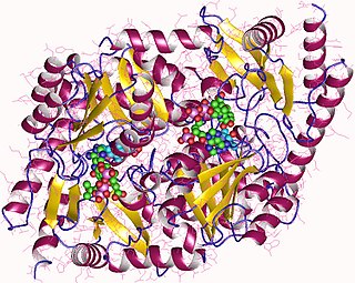
Uroporphyrinogen III synthase is an enzyme involved in the metabolism of the cyclic tetrapyrrole compound porphyrin. It is involved in the conversion of hydroxymethyl bilane into uroporphyrinogen III. This enzyme catalyses the inversion of the final pyrrole unit of the linear tetrapyrrole molecule, linking it to the first pyrrole unit, thereby generating a large macrocyclic structure, uroporphyrinogen III. The enzyme folds into two alpha/beta domains connected by a beta-ladder, the active site being located between the two domains.

<span class="mw-page-title-main">Uroporphyrinogen III</span> Chemical compound

Uroporphyrinogen III is a tetrapyrrole, the first macrocyclic intermediate in the biosynthesis of heme, chlorophyll, vitamin B12, and siroheme. It is a colorless compound, like other porphyrinogens.

Erythropoietic porphyria is a type of porphyria associated with erythropoietic cells. In erythropoietic porphyrias, the enzyme deficiency occurs in the red blood cells.

<span class="mw-page-title-main">Hepatoerythropoietic porphyria</span> Medical condition

Hepatoerythropoietic porphyria is a very rare form of hepatic porphyria caused by a disorder in both genes which code Uroporphyrinogen III decarboxylase (UROD).

<span class="mw-page-title-main">Uroporphyrinogen I</span> Chemical compound

Uroporphyrinogen I is an isomer of uroporphyrinogen III, a metabolic intermediate in the biosynthesis of heme. A type of porphyria is caused by production of uroporphyrinogen I instead of III.

<span class="mw-page-title-main">Coproporphyrinogen I</span> Chemical compound

Coproporphyrinogen I is an isomer of coproporphyrinogen III, a metabolic intermediate in the normal biosynthesis of heme. The compound is not normally produced by the human body; its production and accumulation causes a type of porphyria.

Congenital hemolytic anemia (CHA) is a diverse group of rare hereditary conditions marked by decreased life expectancy and premature removal of erythrocytes from blood flow. Defects in erythrocyte membrane proteins and red cell enzyme metabolism, as well as changes at the level of erythrocyte precursors, lead to impaired bone marrow erythropoiesis. CAH is distinguished by variable anemia, chronic extravascular hemolysis, decreased erythrocyte life span, splenomegaly, jaundice, biliary lithiasis, and iron overload. Immune-mediated mechanisms may play a role in the pathogenesis of these uncommon diseases, despite the paucity of data regarding the immune system's involvement in CHAs.

<span class="mw-page-title-main">Aminolevulinic acid dehydratase deficiency porphyria</span> Medical condition

Aminolevulinic acid dehydratase deficiency porphyria is an extremely rare autosomal recessive metabolic disorder that results from inappropriately low levels of the enzyme delta-aminolevulinic acid dehydratase (ALAD), which is required for normal heme synthesis. This deficiency results in the accumulation of a toxic metabolic precursor in the heme synthesis pathway called aminolevulinic acid (ALA). Lead poisoning can also disrupt ALAD and result in elevated ALA causing the same symptoms. Heme is a component of hemoglobin which carries oxygen in red blood cells.

<span class="mw-page-title-main">Harderoporphyria</span> Medical condition

Harderoporphyria is a rare disorder of heme biosynthesis, inherited in an autosomal recessive manner caused by specific mutations in the CPOX gene. Mutations in CPOX usually cause hereditary coproporphyria, an acute hepatic porphyria, however the K404E mutation in a homozygous or compound heterozygous state with a null allele cause the more severe harderoporphyria. Harderoporphyria is the first known metabolic disorder where the disease phenotype depended on the type and location of the mutations in a gene associated with multiple disorders.

References

  1. Online Mendelian Inheritance in Man (OMIM): 263700
  2. James, William D.; Berger, Timothy G.; et al. (2006). Andrews' Diseases of the Skin: clinical Dermatology. Saunders Elsevier. p. 526. ISBN   978-0-7216-2921-6.
  3. King, Michael. "Introduction to Congenital Erythropoietic Porphyria". Medical Biochemistry Page. Retrieved 30 November 2012.
  4. 1 2 3 Deybach JC, De Verneuil H, Boulechfar S, Grandchamp B, Nordmann Y (1990). "Point mutations in the uroporphyrinogen III synthase gene in congenital erythropoietic porphyria (Gunther's disease)". Blood. 75 (9): 1763–5. doi: 10.1182/blood.V75.9.1763.1763 . ISSN   0006-4971. PMID   2331520.
  5. Robert-Richard E, Moreau-Gaudry F, Lalanne M, et al. (January 2008). "Effective gene therapy of mice with congenital erythropoietic porphyria is facilitated by a survival advantage of corrected erythroid cells". Am. J. Hum. Genet. 82 (1): 113–24. doi:10.1016/j.ajhg.2007.09.007. PMC   2253957 . PMID   18179890.
  6. Thadani, H.; Deacon, A.; Peters, T. (2000). "Diagnosis and management of porphyria". BMJ. 320 (7250): 1647–1651. doi:10.1136/bmj.320.7250.1647. PMC   1127427 . PMID   10856069.
  7. Burzio, Chiara. "Gunther's Disease" . Retrieved November 28, 2012.
  8. Gorchein, Abel; Rong Guo; Chang Kee Lim; Ana Raimundo; Humphrey W.H. Pullon; Alastair J. Bellinham (18 January 1999). "Porphyrins in urine, plasma, erythrocytes, bile and faeces in a case of congenital erythropoietic porphyria (Gunther's disease) treated with blood transfusion and iron chelation: lack of benefit from oral charcoal". Biomedical Chromatography. 12 (6): 350–356. doi:10.1002/(SICI)1099-0801(199811/12)12:6<350::AID-BMC761>3.0.CO;2-B. PMID   9861496.
  9. Fortian, A; González, E; Castaño, D; Falcon-Perez, JM; Millet, O (Apr 15, 2011). "Intracellular rescue of the uroporphyrinogen III synthase activity in enzymes carrying the hotspot mutation C73R". The Journal of Biological Chemistry. 286 (15): 13127–33. doi: 10.1074/jbc.m110.205849 . PMC   3075659 . PMID   21343304.
  10. 1 2 3 4 Balwani, M; Desnick, RJ (2012). "The porphyrias: Advances in diagnosis and treatment". Hematology. 2012: 19–27. doi: 10.1182/asheducation.v2012.1.19.3795678 . PMC   3512229 . PMID   23233556.
  11. De, AK; Das, K; Sil, A; Joardar, S (Sep 2013). "A Case of Congenital Erythropoietic Porphyria without Hemolysis". Indian Journal of Dermatology. 58 (5): 407. doi: 10.4103/0019-5154.117336 . PMC   3778801 . PMID   24082206.
  12. Saval, Herrera; Moruno Tirado (24 December 2001). "Congenital erythropoietic porphyria affecting two brothers". British Journal of Dermatology. 141 (3): 547–550. doi:10.1046/j.1365-2133.1999.03057.x. PMID   10583066.
  13. 1 2 Katugampola, RP; Anstey, AV; Finlay, AY; Whatley, S; Woolf, J; Mason, N; Deybach, JC; Puy, H; Ged, C; de Verneuil, H; Hanneken, S; Minder, E; Schneider-Yin, X; Badminton, MN (Oct 2012). "A management algorithm for congenital erythropoietic porphyria derived from a study of 29 cases". The British Journal of Dermatology. 167 (4): 888–900. doi:10.1111/j.1365-2133.2012.11154.x. PMID   22804244.
  14. Online Mendelian Inheritance in Man (OMIM): 606938
  15. "Congenital erythropoietic porphyria". New Zealand Dermatological Society. Retrieved 28 November 2012.
  16. Pannier, E; G. Viot; M.C. Aubry; G. Grange; J. Tantau; C. Fallet-Bianco; F. Muller; D. Cabrol (9 December 2002). "Congenital erythropoietic porphyria (Günther's disease): two cases with very early prenatal manifestation and cystic hygroma". Prenatal Diagnosis. 23 (1): 25–30. doi:10.1002/pd.491. PMID   12533808. S2CID   25995061.
  17. 1 2 3 4 Hebel, Jeanette (2018-05-24). "Congenital Erythropoietic Porphyria Treatment & Management". Medscape. Retrieved 30 November 2012.
  18. Taibjee, S.M; O.E. Steveson; A. Adullah; C.Y. Tan; P. Darbyshire; C. Moss; H. Goodyear; A. Heargerty; S. Wheatley; M.N. Badminton (18 January 2007). "Allogeneic bone marrow transplantation in a 7-year-old girl with congenital erythropoietic porphyria: a treatment dilemma". British Journal of Dermatology. 156 (3): 567–571. doi:10.1111/j.1365-2133.2006.07699.x. PMID   17300251. S2CID   39733402.
  19. Madan P, Schaaf CP, Vardhan P, Bhayana S, Chandra P, Anderson KE (2007). "Hans Gunther and his disease". Photodermatol Photoimmunol Photomed. 23 (6): 261–3. doi:10.1111/j.1600-0781.2007.00323.x. PMID   17986065. S2CID   44372076.