Historadiography

Last updated

Historadiography is a technique formerly utilized in the fields of histology and cellular biology to provide semiquantitative information regarding the density of a tissue sample. It is usually synonymous with microradiography. [1] This is achieved by layering a ground section of mineralized tissue (such as bone) with photographic emulsion on a glass slide and exposing the sample to a beam of X-rays. After developing the emulsion, the resulting radiograph can be viewed with a microscope. A side-by-side comparison with a slide containing radiographs of various substances of known mass can provide a rough mass estimate, and therefore a rough approximation of the concentration of calcium salts in the sample.

Histology study of the microscopic anatomy of cells and tissues of plants and animals

Histology, also microanatomy, is the branch of biology which studies the tissues of animals and plants using microscopy. It is commonly studied using a light microscope or electron microscope, the specimen having been sectioned, stained, and mounted on a microscope slide. Histological studies may be conducted using tissue culture, where live animal cells are isolated and maintained in an artificial environment for various research projects. The ability to visualize or differentially identify microscopic structures is frequently enhanced through the use of staining. Histology is one of the major preclinical subjects in medical school. Medical students are expected to be familiar with the morphological features and function of all cells and tissues of the human body from an early stage of their studies, so histology often stretches over several semesters.

Microscope instrument used to see objects that are too small for the naked eye

A microscope is an instrument used to see objects that are too small to be seen by the naked eye. Microscopy is the science of investigating small objects and structures using such an instrument. Microscopic means invisible to the eye unless aided by a microscope.

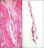
Historadiography has also been used to visualize staining of tissue, such as spinal cord samples with thorotrast, which contains thorium that is opaque to X-rays. [2]

Spinal cord long, thin, tubular bundle of nervous tissue and support cells that extends from the brain

The spinal cord is a long, thin, tubular structure made up of nervous tissue, that extends from the medulla oblongata in the brainstem to the lumbar region of the vertebral column. It encloses the central canal of the spinal cord that contains cerebrospinal fluid. The brain and spinal cord together make up the central nervous system (CNS). In humans, the spinal cord begins at the occipital bone where it passes through the foramen magnum, and meets and enters the spinal canal at the beginning of the cervical vertebrae. The spinal cord extends down to between the first and second lumbar vertebrae where it ends. The enclosing bony vertebral column protects the relatively shorter spinal cord. It is around 45 cm (18 in) in men and around 43 cm (17 in) long in women. Also, the spinal cord has a varying width, ranging from 13 mm thick in the cervical and lumbar regions to 6.4 mm thick in the thoracic area.

Thorotrast

Thorotrast is a suspension containing particles of the radioactive compound thorium dioxide, ThO2, that was used as a radiocontrast agent in medical radiography in the 1930s and 1940s. Use in some countries, such as the U.S., continued into the 1950s.

Thorium Chemical element with atomic number 90

Thorium is a weakly radioactive metallic chemical element with symbol Th and atomic number 90. Thorium is silvery and tarnishes black when it is exposed to air, forming thorium dioxide; it is moderately hard, malleable, and has a high melting point. Thorium is an electropositive actinide whose chemistry is dominated by the +4 oxidation state; it is quite reactive and can ignite in air when finely divided.

Over recent decades researchers have generally lost interest in historadiography. The most recent publication using the term (1998) to be indexed in PubMed [3] referred to autoradiography of tritium incorporated in thymidine. [4]

PubMed online database with abstracts of medical articles, hosted by US National Library of Medicine

PubMed is a free search engine accessing primarily the MEDLINE database of references and abstracts on life sciences and biomedical topics. The United States National Library of Medicine (NLM) at the National Institutes of Health maintains the database as part of the Entrez system of information retrieval.

Tritium isotope of hydrogen with 2 neutrons

Tritium is a radioactive isotope of hydrogen. The nucleus of tritium contains one proton and two neutrons, whereas the nucleus of protium contains one proton and no neutrons. Naturally occurring tritium is extremely rare on Earth, where trace amounts are formed by the interaction of the atmosphere with cosmic rays. It can be produced by irradiating lithium metal or lithium-bearing ceramic pebbles in a nuclear reactor. Tritium is used as a radioactive tracer, in radioluminescent light sources for watches and instruments, and, along with deuterium, as a fuel for nuclear fusion reactions with applications in energy generation and weapons. The name of this isotope is derived from Greek, Modern τρίτος (trítos), meaning 'third'.

Thymidine Chemical compound

Thymidine is a pyrimidine deoxynucleoside. Deoxythymidine is the DNA nucleoside T, which pairs with deoxyadenosine (A) in double-stranded DNA. In cell biology it is used to synchronize the cells in G1/early S phase.

Related Research Articles

Biopsy medical test involving sampling of cells or tissues for examination

A biopsy is a medical test commonly performed by a surgeon, interventional radiologist, or an interventional cardiologist involving extraction of sample cells or tissues for examination to determine the presence or extent of a disease. The tissue is generally examined under a microscope by a pathologist, and can also be analyzed chemically. When an entire lump or suspicious area is removed, the procedure is called an excisional biopsy. An incisional biopsy or core biopsy samples a portion of the abnormal tissue without attempting to remove the entire lesion or tumor. When a sample of tissue or fluid is removed with a needle in such a way that cells are removed without preserving the histological architecture of the tissue cells, the procedure is called a needle aspiration biopsy. Biopsies are most commonly performed for insight into possible cancerous and inflammatory conditions.

Autoradiograph

An autoradiograph is an image on an x-ray film or nuclear emulsion produced by the pattern of decay emissions from a distribution of a radioactive substance. Alternatively, the autoradiograph is also available as a digital image, due to the recent development of scintillation gas detectors or rare earth phosphorimaging systems. The film or emulsion is apposed to the labeled tissue section to obtain the autoradiograph. The auto- prefix indicates that the radioactive substance is within the sample, as distinguished from the case of historadiography or microradiography, in which the sample is marked using an external source. Some autoradiographs can be examined microscopically for localization of silver grains in which the process is termed micro-autoradiography. For example, micro-autoradiography was used to examine whether atrazine was being metabolized by the hornwort plant or by epiphytic microorganisms in the biofilm layer surrounding the plant.

Radiocontrast agents are substances used to enhance the visibility of internal structures in X-ray-based imaging techniques such as computed tomography, projectional radiography, and fluoroscopy. Radiocontrast agents are typically iodine, barium-sulphate or gadolinium based compounds. They absorb external X-rays, resulting in decreased exposure on the X-ray detector. This is different from radiopharmaceuticals used in nuclear medicine which emit radiation.

Radiodensity is opacity to the radio wave and X-ray portion of the electromagnetic spectrum: that is, the relative inability of those kinds of electromagnetic radiation to pass through a particular material. Radiolucency or hypodensity indicates greater passage to X-ray photons and is the analogue of transparency and translucency with visible light. Materials that inhibit the passage of electromagnetic radiation are called radiodense or radiopaque, while those that allow radiation to pass more freely are referred to as radiolucent. Radiopaque volumes of material have white appearance on radiographs, compared with the relatively darker appearance of radiolucent volumes. For example, on typical radiographs, bones look white or light gray (radiopaque), whereas muscle and skin look black or dark gray, being mostly invisible (radiolucent).

Cerebral angiography

Cerebral angiography is a form of angiography which provides images of blood vessels in and around the brain, thereby allowing detection of abnormalities such as arteriovenous malformations and aneurysms. It was pioneered in 1927 by the Portuguese neurologist Egas Moniz at the University of Lisbon, who also helped develop thorotrast for use in the procedure.

Chest radiograph

A chest radiograph, colloquially called a chest X-ray (CXR), or chest film, is a projection radiograph of the chest used to diagnose conditions affecting the chest, its contents, and nearby structures. Chest radiographs are the most common film taken in medicine.

The film badge dosimeter or film badge is a personal dosimeter used for monitoring cumulative radiation dose due to ionizing radiation.

Fine-needle aspiration diagnostic procedure to investigate lumps using a thin needle. can be either for histopathology or cytopathology

Fine-needle aspiration (FNA) is a diagnostic procedure used to investigate lumps or masses. In this technique, a thin, hollow needle is inserted into the mass for sampling of cells that, after being stained, will be examined under a microscope (biopsy). The sampling and biopsy considered together are called fine-needle aspiration biopsy (FNAB) or fine-needle aspiration cytology (FNAC). Fine-needle aspiration biopsies are very safe minor surgical procedures. Often, a major surgical biopsy can be avoided by performing a needle aspiration biopsy instead, eliminating the need for hospitalization. In 1981, the first fine-needle aspiration biopsy in the United States was done at Maimonides Medical Center. Today, this procedure is widely used in the diagnosis of cancer and inflammatory conditions.

Laser capture microdissection

Laser capture microdissection (LCM), also called microdissection, laser microdissection (LMD), or laser-assisted microdissection, is a method for isolating specific cells of interest from microscopic regions of tissue/cells/organisms.

Infantile cortical hyperostosis is a self-limited inflammatory disorder of infants that causes bone changes, soft tissue swelling and irritability. The disease may be present at birth or occur shortly thereafter. The cause is unknown. Both familial and sporadic forms occur. It is also known as Caffey disease or Caffey's disease.

Dental radiography

Dental radiographs are commonly called X-rays. Dentists use radiographs for many reasons: to find hidden dental structures, malignant or benign masses, bone loss, and cavities.

In particle and nuclear physics, a nuclear emulsion plate is a photographic plate with a particularly thick emulsion layer and with a very uniform grain size. Like bubble chambers, cloud chambers, and wire chambers nuclear emulsion plates record the tracks of charged particles passing through. They are compact, have high density and produce a cumulative record, but have the disadvantage that the plates must be developed before the tracks can be observed.

Projectional radiography

Projectional radiography is a form of radiography and medical imaging that produces two-dimensional images by x-ray radiation. The image acquisition is generally performed by radiographers, and the images are often examined by radiologists. Plain radiography generally refers to projectional radiography. Plain radiography can also refer to radiography without a radiocontrast agent or radiography that generates single static images, as contrasted to fluoroscopy, which are technically also projectional.

Abdominal x-ray

An abdominal x-ray is an x-ray of the abdomen. It is sometimes abbreviated to AXR, or KUB.

Lymphography is a medical imaging technique in which a radiocontrast agent is injected, and then an X-ray picture is taken to visualize structures of the lymphatic system, including lymph nodes, lymph ducts, lymphatic tissues, lymph capillaries and lymph vessels. Lymphangiography is the same procedure, used only to visualize the lymph vessels. The X-ray film or image of the vessels and nodes is called a lymphogram or a lymphangiogram.

Panoramic radiograph

A panoramic radiograph is a panoramic scanning dental X-ray of the upper and lower jaw. It shows a two-dimensional view of a half-circle from ear to ear. Panoramic radiography is a form of focal plane tomography; thus, images of multiple planes are taken to make up the composite panoramic image, where the maxilla and mandible are in the focal trough and the structures that are superficial and deep to the trough are blurred.

The D-DIA or deformation-DIA is an apparatus used for high pressure and high temperature deformation experiments. The advantage of this apparatus is the ability to apply pressures up to approximately 15 GPa while independently creating uniaxial strains up to 50%.

Paleoradiology Study of archaeological remains through the use of radiographic techniques

Paleoradiology is the study of archaeological remains through the use of radiographic techniques, such as X-ray, CT and micro-CT scans. It is predominately used by archaeologists and anthropologists to examine mummified remains due to its non-invasive nature. Paleoradiologists can discover post-mortem damage to the body, or any artefacts buried with them, while still keeping the remains intact. Radiological images can also contribute evidence about the person's life, such as their age and cause of death. The first recorded use of paleoradiology was in 1896, just a year after the Rōntgen radiograph was first produced. Although this method of viewing ancient remains is advantageous due to its non-invasive manner, many radiologists lack expertise in archeology and very few radiologists can identify ancient diseases which may be present.

References

  1. "CancerWeb Online Medical Dictionary".
  2. F. P. BOHATIRCHUK; T. F. JELETZKY; L. P. IVAN (1973). "CONTACT HISTORADIOGRAPHY OF NERVOUS TISSUE IMPREGNATED WITH THOROTRAST".
  3. "PubMed search: historadiography".
  4. "Effect of the lipidosterolic extract of Serenoa repens (Permixon) and its major components on basic fibroblast growth factor-induced proliferation of cultures of human prostate biopsies". Eur. Urol. 33 (3): 340–7. 1998. PMID   9555564.