Hyaline cartilage

Last updated

Hyaline cartilage
Hypertrophic Zone of Epiphyseal Plate.jpg
Light micrograph of undecalcified hyaline cartilage showing microanatomy of chondrocytes and organelles, lacunae and matrix.
Identifiers
MeSH D051457
TH H2.00.03.5.00015
FMA 64783
Anatomical terminology

Hyaline cartilage is the glass-like (hyaline) and translucent cartilage found on many joint surfaces. It is also most commonly found in the ribs, nose, larynx, and trachea. [1] Hyaline cartilage is pearl-gray in color, with a firm consistency and has a considerable amount of collagen. It contains no nerves or blood vessels, and its structure is relatively simple.

Contents

Structure

Hyaline cartilage is covered externally by a fibrous membrane known as the perichondrium or, when it's along articulating surfaces, the synovial membrane. This membrane contains vessels that provide the cartilage with nutrition through diffusion.

Hyaline cartilage matrix is primarily made of type II collagen and chondroitin sulphate, both of which are also found in elastic cartilage.

Hyaline cartilage exists on the sternal ends of the ribs, in the larynx, trachea, and bronchi, and on the articulating surfaces of bones. It gives the structures a definite but pliable form. The presence of collagen fibres makes such structures and joints strong, but with limited mobility and flexibility.

Hyaline cartilage is the most prevalent type of cartilage. It also forms the temporary embryonic skeleton, which is gradually replaced by bone, and the skeleton of elasmobranch fish.

Microanatomy

When a slice of hyaline cartilage is examined under the microscope, it is shown to consist of cells (chondrocytes) of a rounded or bluntly angular form, lying in groups of two or more in a granular, or almost homogeneous matrix. When arranged in groups of two or more, the chondrocytes have rounded, but generally straight outlines; where they are in contact with each other, and in the rest of their circumference, they are rounded.

They consist of translucent protoplasm with fine interlacing filaments and minute granules are sometimes present. Embedded in this are one or two round nuclei, having the usual intranuclear network.

The cells are contained in cavities in the matrix, called cartilage lacunae. These cavities are actually artificial gaps formed from the shrinking of the cells during the staining and setting of the tissue for examination. The inter-territorial space between the isogenous cell groups contains relatively more collagen fibres, allowing it to maintain its shape while the actual cells shrink, creating the lacunae. This constitutes the so-called 'capsule' of the space. Each lacuna is usually occupied by a single cell, but during mitosis, it may contain two, four, or even eight cells.

Articular cartilage

Histology of articular cartilage zones. Histology of articular cartilage zones.jpg
Histology of articular cartilage zones.

Articular cartilage is hyaline cartilage on the articular surfaces of bones, [3] and lies inside the joint cavity of synovial joints, bathed in synovial fluid produced by the synovial membrane, which lines the walls of the cavity.

Though it is often found in close contact with menisci and articular disks, articular cartilage is not considered a part of either of these structures, which are made entirely of fibrocartilage.

The articular cartilage extracellular matrix (ECM) has a highly specialized architecture that is zonally organized: the superficial zone consists mostly of collagen II fibers aligned parallel to the articular surface to resist shear forces, whereas the deep zone consists of the same fibers aligned perpendicularly to the bone interface to absorb compressive loads. [2]

The biochemical breakdown of the articular cartilage results in osteoarthritis – the most common type of joint disease. [4] Osteoarthritis affects over 30 million individuals in the United States alone, and is the leading cause of chronic disability amongst the elderly. [5]

Articular cartilage development begins with interzone condensation of a Collagen II positive limb bud at the future joint site. This is followed by definition of specific cellular subtypes (meniscal progenitors, articular progenitors, synovial progenitors, and ligament progenitors) that will eventually form the joint capsule. Finally, the joint capsule matures and forms a cavity, with a central meniscus, and an encasement of synovium. [6] This final structure will form several distinct layers of the articular cartilage found in all synovial joints including the Deep Zone (closest to the bone), Middle Zone, and Superficial Zone (closest to the synovial fluid).

Maintenance of articular cartilage is guided by a balance of anabolic (cartilage generating) [7] [8] and catabolic (cartilage degrading factors), [9] [10] in a manner similar to the maintenance of bone. [11] Over the lifetime of the organism, anabolic factors and catabolic factors are generally in balance, however, as the organism ages, catabolism predominates and cartilage begins to degrade. Eventually, the loss of hyaline cartilage matrix and reduction in the chondrocyte content of the hyaline cartilage matrix results in the development of joint disease such as Osteoarthritis (OA). Overexpression of hyaline-cartilage specific anabolic factors, such as FGF18, or appears to restore the balance between cartilage loss and generation. [12] [13]

Additional images

See also

Related Research Articles

<span class="mw-page-title-main">Cartilage</span> Resilient and smooth elastic tissue in animals

Cartilage is a resilient and smooth type of connective tissue. In tetrapods, it covers and protects the ends of long bones at the joints as articular cartilage, and is a structural component of many body parts including the rib cage, the neck and the bronchial tubes, and the intervertebral discs. In other taxa, such as chondrichthyans, but also in cyclostomes, it may constitute a much greater proportion of the skeleton. It is not as hard and rigid as bone, but it is much stiffer and much less flexible than muscle. The matrix of cartilage is made up of glycosaminoglycans, proteoglycans, collagen fibers and, sometimes, elastin.

<span class="mw-page-title-main">Synovial membrane</span> Connective tissue present within and around synovial joints

The synovial membrane is a specialized connective tissue that lines the inner surface of capsules of synovial joints and tendon sheath. It makes direct contact with the fibrous membrane on the outside surface and with the synovial fluid lubricant on the inside surface. In contact with the synovial fluid at the tissue surface are many rounded macrophage-like synovial cells and also type B cells, which are also known as fibroblast-like synoviocytes (FLS). Type A cells maintain the synovial fluid by removing wear-and-tear debris. As for the FLS, they produce hyaluronan, as well as other extracellular components in the synovial fluid.

<span class="mw-page-title-main">Synovial joint</span> Articulation which admits free motion in the joint; the most common type of articulation

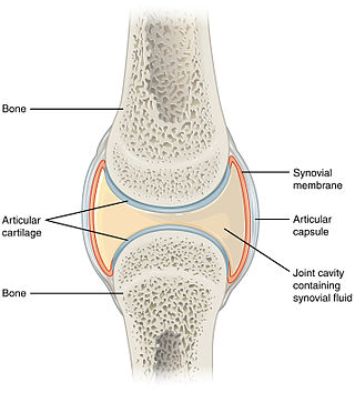
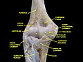
A synovial joint, also known as diarthrosis, joins bones or cartilage with a fibrous joint capsule that is continuous with the periosteum of the joined bones, constitutes the outer boundary of a synovial cavity, and surrounds the bones' articulating surfaces. This joint unites long bones and permits free bone movement and greater mobility. The synovial cavity/joint is filled with synovial fluid. The joint capsule is made up of an outer layer of fibrous membrane, which keeps the bones together structurally, and an inner layer, the synovial membrane, which seals in the synovial fluid.

<span class="mw-page-title-main">Periosteum</span> Membrane covering outer surface of bones

The periosteum is a membrane that covers the outer surface of all bones, except at the articular surfaces of long bones. Endosteum lines the inner surface of the medullary cavity of all long bones.

<span class="mw-page-title-main">Synovial fluid</span> Fluid found in the cavities of synovial joints

Synovial fluid, also called synovia,[help 1] is a viscous, non-Newtonian fluid found in the cavities of synovial joints. With its egg white–like consistency, the principal role of synovial fluid is to reduce friction between the articular cartilage of synovial joints during movement. Synovial fluid is a small component of the transcellular fluid component of extracellular fluid.

<span class="mw-page-title-main">Chondrocyte</span> Cell that makes up cartilage

Chondrocytes are the only cells found in healthy cartilage. They produce and maintain the cartilaginous matrix, which consists mainly of collagen and proteoglycans. Although the word chondroblast is commonly used to describe an immature chondrocyte, the term is imprecise, since the progenitor of chondrocytes can differentiate into various cell types, including osteoblasts.

<span class="mw-page-title-main">Chondroblast</span> Mesenchymal progenitor cell that forms a chondrocyte

Chondroblasts, or perichondrial cells, is the name given to mesenchymal progenitor cells in situ which, from endochondral ossification, will form chondrocytes in the growing cartilage matrix. Another name for them is subchondral cortico-spongious progenitors. They have euchromatic nuclei and stain by basic dyes.

<span class="mw-page-title-main">Chondrogenesis</span> Process by which cartilage is developed

Chondrogenesis is the process by which cartilage is developed.

Articular cartilage, most notably that which is found in the knee joint, is generally characterized by very low friction, high wear resistance, and poor regenerative qualities. It is responsible for much of the compressive resistance and load bearing qualities of the knee joint and, without it, walking is painful to impossible. Osteoarthritis is a common condition of cartilage failure that can lead to limited range of motion, bone damage and invariably, pain. Due to a combination of acute stress and chronic fatigue, osteoarthritis directly manifests itself in a wearing away of the articular surface and, in extreme cases, bone can be exposed in the joint. Some additional examples of cartilage failure mechanisms include cellular matrix linkage rupture, chondrocyte protein synthesis inhibition, and chondrocyte apoptosis. There are several different repair options available for cartilage damage or failure.

<span class="mw-page-title-main">Isogenous group</span>

An isogenous group is a cluster of up to eight chondrocytes found in hyaline and elastic cartilage.

<span class="mw-page-title-main">FGF18</span> Mammalian protein found in Homo sapiens

Fibroblast growth factor 18 (FGF18) is a protein that is encoded by the Fgf18 gene in humans. The protein was first discovered in 1998, when two newly-identified murine genes Fgf17 and Fgf18 were described and confirmed as being closely related by sequence homology to Fgf8. The three proteins were eventually grouped into the FGF8 subfamily, which contains several of the endocrine FGF superfamily members FGF8, FGF17, and FGF18. Subsequent studies identified FGF18's role in promoting chondrogenesis, and an apparent specific activity for the generation of the hyaline cartilage in articular joints.

<span class="mw-page-title-main">Proteoglycan 4</span> Proteoglycan; lubricant; gene

Proteoglycan 4 or lubricin is a proteoglycan that in humans is encoded by the PRG4 gene. It acts as a joint/boundary lubricant.

Articular cartilage repair treatment involves the repair of the surface of an articular joint's hyaline cartilage. Over the last few decades, surgeons and researchers have made progress in elaborating surgical cartilage repair interventions. Though these solutions do not perfectly restore the articular cartilage, some of the latest technologies have started to bring very promising results in repairing cartilages from traumatic injuries or chondropathies. These treatments have been shown to be especially beneficial for patients who have articular cartilage damage. They can provide some measure of pain relief, while at the same time slowing down the accumulation of damage, or considerably delaying the need for joint replacement surgery.

Autologous chondrocyte implantation is a biomedical treatment that repairs damages in articular cartilage. ACI provides pain relief while at the same time slowing down the progression or considerably delaying partial or total joint replacement surgery. The goal of ACI is to allow people suffering from articular cartilage damage to return to their old lifestyle; regaining mobility, going back to work and even practicing sports again.

Mesenchymal stem cells (MSCs) are multipotent cells found in multiple human adult tissues including bone marrow, synovial tissues, and adipose tissues. Since they are derived from the mesoderm, they have been shown to differentiate into bone, cartilage, muscle, and adipose tissue. MSCs from embryonic sources have shown promise scientifically while creating significant controversy. As a result, many researchers have focused on adult stem cells, or stem cells isolated from adult humans that can be transplanted into damaged tissue.

Autologous matrix-induced chondrogenesis (AMIC) is a treatment for articular cartilage damage. It combines microfracture surgery with the application of a bi-layer collagen I/III membrane. There is tentative short to medium term benefits as of 2017.

Gene therapy for osteoarthritis is the application of gene therapy to treat osteoarthritis (OA). Unlike pharmacological treatments which are administered locally or systemically as a series of interventions, gene therapy aims to establish sustained therapeutic effect after a single, local injection.

Sprifermin (INN), is a recombinant human fibroblast growth factor 18 (rhFGF18) analog, which is under development by TrialSpark for the treatment of osteoarthritis. FGF18 and sprifermin act via the Fibroblast Growth Factor Receptor (FGFR) family, with preferential activity via FGFR3c.

Nasal chondrocytes (NC) are present in the hyaline cartilage of the nasal septum and in fact are the only cell type within the tissue. Similar to chondrocytes present in articular cartilage, NC express extracellular matrix proteins such as glycosaminoglycans and collagen.

Artificial cartilage is a synthetic material made of hydrogels or polymers that aims to mimic the functional properties of natural cartilage in the human body. Tissue engineering principles are used in order to create a non-degradable and biocompatible material that can replace cartilage. While creating a useful synthetic cartilage material, certain challenges need to be overcome. First, cartilage is an avascular structure in the body and therefore does not repair itself. This creates issues in regeneration of the tissue. Synthetic cartilage also needs to be stably attached to its underlying surface i.e. the bone. Lastly, in the case of creating synthetic cartilage to be used in joint spaces, high mechanical strength under compression needs to be an intrinsic property of the material.

References

  1. Adele, Knibbs (2003). "The Leeds Histology Guide" . Retrieved 27 October 2018.
  2. 1 2 Zhao, Feng; Bautista, Catherine A.; Park, Hee Jun; Mazur, Courtney M.; Aaron, Roy K.; Bilgen, Bahar (2016). "Effects of Chondroitinase ABC-Mediated Proteoglycan Digestion on Decellularization and Recellularization of Articular Cartilage". PLOS ONE. 11 (7): e0158976. Bibcode:2016PLoSO..1158976B. doi: 10.1371/journal.pone.0158976 . ISSN   1932-6203. PMC   4938233 . PMID   27391810.
    -"The work is made available under the Creative Commons CC0 public domain dedication."
  3. "Wheeless' Textbook of Orthopaedics". 22 July 2020.
  4. Brown, Angelina. "Coping with Osteoarthritis". Archived from the original on 1 December 2017. Retrieved 24 July 2017.
  5. "Osteoarthritis Fact Sheet". Center for Disease Control and Prevention. Retrieved 24 July 2017.
  6. Wilkinson, J. Mark; Zeggini, Eleftheria (1 September 2021). "The Genetic Epidemiology of Joint Shape and the Development of Osteoarthritis". Calcified Tissue International. 109 (3): 257–276. doi:10.1007/s00223-020-00702-6. ISSN   1432-0827. PMC   8403114 . PMID   32393986.
  7. Davidson, David; Blanc, Antoine; Filion, Dominic; Wang, Huifen; Plut, Paul; Pfeffer, Gerald; Buschmann, Michael D.; Henderson, Janet E. (27 May 2005). "Fibroblast growth factor (FGF) 18 signals through FGF receptor 3 to promote chondrogenesis". The Journal of Biological Chemistry. 280 (21): 20509–20515. doi: 10.1074/jbc.M410148200 . ISSN   0021-9258. PMID   15781473.
  8. Takahata, Yoshifumi; Hagino, Hiromasa; Kimura, Ayaka; Urushizaki, Mitsuki; Yamamoto, Shiori; Wakamori, Kanta; Murakami, Tomohiko; Hata, Kenji; Nishimura, Riko (23 April 2022). "Regulatory Mechanisms of Prg4 and Gdf5 Expression in Articular Cartilage and Functions in Osteoarthritis". International Journal of Molecular Sciences. 23 (9): 4672. doi: 10.3390/ijms23094672 . ISSN   1422-0067. PMC   9105027 . PMID   35563063.
  9. Alvaro-Gracia, J. M. (February 2004). "Licofelone--clinical update on a novel LOX/COX inhibitor for the treatment of osteoarthritis". Rheumatology. 43 (Suppl 1): i21–25. doi:10.1093/rheumatology/keh105. ISSN   1462-0324. PMID   14752172.
  10. Li, Ting; Peng, Jie; Li, Qingqing; Shu, Yuan; Zhu, Peijun; Hao, Liang (8 July 2022). "The Mechanism and Role of ADAMTS Protein Family in Osteoarthritis". Biomolecules. 12 (7): 959. doi: 10.3390/biom12070959 . ISSN   2218-273X. PMC   9313267 . PMID   35883515.
  11. Maeda, Kazuhiro; Kobayashi, Yasuhiro; Koide, Masanori; Uehara, Shunsuke; Okamoto, Masanori; Ishihara, Akihiro; Kayama, Tomohiro; Saito, Mitsuru; Marumo, Keishi (6 November 2019). "The Regulation of Bone Metabolism and Disorders by Wnt Signaling". International Journal of Molecular Sciences. 20 (22): 5525. doi: 10.3390/ijms20225525 . ISSN   1422-0067. PMC   6888566 . PMID   31698687.
  12. Hollander, Judith M.; Goraltchouk, Alex; Rawal, Miraj; Liu, Jingshu; Luppino, Francesco; Zeng, Li; Seregin, Alexey (6 March 2023). "Adeno-Associated Virus-Delivered Fibroblast Growth Factor 18 Gene Therapy Promotes Cartilage Anabolism". Cartilage: 19476035231158774. doi:10.1177/19476035231158774. ISSN   1947-6043. PMID   36879540. S2CID   257376179.
  13. Moore, E. E.; Bendele, A. M.; Thompson, D. L.; Littau, A.; Waggie, K. S.; Reardon, B.; Ellsworth, J. L. (July 2005). "Fibroblast growth factor-18 stimulates chondrogenesis and cartilage repair in a rat model of injury-induced osteoarthritis". Osteoarthritis and Cartilage. 13 (7): 623–631. doi:10.1016/j.joca.2005.03.003. ISSN   1063-4584. PMID   15896984.