Interatrial septum

Last updated
Interatrial septum
Gray493.png
Interior of right side of heart. (labeled at left of image, as 'atrial septum')
Gray467.png
Interior of dorsal half of heart of human embryo of about thirty-five days.
Details
Identifiers
Latin septum interatriale
MeSH D054087
TA98 A12.1.00.019
TA2 3964
FMA 7108
Anatomical terminology

The interatrial septum is the wall of tissue that separates the right and left atria of the heart.

Contents

Structure

The interatrial septum is a septum that lies between the left atrium and right atrium of the human heart. The interatrial septum lies at angle of 65 degrees from right posterior to left anterior because right atrium is located at the right side of the body while left atrium is located at the left side of the body. [1] The interatrial septum represents the posterior wall of the right atrium. [2]

Development

The interatrial septum forms during the first and second months of fetal development. Formation of the septum occurs in several stages. The first is the development of the septum primum, a crescent-shaped piece of tissue forming the initial divider between the right and left atria. Because of its crescent shape, the septum primum does not fully occlude the space between the left and right atria; the opening that remains is called the ostium primum. During fetal development, this opening allows blood to be shunted from the right atrium to the left.

As the septum primum grows, the ostium primum progressively narrows. Before the ostium primum is completely occluded, a second opening called the ostium secundum begins to form in the septum primum. The ostium secundum allows continued shunting of blood from the right atrium to the left.

To the right of the septum primum, the septum secundum begins to form. This thick, muscular structure initially takes on the same crescent shape as the septum primum, except that it originates anteriorly, whereas the septum primum originates posteriorly. As the septum secundum grows, it leaves a small opening called the foramen ovale. The foramen ovale is continuous with the ostium secundum, again providing for continued shunting of blood.

The ostium secundum progressively enlarges and the size of the septum primum diminishes. Eventually, the septum primum is nothing more than a small flap that covers the foramen ovale on its left side. This flap of tissue is called the valve of the foramen ovale. It opens and closes in response to pressure gradients between the left and right atria. When the pressure is greater in the right atrium, the valve opens; when the pressure is greater in the left atrium, the valve closes. Because the lungs are nonfunctional in fetal life, pressure in the pulmonary circulation is greater than that of the systemic circulation. Consequently, the right atrium is generally under higher pressures than the left atrium, and the valve of the foramen ovale is normally open.

At birth

At birth, there is a reversal in the pressure gradient between the atria, resulting in functional closure of the valve of the foramen ovale. Permanent anatomical closure of the foramen ovale occurs with time in normal infants. Inappropriate closure of the foramen ovale results in patent foramen ovale.

Clinical significance

An Atrial septal defect is a relatively common heart malformation that occurs when the interatrial septum fails to develop properly.

Persistence of the ostium secundum is the most common atrial septal defect. [3] Additionally, in a subset of the population, the foramen ovale is not overtly patent but the two septa have not fused. In normal physiologic circumstances, the septum primum acts as a one-way valve preventing blood flow as described above; but, if pathologic conditions cause right atrial pressure to exceed left atrial pressure, blood may flow through the foramen ovale from right to left.

Failure of the septum primum to fuse with the endocardial cushion can lead to an ostium primum atrial septal defect. [4] This is the second most common type of atrial septal defect [3] and is commonly seen in Down syndrome. Typically, this defect will cause a shunt to occur from the left atrium to the right atrium. Children born with this defect may be asymptomatic, however, over time pulmonary hypertension and the resulting hypertrophy of the right side of the heart will lead to a reversal of this shunt. This reversal is called Eisenmenger syndrome.

Lipomatous atrial septal hypertrophy (LASH) is the fat deposition in the infoldings of the interatrial septum adjacent to the true atrial septum. It is shaped like a “dumbbell” because the deposition is located at the above and the below the fossa ovalis, sparing the fossa itself. The incidence of LASH increases with older age and obesity. On CT scan, it shows homogeneous, dumbbell mass which is non-enhancing at the interatrial septum. [5] MRI shows extension of the mass into interventricular septum and ventricular wall. [6]

See also

Related Research Articles

dextro-Transposition of the great arteries Medical condition

dextro-Transposition of the great arteries is a potentially life-threatening birth defect in the large arteries of the heart. The primary arteries are transposed.

<span class="mw-page-title-main">Atrial septal defect</span> Human heart defect present at birth

Atrial septal defect (ASD) is a congenital heart defect in which blood flows between the atria of the heart. Some flow is a normal condition both pre-birth and immediately post-birth via the foramen ovale; however, when this does not naturally close after birth it is referred to as a patent (open) foramen ovale (PFO). It is common in patients with a congenital atrial septal aneurysm (ASA).

<span class="mw-page-title-main">Ostium primum atrial septal defect</span> Medical condition

The ostium primum atrial septal defect is a defect in the atrial septum at the level of the tricuspid and mitral valves. This is sometimes known as an endocardial cushion defect because it often involves the endocardial cushion, which is the portion of the heart where the atrial septum meets the ventricular septum and the mitral valve meets the tricuspid valve.

<span class="mw-page-title-main">Congenital heart defect</span> Defect in the structure of the heart that is present at birth

A congenital heart defect (CHD), also known as a congenital heart anomaly, congenital cardiovascular malformation, and congenital heart disease, is a defect in the structure of the heart or great vessels that is present at birth. A congenital heart defect is classed as a cardiovascular disease. Signs and symptoms depend on the specific type of defect. Symptoms can vary from none to life-threatening. When present, symptoms are variable and may include rapid breathing, bluish skin (cyanosis), poor weight gain, and feeling tired. CHD does not cause chest pain. Most congenital heart defects are not associated with other diseases. A complication of CHD is heart failure.

<span class="mw-page-title-main">Atrium (heart)</span> Part of the human heart

The atrium is one of the two upper chambers in the heart that receives blood from the circulatory system. The blood in the atria is pumped into the heart ventricles through the atrioventricular mitral and tricuspid heart valves.

<span class="mw-page-title-main">Atrioventricular septal defect</span> Medical condition

Atrioventricular septal defect (AVSD) or atrioventricular canal defect (AVCD), also known as "common atrioventricular canal" or "endocardial cushion defect" (ECD), is characterized by a deficiency of the atrioventricular septum of the heart that creates connections between all four of its chambers. It is a very specific combination of 3 defects:

<span class="mw-page-title-main">Fossa ovalis (heart)</span> Feature of the right atrium in the human heart

The fossa ovalis is a depression in the right atrium of the heart, at the level of the interatrial septum, the wall between right and left atrium. The fossa ovalis is the remnant of a thin fibrous sheet that covered the foramen ovale during fetal development.

<span class="mw-page-title-main">Foramen ovale (heart)</span> Passageway between the atria of the human heart

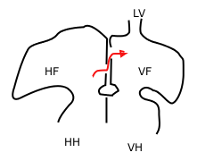
In the fetal heart, the foramen ovale, also foramen Botalli, or the ostium secundum of Born, allows blood to enter the left atrium from the right atrium. It is one of two fetal cardiac shunts, the other being the ductus arteriosus. Another similar adaptation in the fetus is the ductus venosus. In most individuals, the foramen ovale closes at birth. It later forms the fossa ovalis.

<span class="mw-page-title-main">Interventricular septum</span> Wall of tissue separating ventricles of human heart

The interventricular septum is the stout wall separating the ventricles, the lower chambers of the heart, from one another.

<span class="mw-page-title-main">Primitive atrium</span>

The primitive atrium is a stage in the embryonic development of the human heart. It grows rapidly and partially encircles the bulbus cordis; the groove against which the bulbus cordis lies is the first indication of a division into right and left atria.

<span class="mw-page-title-main">Septum primum</span>

During heart development of a human embryo, the single primitive atrium becomes divided into right and left by a septum, the septum primum. The septum primum grows downward into the single atrium.

<span class="mw-page-title-main">Septum secundum</span>

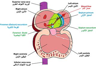
The septum secundum is a muscular flap that is important in heart development. It is semilunar in shape, and grows downward from the upper wall of the atrium immediately to the right of the septum primum and ostium secundum. It is important in the closure of the foramen ovale after birth.

<span class="mw-page-title-main">Foramen secundum</span>

The foramen secundum, or ostium secundum is a foramen in the septum primum, a precursor to the interatrial septum of the human heart.

<span class="mw-page-title-main">Primary interatrial foramen</span>

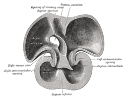
In the developing heart, the atria are initially open to each other, with the opening known as the primary interatrial foramen or ostium primum. The foramen lies beneath the edge of septum primum and the endocardial cushions. It progressively decreases in size as the septum grows downwards, and disappears with the formation of the atrial septum.

<span class="mw-page-title-main">Valve of inferior vena cava</span>

The valve of the inferior vena cava is a venous valve that lies at the junction of the inferior vena cava and right atrium.

The heart is the first functional organ in a vertebrate embryo. There are 5 stages to heart development.

A cardiac shunt is a pattern of blood flow in the heart that deviates from the normal circuit of the circulatory system. It may be described as right-left, left-right or bidirectional, or as systemic-to-pulmonary or pulmonary-to-systemic. The direction may be controlled by left and/or right heart pressure, a biological or artificial heart valve or both. The presence of a shunt may also affect left and/or right heart pressure either beneficially or detrimentally.

<span class="mw-page-title-main">Lutembacher's syndrome</span> Medical condition

Lutembacher's syndrome is a very rare form of congenital heart disease that affects one of the chambers of the heart as well as a valve. It is commonly known as both congenital atrial septal defect (ASD) and acquired mitral stenosis (MS). Congenital atrial septal defect refers to a hole being in the septum or wall that separates the two atria; this condition is usually seen in fetuses and infants. Mitral stenosis refers to mitral valve leaflets sticking to each other making the opening for blood to pass from the atrium to the ventricles very small. With the valve being so small, blood has difficulty passing from the left atrium into the left ventricle. Septal defects that may occur with Lutembacher's syndrome include: Ostium primum atrial septal defect or ostium secundum which is more prevalent.

<span class="mw-page-title-main">Heart development</span> Prenatal development of the heart

Heart development, also known as cardiogenesis, refers to the prenatal development of the heart. This begins with the formation of two endocardial tubes which merge to form the tubular heart, also called the primitive heart tube. The heart is the first functional organ in vertebrate embryos.

The heart is a muscular organ situated in the mediastinum. It consists of four chambers, four valves, two main arteries, and the conduction system. The left and right sides of the heart have different functions: the right side receives de-oxygenated blood through the superior and inferior venae cavae and pumps blood to the lungs through the pulmonary artery, and the left side receives saturated blood from the lungs.

References

  1. Naqvi N, McCarthy KP, Ho SY (September 2018). "Anatomy of the atrial septum and interatrial communications". Journal of Thoracic Disease. 10 (Suppl 24): S2837–S2847. doi: 10.21037/jtd.2018.02.18 . PMC   6174145 . PMID   30305943.
  2. Sinnatamby, Chummy S. (2011). Last's Anatomy (12th ed.). Elsevier Australia. p. 200. ISBN   978-0-7295-3752-0.
  3. 1 2 Diagram of Ostium Secundum Atrial Septal Defect at Mayo Clinic
  4. "ostium primum atrial septal defect" . GPnotebook.
  5. Kotaru VPK, Martin D, Tokala H, Kalavakunta JK. Right atrial mass-multimodality imaging—Massive lipomatous hypertrophy of the atrial septum. Clin Case Rep. 2020;00:1–2. https://doi.org/10.1002/ccr3.3431
  6. Laura, Diana M.; Donnino, Robert; Kim, Eugene E.; Benenstein, Ricardo; Freedberg, Robin S.; Saric, Muhamed (August 2016). "Lipomatous Atrial Septal Hypertrophy: A Review of Its Anatomy, Pathophysiology, Multimodality Imaging, and Relevance to Percutaneous Interventions". Journal of the American Society of Echocardiography. 29 (8): 717–723. doi:10.1016/j.echo.2016.04.014. PMID   27288088.