Intracellular transport

Last updated

Intracellular transport is the movement of vesicles and substances within a cell. Intracellular transport is required for maintaining homeostasis within the cell by responding to physiological signals. [1] Proteins synthesized in the cytosol are distributed to their respective organelles, according to their specific amino acid’s sorting sequence. [2] Eukaryotic cells transport packets of components to particular intracellular locations by attaching them to molecular motors that haul them along microtubules and actin filaments. Since intracellular transport heavily relies on microtubules for movement, the components of the cytoskeleton play a vital role in trafficking vesicles between organelles and the plasma membrane by providing mechanical support. Through this pathway, it is possible to facilitate the movement of essential molecules such as membrane‐bounded vesicles and organelles, mRNA, and chromosomes.

Contents

Intracellular transport between the Golgi apparatus and the endoplasmic reticulum Intracellular Signaling Clip Art.jpg
Intracellular transport between the Golgi apparatus and the endoplasmic reticulum

Intracellular transport is unique to eukaryotic cells because they possess organelles enclosed in membranes that need to be mediated for exchange of cargo to take place. [3] Conversely, in prokaryotic cells, there is no need for this specialized transport mechanism because there are no membranous organelles and compartments to traffic between. Prokaryotes are able to subsist by allowing materials to enter the cell via simple diffusion. Intracellular transport is more specialized than diffusion; it is a multifaceted process which utilizes transport vesicles. Transport vesicles are small structures within the cell consisting of a fluid enclosed by a lipid bilayer that hold cargo. These vesicles will typically execute cargo loading and vesicle budding, vesicle transport, the binding of the vesicle to a target membrane and the fusion of the vesicle membranes to target membrane. To ensure that these vesicles embark in the right direction and to further organize the cell, special motor proteins attach to cargo-filled vesicles and carry them along the cytoskeleton. For example, they have to ensure that lysosomal enzymes are transferred specifically to the golgi apparatus and not to another part of the cell which could lead to deleterious effects.

Fusion

Small membrane bound vesicles responsible for transporting proteins from one organelle to another are commonly found in endocytic and secretory pathways. Vesicles bud from their donor organelle and release the contents of their vesicle by a fusion event in a particular target organelle. [4] :634 The endoplasmic reticulum serves as a channel that proteins will pass through bound for their final destination. [3] Outbound proteins from the endoplasmic reticulum will bud off into transport vesicles that travel along the cell cortex to reach their specific destinations. [3] Since the ER is the site of protein synthesis, it would serve as the parent organelle, and the cis face of the golgi, where proteins and signals are received, would be the acceptor. In order for the transport vesicle to accurately undergo a fusion event, it must first recognize the correct target membrane then fuse with that membrane.

How SNARE proteins play a role in intracellular transport SNAREs.png
How SNARE proteins play a role in intracellular transport

Rab proteins on the surface of the transport vesicle are responsible for aligning with the complementary tethering proteins found on the respective organelle's cytosolic surface. [3] This fusion event allows for the delivery of the vesicles contents mediated by proteins such as SNARE proteins. SNAREs are small, tail-anchored proteins which are often post-translationally inserted into membranes that are responsible for the fusion event necessary for vesicles to transport between organelles in the cytosol. There are two forms of SNARES, the t-SNARE and v-SNARE, which fit together similar to a lock and key. The t-SNAREs function by binding to the membranes of the target organelles, while the v-SNAREs function by binding to the vesicle membranes.

Role of endocytosis

Intracellular transport is an overarching category of how cells obtain nutrients and signals. One very well understood form of intracellular transport is known as endocytosis. Endocytosis is defined as the uptake of material by the invagination of the plasma membrane. [4] More specifically, eukaryotic cells use endocytosis of the uptake of nutrients, down regulation of growth factor receptors’ and as a mass regulator of the signaling circuit. This method of transport is largely intercellular in lieu of uptake of large particles such as bacteria via phagocytosis in which a cell engulfs a solid particle to form an internal vesicle called a phagosome. However, many of these processes have an intracellular component. Phagocytosis is of great importance to intracellular transport because once a substance is deemed harmful and engulfed in a vesicle, it can be trafficked to the appropriate location for degradation. These endocytosed molecules are sorted into early endosomes within the cell, which serves to further sort these substances to the correct final destination (in the same way the Golgi does in the secretory pathway). From here, the early endosome starts a cascade of transport where the cargo is eventually hydrolyzed inside the lysosome for degradation. This capability is necessary for the degradation of any cargo that is harmful or unnecessary for the cell; this is commonly seen in response to foreign material. Phagocytosis has an immunologic function and role in apoptosis. Additionally, endocytosis can be observed through the nonspecific internalization of fluid droplets via pinocytosis and in receptor mediated endocytosis.

Role of microtubules

A cytoplasmic dynein motor bound to a microtubule. CytoplasmicDyneinOnMT noLabels.png
A cytoplasmic dynein motor bound to a microtubule.
A kinesin molecule bound to a microtubule. Kinesin cartoon.png
A kinesin molecule bound to a microtubule.

The transport mechanism depends on the material being moved. Intracellular transport that requires quick movement will use an actin-myosin mechanism while more specialized functions require microtubules for transport. [5] Microtubules function as tracks in the intracellular transport of membrane-bound vesicles and organelles. This process is propelled by motor proteins such as dynein. Motor proteins connect the transport vesicles to microtubules and actin filaments to facilitate intracellular movement. [1] Microtubules are organized so their plus ends extend through the periphery of the cells and their minus ends are anchored within the centrosome, so they utilize the motor proteins kinesin’s (positive end directed) and dynein’s (negative end directed) to transport vesicles and organelles in opposite directions through the cytoplasm. [6] Each type of membrane vesicle is specifically bound to its own kinesin motor protein via binding within the tail domain. One of the major roles of microtubules is to transport membrane vesicles and organelles through the cytoplasm of eukaryotic cells. It is speculated that areas within the cell considered "microtubule-poor" are probably transported along microfilaments aided by a myosin motor protein. In this manner, microtubules assist the transport of chromosomes towards the spindle poles by utilizing the dynein motor proteins during anaphase.

Diseases

By understanding the components and mechanisms of intracellular transport it is possible to see its implication in diseases. Defects encompass improper sorting of cargo into transport carriers, vesicle budding, issues in movement of vesicles along cytoskeletal tracks, and fusion at the target membrane. Since the life cycle of the cell is a highly regulated and important process, if any component goes awry there is the possibility for deleterious effects. If the cell is unable to correctly execute components of the intracellular pathway there is the impending possibility for protein aggregates to form. Growing evidence supports the concept that deficits in axonal transport contributes to pathogenesis in multiple neurodegenerative diseases. It is proposed that protein aggregations due to faulty transport is a leading cause of the development of ALS, Alzheimer’s and dementia. [7]

How microtubules play a role in intracellular transport Microtubules and intracellular transport.jpg
How microtubules play a role in intracellular transport

On the other hand, targeting the intracellular transport processes of these motor proteins constitutes the possibility for pharmacological targeting of drugs. By understanding the method in which substances move along neurons or microtubules it is possible to target specific pathways for disease. Currently, many drug companies are aiming to utilize the trajectory of intracellular transport mechanisms to deliver drugs to localized regions and target cells without harming healthy neighboring cells. The potential for this type of treatment in anti-cancer drugs is an exciting, promising area of research.

See also

Related Research Articles

<span class="mw-page-title-main">Endomembrane system</span> Membranes in the cytoplasm of a eukaryotic cell

The endomembrane system is composed of the different membranes (endomembranes) that are suspended in the cytoplasm within a eukaryotic cell. These membranes divide the cell into functional and structural compartments, or organelles. In eukaryotes the organelles of the endomembrane system include: the nuclear membrane, the endoplasmic reticulum, the Golgi apparatus, lysosomes, vesicles, endosomes, and plasma (cell) membrane among others. The system is defined more accurately as the set of membranes that forms a single functional and developmental unit, either being connected directly, or exchanging material through vesicle transport. Importantly, the endomembrane system does not include the membranes of plastids or mitochondria, but might have evolved partially from the actions of the latter.

<span class="mw-page-title-main">Endocytosis</span> Cellular process

Endocytosis is a cellular process in which substances are brought into the cell. The material to be internalized is surrounded by an area of cell membrane, which then buds off inside the cell to form a vesicle containing the ingested material. Endocytosis includes pinocytosis and phagocytosis. It is a form of active transport.

<span class="mw-page-title-main">Golgi apparatus</span> Cell organelle that packages proteins for export

The Golgi apparatus, also known as the Golgi complex, Golgi body, or simply the Golgi, is an organelle found in most eukaryotic cells. Part of the endomembrane system in the cytoplasm, it packages proteins into membrane-bound vesicles inside the cell before the vesicles are sent to their destination. It resides at the intersection of the secretory, lysosomal, and endocytic pathways. It is of particular importance in processing proteins for secretion, containing a set of glycosylation enzymes that attach various sugar monomers to proteins as the proteins move through the apparatus.

<span class="mw-page-title-main">Microtubule</span> Polymer of tubulin that forms part of the cytoskeleton

Microtubules are polymers of tubulin that form part of the cytoskeleton and provide structure and shape to eukaryotic cells. Microtubules can be as long as 50 micrometres, as wide as 23 to 27 nm and have an inner diameter between 11 and 15 nm. They are formed by the polymerization of a dimer of two globular proteins, alpha and beta tubulin into protofilaments that can then associate laterally to form a hollow tube, the microtubule. The most common form of a microtubule consists of 13 protofilaments in the tubular arrangement.

<span class="mw-page-title-main">Vesicle (biology and chemistry)</span> Any small, fluid-filled, spherical organelle enclosed by a membrane

In cell biology, a vesicle is a structure within or outside a cell, consisting of liquid or cytoplasm enclosed by a lipid bilayer. Vesicles form naturally during the processes of secretion (exocytosis), uptake (endocytosis), and the transport of materials within the plasma membrane. Alternatively, they may be prepared artificially, in which case they are called liposomes. If there is only one phospholipid bilayer, the vesicles are called unilamellar liposomes; otherwise they are called multilamellar liposomes. The membrane enclosing the vesicle is also a lamellar phase, similar to that of the plasma membrane, and intracellular vesicles can fuse with the plasma membrane to release their contents outside the cell. Vesicles can also fuse with other organelles within the cell. A vesicle released from the cell is known as an extracellular vesicle.

<span class="mw-page-title-main">Exocytosis</span> Active transport and bulk transport in which a cell transports molecules out of the cell

Exocytosis is a form of active transport and bulk transport in which a cell transports molecules out of the cell. As an active transport mechanism, exocytosis requires the use of energy to transport material. Exocytosis and its counterpart, endocytosis, are used by all cells because most chemical substances important to them are large polar molecules that cannot pass through the hydrophobic portion of the cell membrane by passive means. Exocytosis is the process by which a large amount of molecules are released; thus it is a form of bulk transport. Exocytosis occurs via secretory portals at the cell plasma membrane called porosomes. Porosomes are permanent cup-shaped lipoprotein structure at the cell plasma membrane, where secretory vesicles transiently dock and fuse to release intra-vesicular contents from the cell.

<span class="mw-page-title-main">Cytoskeleton</span> Network of filamentous proteins that forms the internal framework of cells

The cytoskeleton is a complex, dynamic network of interlinking protein filaments present in the cytoplasm of all cells, including those of bacteria and archaea. In eukaryotes, it extends from the cell nucleus to the cell membrane and is composed of similar proteins in the various organisms. It is composed of three main components, microfilaments, intermediate filaments and microtubules, and these are all capable of rapid growth or disassembly dependent on the cell's requirements.

<span class="mw-page-title-main">Cytokinesis</span> Part of the cell division process

Cytokinesis is the part of the cell division process during which the cytoplasm of a single eukaryotic cell divides into two daughter cells. Cytoplasmic division begins during or after the late stages of nuclear division in mitosis and meiosis. During cytokinesis the spindle apparatus partitions and transports duplicated chromatids into the cytoplasm of the separating daughter cells. It thereby ensures that chromosome number and complement are maintained from one generation to the next and that, except in special cases, the daughter cells will be functional copies of the parent cell. After the completion of the telophase and cytokinesis, each daughter cell enters the interphase of the cell cycle.

<span class="mw-page-title-main">Endosome</span> Vacuole to which materials ingested by endocytosis are delivered

Endosomes are a collection of intracellular sorting organelles in eukaryotic cells. They are parts of endocytic membrane transport pathway originating from the trans Golgi network. Molecules or ligands internalized from the plasma membrane can follow this pathway all the way to lysosomes for degradation or can be recycled back to the cell membrane in the endocytic cycle. Molecules are also transported to endosomes from the trans Golgi network and either continue to lysosomes or recycle back to the Golgi apparatus.

<span class="mw-page-title-main">Kinesin</span> Eukaryotic motor protein

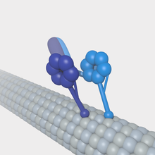
A kinesin is a protein belonging to a class of motor proteins found in eukaryotic cells. Kinesins move along microtubule (MT) filaments and are powered by the hydrolysis of adenosine triphosphate (ATP). The active movement of kinesins supports several cellular functions including mitosis, meiosis and transport of cellular cargo, such as in axonal transport, and intraflagellar transport. Most kinesins walk towards the plus end of a microtubule, which, in most cells, entails transporting cargo such as protein and membrane components from the center of the cell towards the periphery. This form of transport is known as anterograde transport. In contrast, dyneins are motor proteins that move toward the minus end of a microtubule in retrograde transport.

<span class="mw-page-title-main">Dynein</span> Class of enzymes

Dyneins are a family of cytoskeletal motor proteins that move along microtubules in cells. They convert the chemical energy stored in ATP to mechanical work. Dynein transports various cellular cargos, provides forces and displacements important in mitosis, and drives the beat of eukaryotic cilia and flagella. All of these functions rely on dynein's ability to move towards the minus-end of the microtubules, known as retrograde transport; thus, they are called "minus-end directed motors". In contrast, most kinesin motor proteins move toward the microtubules' plus-end, in what is called anterograde transport.

<span class="mw-page-title-main">Phagosome</span>

In cell biology, a phagosome is a vesicle formed around a particle engulfed by a phagocyte via phagocytosis. Professional phagocytes include macrophages, neutrophils, and dendritic cells (DCs).

<span class="mw-page-title-main">Molecular motor</span> Biological molecular machines

Molecular motors are natural (biological) or artificial molecular machines that are the essential agents of movement in living organisms. In general terms, a motor is a device that consumes energy in one form and converts it into motion or mechanical work; for example, many protein-based molecular motors harness the chemical free energy released by the hydrolysis of ATP in order to perform mechanical work. In terms of energetic efficiency, this type of motor can be superior to currently available man-made motors. One important difference between molecular motors and macroscopic motors is that molecular motors operate in the thermal bath, an environment in which the fluctuations due to thermal noise are significant.

<span class="mw-page-title-main">Axonal transport</span> Movement of organelles

Axonal transport, also called axoplasmic transport or axoplasmic flow, is a cellular process responsible for movement of mitochondria, lipids, synaptic vesicles, proteins, and other organelles to and from a neuron's cell body, through the cytoplasm of its axon called the axoplasm. Since some axons are on the order of meters long, neurons cannot rely on diffusion to carry products of the nucleus and organelles to the end of their axons. Axonal transport is also responsible for moving molecules destined for degradation from the axon back to the cell body, where they are broken down by lysosomes.

<span class="mw-page-title-main">Motor protein</span> Class of molecular proteins

Motor proteins are a class of molecular motors that can move along the cytoplasm of cells. They convert chemical energy into mechanical work by the hydrolysis of ATP. Flagellar rotation, however, is powered by a proton pump.

<span class="mw-page-title-main">Dynactin</span>

Dynactin is a 23 subunit protein complex that acts as a co-factor for the microtubule motor cytoplasmic dynein-1. It is built around a short filament of actin related protein-1 (Arp1).

<span class="mw-page-title-main">Cell membrane</span> Biological membrane that separates the interior of a cell from its outside environment

The cell membrane is a biological membrane that separates and protects the interior of a cell from the outside environment. The cell membrane consists of a lipid bilayer, made up of two layers of phospholipids with cholesterols interspersed between them, maintaining appropriate membrane fluidity at various temperatures. The membrane also contains membrane proteins, including integral proteins that span the membrane and serve as membrane transporters, and peripheral proteins that loosely attach to the outer (peripheral) side of the cell membrane, acting as enzymes to facilitate interaction with the cell's environment. Glycolipids embedded in the outer lipid layer serve a similar purpose. The cell membrane controls the movement of substances in and out of a cell, being selectively permeable to ions and organic molecules. In addition, cell membranes are involved in a variety of cellular processes such as cell adhesion, ion conductivity, and cell signalling and serve as the attachment surface for several extracellular structures, including the cell wall and the carbohydrate layer called the glycocalyx, as well as the intracellular network of protein fibers called the cytoskeleton. In the field of synthetic biology, cell membranes can be artificially reassembled.

Membrane vesicle trafficking in eukaryotic animal cells involves movement of biochemical signal molecules from synthesis-and-packaging locations in the Golgi body to specific release locations on the inside of the plasma membrane of the secretory cell. It takes place in the form of Golgi membrane-bound micro-sized vesicles, termed membrane vesicles (MVs).

<span class="mw-page-title-main">Anna Akhmanova</span> Russian cell biologist

Anna Sergeevna Akhmanova is a Russian-born professor of Cell Biology at Utrecht University in the Netherlands. She is best known for her research regarding microtubules and the proteins, called TIPs, that stabilize one specific end of the tubules. Among the awards she has won, she was one of the recipients of the 2018 Spinoza Prize, the highest honor for Dutch scientists.

<span class="mw-page-title-main">Neurotubule</span>

Neurotubules are microtubules found in neurons in nervous tissues. Along with neurofilaments and microfilaments, they form the cytoskeleton of neurons. Neurotubules are undivided hollow cylinders that are made up of tubulin protein polymers and arrays parallel to the plasma membrane in neurons. Neurotubules have an outer diameter of about 23 nm and an inner diameter, also known as the central core, of about 12 nm. The wall of the neurotubules is about 5 nm in width. There is a non-opaque clear zone surrounding the neurotubule and it is about 40 nm in diameter. Like microtubules, neurotubules are greatly dynamic and the length of them can be adjusted by polymerization and depolymerization of tubulin.

References

  1. 1 2 Barlan K, Gelfand VI (May 2017). "Microtubule-Based Transport and the Distribution, Tethering, and Organization of Organelles". Cold Spring Harbor Perspectives in Biology. 9 (5): a025817. doi:10.1101/cshperspect.a025817. PMC   5411697 . PMID   28461574.
  2. Mellman I, Nelson WJ (November 2008). "Coordinated protein sorting, targeting and distribution in polarized cells". Nature Reviews. Molecular Cell Biology. 9 (11): 833–45. doi:10.1038/nrm2525. PMC   3369829 . PMID   18946473.
  3. 1 2 3 4 Alberts, Bruce (November 2018). Essential cell biology (Fifth ed.). New York. ISBN   978-0-393-67953-3. OCLC   1048014962.{{cite book}}: CS1 maint: location missing publisher (link)
  4. 1 2 Lodish HF, Berk A, Zipursky SL, Matsudaira P, Baltimore D, Darnell J (2000). Molecular cell biology (4th ed.). New York: W.H. Freeman. ISBN   978-0-7167-3136-8.
  5. Geitmann A, Nebenführ A (October 2015). Kozminski KG (ed.). "Navigating the plant cell: intracellular transport logistics in the green kingdom". Molecular Biology of the Cell. 26 (19): 3373–8. doi:10.1091/mbc.E14-10-1482. PMC   4591683 . PMID   26416952.
  6. The Cell: A Molecular Approach.
  7. Chevalier-Larsen E, Holzbaur EL (2006). "Axonal transport and neurodegenerative disease". Biochimica et Biophysica Acta (BBA) - Molecular Basis of Disease. 1762 (11–12): 1094–108. doi:10.1016/j.bbadis.2006.04.002. PMID   16730956.