L-form bacteria

Last updated
Transmission electron micrograph of L-form Bacillus subtilis. The cells lack the electron-dense cell wall of normal bacteria. Scale bar is 500 nanometers. TEM of L-form bacteria-Mark Leaver Newcastle University.jpeg
Transmission electron micrograph of L-form Bacillus subtilis . The cells lack the electron-dense cell wall of normal bacteria. Scale bar is 500 nanometers.

L-form bacteria, also known as L-phase bacteria, L-phase variants or cell wall-deficient bacteria (CWDB), are growth forms derived from different bacteria. They lack cell walls. [1] Two types of L-forms are distinguished: unstable L-forms, spheroplasts that are capable of dividing, but can revert to the original morphology, and stable L-forms, L-forms that are unable to revert to the original bacteria.

Contents

Discovery and early studies

L-form bacteria were first isolated in 1935 by Emmy Klieneberger-Nobel, who named them "L-forms" after the Lister Institute in London where she was working. [2]

She first interpreted these growth forms as symbionts related to pleuropneumonia-like organisms (PPLOs, later commonly called mycoplasmas). [3] Mycoplasmas (now in scientific classification called Mollicutes ), parasitic or saprotrophic species of bacteria, also lack a cell wall (peptidoglycan/murein is absent). [4] [5] Morphologically, they resemble L-form bacteria. Therefore, mycoplasmas formerly were sometimes considered stable L-forms or, because of their small size, even viruses, but phylogenetic analysis has identified them as bacteria that have lost their cell walls in the course of evolution. [6] Both, mycoplasmas and L-form bacteria are resistant against penicillin.

After the discovery of PPLOs (mycoplasmas/Mollicutes) and L-form bacteria, their mode of reproduction (proliferation) became a major subject of discussion. In 1954, using phase-contrast microscopy, continual observations of live cells have shown that L-form bacteria (previously also called L-phase bacteria) and pleuropneumonia-like organisms (PPLOs, now mycoplasmas/Mollicutes) ) do not proliferate by binary fission, but by a uni- or multi-polar budding mechanism. Microphotograph series of growing microcultures of different strains of L-form bacteria, PPLOs and, as a control, a Micrococcus species (dividing by binary fission) have been presented. [3] Additionally, electron microscopic studies have been performed. [7]

Appearance and cell division

Transmission electron micrograph of a population of L-form Bacillus subtilis, showing a range of sizes. Scale bar is 10 micrometers. Wide field EM of L-form bacteria-Mark Leaver Newcastle University.jpeg
Transmission electron micrograph of a population of L-form Bacillus subtilis, showing a range of sizes. Scale bar is 10 micrometers.

Bacterial morphology is determined by the cell wall. Since the L-form has no cell wall, its morphology is different from that of the strain of bacteria from which it is derived. Typical L-form cells are spheres or spheroids. For example, L-forms of the rod-shaped bacterium Bacillus subtilis appear round when viewed by phase contrast microscopy or by transmission electron microscopy. [8]

Although L-forms can develop from Gram-positive as well as from Gram-negative bacteria, in a Gram stain test, the L-forms always colour Gram-negative, due to the lack of a cell wall.

The cell wall is important for cell division, which, in most bacteria, occurs by binary fission. This process usually requires a cell wall and components of the bacterial cytoskeleton such as FtsZ. The ability of L-form bacteria and mycoplasmas to grow and divide in the absence of both of these structures is highly unusual, and may represent a form of cell division that was important in early forms of life. This mode of division seems to involve the extension of thin protrusions from the cell's surface and these protrusions then pinching off to form new cells. The lack of cell wall in L-forms means that division is disorganised, giving rise to a variety of cell sizes, from very tiny to very big. [1]

Phase contrast image of L-form cells from Bacillus subtilis showing a range of sizes. Scale bar is 5 micrometers. Phase contrast image of L-form bacteria-Mark Leaver Newcastle University.jpeg
Phase contrast image of L-form cells from Bacillus subtilis showing a range of sizes. Scale bar is 5 micrometers.

Generation in cultures

L-forms can be generated in the laboratory from many bacterial species that usually have cell walls, such as Bacillus subtilis or Escherichia coli . This is done by inhibiting peptidoglycan synthesis with antibiotics or treating the cells with lysozyme, an enzyme that digests cell walls. The L-forms are generated in a culture medium that is the same osmolarity as the bacterial cytosol (an isotonic solution), which prevents cell lysis by osmotic shock. [2] L-form strains can be unstable, tending to revert to the normal form of the bacteria by regrowing a cell wall, but this can be prevented by long-term culture of the cells under the same conditions that were used to produce them letting the wall-disabling mutations to accumulate by genetic drift. [9]

Some studies have identified mutations that occur, as these strains are derived from normal bacteria. [1] [2] One such point mutation D92E is in an enzyme yqiD/ispA ( P54383 ) involved in the mevalonate pathway of lipid metabolism that increased the frequency of L-form formation 1,000-fold. [1] The reason for this effect is not known, but it is presumed that the increase is related to this enzyme's role in making a lipid important in peptidoglycan synthesis.

Another methodology of induction relies on nanotechnology and landscape ecology. Microfluidics devices can be built in order to challenge peptidoglycan synthesis by extreme spatial confinement. After biological dispersal through a constricted (sub-micrometre scale) biological corridor connecting adjacent micro habitat patches, L-form-like cells can be derived [10] using a microfluifics-based (synthetic) ecosystem implementing an adaptive landscape [11] selecting for shape-shifting phenotypes similar to L-forms.

Significance and applications

Some publications have suggested that L-form bacteria might cause diseases in humans, [12] and other animals [13] but, as the evidence that links these organisms to disease is fragmentary and frequently contradictory, this hypothesis remains controversial. [14] [15] The two extreme viewpoints on this question are that L-form bacteria are either laboratory curiosities of no clinical significance or important but unappreciated causes of disease. [5] Research on L-form bacteria is continuing. For example, L-form organisms have been observed in mouse lungs after experimental inoculation with Nocardia caviae , [16] [17] and a recent study suggested that these organisms may infect immunosuppressed patients having undergone bone marrow transplants. [18] The formation of strains of bacteria lacking cell walls has also been proposed to be important in the acquisition of bacterial antibiotic resistance. [19] [20]

L-form bacteria may be useful in research on early forms of life, and in biotechnology. These strains are being examined for possible uses in biotechnology as host strains for recombinant protein production. [21] [22] [23] Here, the absence of a cell wall can allow production of large amounts of secreted proteins that would otherwise accumulate in the periplasmic space of bacteria. [24] [25]

L-form bacteria are seen as a persister cells, and a source of recurrent infection that has become of medical interest. [26]

See also

Related Research Articles

<i>Bacillus</i> Genus of bacteria

Bacillus is a genus of Gram-positive, rod-shaped bacteria, a member of the phylum Bacillota, with 266 named species. The term is also used to describe the shape (rod) of other so-shaped bacteria; and the plural Bacilli is the name of the class of bacteria to which this genus belongs. Bacillus species can be either obligate aerobes which are dependent on oxygen, or facultative anaerobes which can survive in the absence of oxygen. Cultured Bacillus species test positive for the enzyme catalase if oxygen has been used or is present.

<span class="mw-page-title-main">Gram stain</span> Investigative procedure in microbiology

Gram stain, is a method of staining used to classify bacterial species into two large groups: gram-positive bacteria and gram-negative bacteria. It may also be used to diagnose a fungal infection. The name comes from the Danish bacteriologist Hans Christian Gram, who developed the technique in 1884.

<span class="mw-page-title-main">Gram-positive bacteria</span> Bacteria that give a positive result in the Gram stain test

In bacteriology, gram-positive bacteria are bacteria that give a positive result in the Gram stain test, which is traditionally used to quickly classify bacteria into two broad categories according to their type of cell wall.

Peptidoglycan or murein is a unique large macromolecule, a polysaccharide, consisting of sugars and amino acids that forms a mesh-like peptidoglycan layer (sacculus) that surrounds the bacterial cytoplasmic membrane. The sugar component consists of alternating residues of β-(1,4) linked N-acetylglucosamine (NAG) and N-acetylmuramic acid (NAM). Attached to the N-acetylmuramic acid is an oligopeptide chain made of three to five amino acids. The peptide chain can be cross-linked to the peptide chain of another strand forming the 3D mesh-like layer. Peptidoglycan serves a structural role in the bacterial cell wall, giving structural strength, as well as counteracting the osmotic pressure of the cytoplasm. This repetitive linking results in a dense peptidoglycan layer which is critical for maintaining cell form and withstanding high osmotic pressures, and it is regularly replaced by peptidoglycan production. Peptidoglycan hydrolysis and synthesis are two processes that must occur in order for cells to grow and multiply, a technique carried out in three stages: clipping of current material, insertion of new material, and re-crosslinking of existing material to new material.

Autolysins are endogenous lytic enzymes that break down the peptidoglycan components of biological cells which enables the separation of daughter cells following cell division. They are involved in cell growth, cell wall metabolism, cell division and separation, as well as peptidoglycan turnover and have similar functions to lysozymes.

<i>Mycoplasma</i> Genus of bacteria

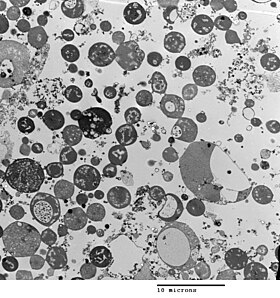
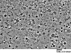
Mycoplasma is a genus of bacteria that, like the other members of the class Mollicutes, lack a cell wall around their cell membranes. Peptidoglycan (murein) is absent. This characteristic makes them naturally resistant to antibiotics that target cell wall synthesis. They can be parasitic or saprotrophic. Several species are pathogenic in humans, including M. pneumoniae, which is an important cause of "walking" pneumonia and other respiratory disorders, and M. genitalium, which is believed to be involved in pelvic inflammatory diseases. Mycoplasma species are among the smallest organisms yet discovered, can survive without oxygen, and come in various shapes. For example, M. genitalium is flask-shaped, while M. pneumoniae is more elongated, many Mycoplasma species are coccoid. Hundreds of Mycoplasma species infect animals.

Mycoplasma pneumoniae is a very small bacterium in the class Mollicutes. It is a human pathogen that causes the disease mycoplasma pneumonia, a form of atypical bacterial pneumonia related to cold agglutinin disease. M. pneumoniae is characterized by the absence of a peptidoglycan cell wall and resulting resistance to many antibacterial agents. The persistence of M. pneumoniae infections even after treatment is associated with its ability to mimic host cell surface composition.

<span class="mw-page-title-main">FtsZ</span> Protein encoded by the ftsZ gene

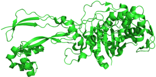
FtsZ is a protein encoded by the ftsZ gene that assembles into a ring at the future site of bacterial cell division. FtsZ is a prokaryotic homologue of the eukaryotic protein tubulin. The initials FtsZ mean "Filamenting temperature-sensitive mutant Z." The hypothesis was that cell division mutants of E. coli would grow as filaments due to the inability of the daughter cells to separate from one another. FtsZ is found in almost all bacteria, many archaea, all chloroplasts and some mitochondria, where it is essential for cell division. FtsZ assembles the cytoskeletal scaffold of the Z ring that, along with additional proteins, constricts to divide the cell in two.

The periplasm is a concentrated gel-like matrix in the space between the inner cytoplasmic membrane and the bacterial outer membrane called the periplasmic space in gram-negative bacteria. Using cryo-electron microscopy it has been found that a much smaller periplasmic space is also present in gram-positive bacteria, between cell wall and the plasma membrane. The periplasm may constitute up to 40% of the total cell volume of gram-negative bacteria, but is a much smaller percentage in gram-positive bacteria.

The cell envelope comprises the inner cell membrane and the cell wall of a bacterium. In gram-negative bacteria an outer membrane is also included. This envelope is not present in the Mollicutes where the cell wall is absent.

<span class="mw-page-title-main">Mesosome</span> Mesosome

Mesosomes or chondrioids are folded invaginations in the plasma membrane of bacteria that are produced by the chemical fixation techniques used to prepare samples for electron microscopy. Although several functions were proposed for these structures in the 1960s, they were recognized as artifacts by the late 1970s and are no longer considered to be part of the normal structure of bacterial cells. These extensions are in the form of vesicles, tubules and lamellae.

<i>Bacillus subtilis</i> Catalase-positive bacterium

Bacillus subtilis, known also as the hay bacillus or grass bacillus, is a Gram-positive, catalase-positive bacterium, found in soil and the gastrointestinal tract of ruminants, humans and marine sponges. As a member of the genus Bacillus, B. subtilis is rod-shaped, and can form a tough, protective endospore, allowing it to tolerate extreme environmental conditions. B. subtilis has historically been classified as an obligate aerobe, though evidence exists that it is a facultative anaerobe. B. subtilis is considered the best studied Gram-positive bacterium and a model organism to study bacterial chromosome replication and cell differentiation. It is one of the bacterial champions in secreted enzyme production and used on an industrial scale by biotechnology companies.

Mollicutes is a class of bacteria distinguished by the absence of a cell wall. The word "Mollicutes" is derived from the Latin mollis, and cutis. Individuals are very small, typically only 0.2–0.3 μm in size and have a very small genome size. They vary in form, although most have sterols that make the cell membrane somewhat more rigid. Many are able to move about through gliding, but members of the genus Spiroplasma are helical and move by twisting. The best-known genus in the Mollicutes is Mycoplasma. Colonies show the typical "fried-egg" appearance.

<span class="mw-page-title-main">Penicillin-binding proteins</span> Class of proteins

Penicillin-binding proteins (PBPs) are a group of proteins that are characterized by their affinity for and binding of penicillin. They are a normal constituent of many bacteria; the name just reflects the way by which the protein was discovered. All β-lactam antibiotics bind to PBPs, which are essential for bacterial cell wall synthesis. PBPs are members of a subgroup of enzymes called transpeptidases. Specifically, PBPs are DD-transpeptidases.

The bacterium, despite its simplicity, contains a well-developed cell structure which is responsible for some of its unique biological structures and pathogenicity. Many structural features are unique to bacteria and are not found among archaea or eukaryotes. Because of the simplicity of bacteria relative to larger organisms and the ease with which they can be manipulated experimentally, the cell structure of bacteria has been well studied, revealing many biochemical principles that have been subsequently applied to other organisms.

Emmy Klieneberger-Nobel was a German Jewish microbiologist and a founder of mycoplasma bacterial research. She performed most of her research at the Lister Institute in London, England, after having been expelled from Germany by the Nazis.

<span class="mw-page-title-main">Otto Kandler</span>

Otto Kandler was a German botanist and microbiologist. Until his retirement in 1986 he was professor of botany at the Ludwig Maximilian University of Munich.

Mycoplasma fermentans is a very small bacterium in the class Mollicutes. Like other mycoplasmas M. fermentans is characterized by the absence of a peptidoglycan cell wall and resulting resistance to many antibacterial agents. It is a possible human pathogen with roles suggested in many illness such as respiratory, genital, and rheumatoid diseases among others. Investigations have focused on a possible link to it being a cofactor in HIV infection as well as fibromyalgia, Gulf War syndrome and Chronic fatigue syndrome, however the belief that M. fermentans is pathogenic in such conditions has largely been disregarded after the failure of several large scale studies to find a link. Due to its incredibly small size it is difficult to determine the full extent of its role in human diseases, while M. fermentans has been implicated in a myriad of diseases, research at the current point has not conclusively proven its pathogenicity in humans outside of opportunistic infections.

<span class="mw-page-title-main">Cyclic di-AMP</span> Chemical compound

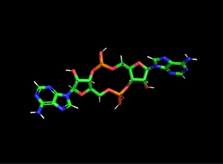
Cyclic di-AMP is a second messenger used in signal transduction in bacteria and archaea. It is present in many Gram-positive bacteria, some Gram-negative species, and archaea of the phylum euryarchaeota.

Mycoplasma orale is a small bacterium found in the class Mollicutes. It belongs to the genus Mycoplasma, a well-known group of bacterial parasites that inhabit humans. It also is known to be an opportunistic pathogen in immunocompromised humans. As with other Mycoplasma species, M. orale is not readily treated with many antibiotics due to its lack of a peptidoglycan cell wall. Therefore, this species is relevant to the medical field as physicians face the task of treating patients infected with this microbe. It is characterized by a small physical size, a small genome size, and a limited metabolism. It is also known to frequently contaminate laboratory experiments. This bacteria is very similar physiologically and morphologically to its sister species within the genus Mycoplasma; however, its recent discovery leaves many questions still unanswered about this microbe.

References

  1. 1 2 3 4 Leaver M, Domínguez-Cuevas P, Coxhead JM, Daniel RA, Errington J (February 2009). "Life without a wall or division machine in Bacillus subtilis". Nature. 457 (7231): 849–53. Bibcode:2009Natur.457..849L. doi:10.1038/nature07742. PMID   19212404. S2CID   4413852.
  2. 1 2 3 Joseleau-Petit D, Liébart JC, Ayala JA, D'Ari R (September 2007). "Unstable Escherichia coli L Forms Revisited: Growth Requires Peptidoglycan Synthesis". J. Bacteriol. 189 (18): 6512–20. doi:10.1128/JB.00273-07. PMC   2045188 . PMID   17586646.
  3. 1 2 Kandler, Gertraud; Kandler, Otto (1954). (Article in English available). "Untersuchungen über die Morphologie und die Vermehrung der pleuropneumonie-ähnlichen Organismen und der L-Phase der Bakterien. I. Lichtmikroskopische Untersuchungen" [Studies on morphology and multiplication (proliferation) of pleuropneumonia-like organisms and on bacterial L-phase, I. Light microscopy (now mycoplasmas and L-form bacteria)](PDF). Archiv für Mikrobiologie (in German). 21 (2): 178–201. doi:10.1007/BF01816378. PMID   14350641. S2CID   21257985.
  4. Razin S, Yogev D, Naot Y (December 1998). "Molecular Biology and Pathogenicity of Mycoplasmas". Microbiol. Mol. Biol. Rev. 62 (4): 1094–156. doi:10.1128/MMBR.62.4.1094-1156.1998. PMC   98941 . PMID   9841667.
  5. 1 2 Domingue GJ, Woody HB (April 1997). "Bacterial persistence and expression of disease". Clin. Microbiol. Rev. 10 (2): 320–44. doi:10.1128/CMR.10.2.320. PMC   172922 . PMID   9105757.  Full PDF
  6. Woese, Carl R.; Maniloff, J.; Zablen, L. B. (1980). "Phylogenetic analysis of the mycoplasmas" (PDF). Proceedings of the National Academy of Sciences of the United States of America. 77 (1): 494–498. Bibcode:1980PNAS...77..494W. doi: 10.1073/pnas.77.1.494 . PMC   348298 . PMID   6928642.
  7. Kandler, Gertraud; Kandler, Otto; Huber, Oskar (1954). (Article in English available). "Untersuchungen über die Morphologie und die Vermehrung der pleuropneumonie-ähnlichen Organismen und der L-Phase der Bakterien. II. Elektronenmikroskopische Untersuchungen" [Studies on morphology and multiplication (proliferation) of pleuropneumonia-like organisms and on bacterial L-phase, II. Electron microscopy (now mycoplasmas and L-form bacteria)](PDF). Archiv für Mikrobiologie (in German). 21 (2): 202–216. doi:10.1007/BF01816379. PMID   1435064. S2CID   45546531.
  8. Gilpin RW, Young FE, Chatterjee AN (January 1973). "Characterization of a Stable L-Form of Bacillus subtilis 168". J. Bacteriol. 113 (1): 486–99. doi:10.1128/JB.113.1.486-499.1973. PMC   251652 . PMID   4631836.
  9. Allan EJ (April 1991). "Induction and cultivation of a stable L-form of Bacillus subtilis". Journal of Applied Bacteriology. 70 (4): 339–43. doi:10.1111/j.1365-2672.1991.tb02946.x. PMID   1905284.
  10. Männik J.; R. Driessen; P. Galajda; J.E. Keymer; C. Dekker (September 2009). "Bacterial growth and motility in sub-micron constrictions". PNAS. 106 (35): 14861–14866. Bibcode:2009PNAS..10614861M. doi: 10.1073/pnas.0907542106 . PMC   2729279 . PMID   19706420.
  11. Keymer J.E.; P. Galajda; C. Muldoon R.; R. Austin (November 2006). "Bacterial metapopulations in nanofabricated landscapes". PNAS. 103 (46): 17290–295. Bibcode:2006PNAS..10317290K. doi: 10.1073/pnas.0607971103 . PMC   1635019 . PMID   17090676.
  12. Wall S, Kunze ZM, Saboor S, Soufleri I, Seechurn P, Chiodini R, McFadden JJ (1993). "Identification of spheroplast-like agents isolated from tissues of patients with Crohn's disease and control tissues by polymerase chain reaction". J. Clin. Microbiol. 31 (5): 1241–5. doi:10.1128/JCM.31.5.1241-1245.1993. PMC   262911 . PMID   8501224.
  13. Hulten K, Karttunen TJ, El-Zimaity HM, Naser SA, Collins MT, Graham DY, El-Zaatari FA (2000). "Identification of cell wall deficient forms of M. avium subsp. paratuberculosis in paraffin embedded tissues from animals with Johne's disease by in situ hybridization". J. Microbiol. Methods. 42 (2): 185–95. doi:10.1016/S0167-7012(00)00185-8. PMID   11018275.
  14. Onwuamaegbu ME, Belcher RA, Soare C (2005). "Cell wall-deficient bacteria as a cause of infections: a review of the clinical significance" (PDF). J. Int. Med. Res. 33 (1): 1–20. doi:10.1177/147323000503300101. PMID   15651712. S2CID   24781904. Archived from the original (PDF) on 24 August 2009.
  15. Casadesús J (December 2007). "Bacterial L-forms require peptidoglycan synthesis for cell division". BioEssays. 29 (12): 1189–91. doi:10.1002/bies.20680. PMID   18008373. S2CID   9863534.
  16. Beaman BL (July 1980). "Induction of L-phase variants of Nocardia caviae within intact murine lungs". Infect. Immun. 29 (1): 244–51. doi:10.1128/IAI.29.1.244-251.1980. PMC   551102 . PMID   7399704.
  17. Beaman BL, Scates SM (September 1981). "Role of L-forms of Nocardia caviae in the development of chronic mycetomas in normal and immunodeficient murine models". Infect. Immun. 33 (3): 893–907. doi:10.1128/IAI.33.3.893-907.1981. PMC   350795 . PMID   7287189.
  18. Woo PC, Wong SS, Lum PN, Hui WT, Yuen KY (March 2001). "Cell-wall-deficient bacteria and culture-negative febrile episodes in bone-marrow-transplant recipients". Lancet. 357 (9257): 675–9. doi:10.1016/S0140-6736(00)04131-3. PMID   11247551. S2CID   1295920.
  19. Fuller E, Elmer C, Nattress F, et al. (December 2005). "β-Lactam Resistance in Staphylococcus aureus Cells That Do Not Require a Cell Wall for Integrity". Antimicrob. Agents Chemother. 49 (12): 5075–80. doi:10.1128/AAC.49.12.5075-5080.2005. PMC   1315936 . PMID   16304175.
  20. Mickiewicz, Katarzyna M.; Kawai, Yoshikazu; Drage, Lauren; Gomes, Margarida C.; Davison, Frances; Pickard, Robert; Hall, Judith; Mostowy, Serge; Aldridge, Phillip D.; Errington, Jeff (2019). "Possible role of L-form switching in recurrent urinary tract infection". Nature Communications. 10 (1): 4379. Bibcode:2019NatCo..10.4379M. doi:10.1038/s41467-019-12359-3. PMC   6763468 . PMID   31558767.
  21. Sieben, Stefan (April 1998). "Die stabilen Protoplasten-Typ L-Formen von Proteus mirabilis als neues Expressionssystem für sekretorische Proteine und integrale Mempranproteine". Dissertation Universität Jena. OCLC   246350676.
  22. Sieben S, Hertle R, Gumpert J, Braun V (October 1998). "The Serratia marcescens hemolysin is secreted but not activated by stable protoplast-type L-forms of Proteus mirabilis". Arch. Microbiol. 170 (4): 236–42. Bibcode:1998ArMic.170..236S. doi:10.1007/s002030050638. PMID   9732437. S2CID   23295806.
  23. Gumpert J, Hoischen C (October 1998). "Use of cell wall-less bacteria (L-forms) for efficient expression and secretion of heterologous gene products". Current Opinion in Biotechnology. 9 (5): 506–9. doi:10.1016/S0958-1669(98)80037-2. PMID   9821280.
  24. Rippmann JF, Klein M, Hoischen C, et al. (1 December 1998). "Procaryotic Expression of Single-Chain Variable-Fragment (scFv) Antibodies: Secretion in L-Form Cells of Proteus mirabilis Leads to Active Product and Overcomes the Limitations of Periplasmic Expression in Escherichia coli". Appl. Environ. Microbiol. 64 (12): 4862–9. Bibcode:1998ApEnM..64.4862R. doi:10.1128/AEM.64.12.4862-4869.1998. PMC   90935 . PMID   9835575.
  25. Choi JH, Lee SY (June 2004). "Secretory and extracellular production of recombinant proteins using Escherichia coli". Appl. Microbiol. Biotechnol. 64 (5): 625–35. doi:10.1007/s00253-004-1559-9. PMID   14966662. S2CID   9923116.
  26. Emami K, Banks P, Wu LJ, Errington J (2023). "Repurposing drugs with specific activity against L-form bacteria". Front Microbiol. 14: 1097413. doi: 10.3389/fmicb.2023.1097413 . PMC   10110866 . PMID   37082179.

Further reading