Laryngeal paralysis

Last updated

Laryngeal paralysis in animals is a condition in which the nerves and muscles that control the movements of one or both arytenoid cartilages of the larynx cease to function, and instead of opening during aspiration and closing during swallowing, the arytenoids remain stationary in a somewhat neutral position. Specifically, the muscle that causes abduction of the arytenoid cartilage, the cricoarytenoideus dorsalis muscle, ceases to function. This leads to inadequate ventilation during exercise and during thermoregulatory panting as well as incomplete protection of the airway during swallowing.

Contents

One of the most common forms of laryngeal paralysis develops in geriatric medium to large breed dogs, in particular the Labrador retriever, but also some other breeds. This had been traditionally known as idiopathic laryngeal paralysis ("ILP": idiopathic means "of unknown cause"), and was believed to be a result of a condition affecting the nerves of the larynx (bilateral mononeuropathy of the recurrent laryngeal nerves). However investigations into ILP by two groups in Michigan and Tennessee between 2005 - 2013 showed that the condition was not limited to, or specifically a result of, dysfunction of the laryngeal nerves. Instead it was the most visible symptom of a slowly progressing polyneuropathy of old age, which also affected other nerves in the body. This finding, now generally believed correct following further research, has led to the proposed renaming of this type of laryngeal paralysis from "Idiopathic laryngeal paralysis" ("ILP") to "Geriatric onset laryngeal paralysis polyneuropathy" ("GOLPP").

Animals affected by laryngeal paralysis have reduced tolerance for exercise and heat and an increased risk of aspiration pneumonia. The condition is not generally regarded as causing pain, other than physical distress and anxiety caused by any difficulty in breathing or emotional distress from any difficulty with physical movement. Where laryngeal paralysis is related to a general progressive polyneuropathy, as in GOLPP, the nervous system will gradually degenerate causing increasing difficulty in management of the limbs (especially rear limbs), swallowing and breathing, and eventually in most cases euthanasia. Laryngeal paralysis is fairly common in large breed and geriatric dogs, particularly in the Labrador retriever, is rarely found in cats, and can also occur in horses where it is referred to as roaring, roarer's syndrome, or medically as laryngeal hemiplegia or recurrent laryngeal neuropathy (RLN). [1] Laryngeal paralysis can be unilateral or bilateral depending upon dysfunction of one or both arytenoid cartilages.

Causes

In most cases, the cause of laryngeal paralysis is unknown or idiopathic. However, the disorder may arise secondary to general neuropathies, generalized neuromuscular diseases, muscular diseases, neoplasia either in the cervical (neck) region or the cranial mediastinum, or trauma. This acquired form occurs predominantly in middle-aged to old large breed or giant breed dogs such as the Labrador Retriever, golden retriever, Siberian Husky, Newfoundland, and St. Bernard. Usually these dogs are born with a normal larynx, but over time the nerves and muscles that control the laryngeal cartilages lose function. [2]

Laryngeal paralysis may also be congenital in some breeds (e.g. Bouvier des Flandres, Dalmatians, Siberian huskies, and bulldogs), appearing in dogs between two and six months of age. Affected puppies may have difficulty swallowing and breathing, they may gag frequently, and their bark often sounds abnormal. In Dalmatians it is part of another condition called 'laryngeal paralysis-polyneuropathy complex.' Affected puppies should not be used for breeding. [2]

Choke collars are not thought to be a significant risk factor for this disorder. However, after LP is diagnosed it is usually recommended to stop using a collar or anything else around the dog's neck and to switch to a harness instead.

Signs

Signs of laryngeal paralysis include voice change (the dog's bark becomes hoarse-sounding), gagging or coughing (often during or after eating or drinking), exercise intolerance, inspiratory stridor (noisy breathing on inspiration), difficulty breathing, and in severe cases cyanosis or syncope (fainting). Secondary problems may also occur, including aspiration or edema in the lungs, though often the problem remains an upper respiratory problem. Affected dogs are vulnerable to heat stroke and heat exhaustion due to their limited ability to cool themselves down by panting, but the disorder itself can be mistaken for heat stroke.

Signs may occur at any time, but initially owners may only notice that their dog's bark sounds different, that their dog can't run as much as before, or that the dog has trouble in hot weather in unilateral cases because the unaffected side can compensate for the paralysed side. However most unilateral cases will eventually progress to include both sides of the larynx, a more serious problem with symptoms appearing more often.

Signs are usually worse in hot and humid weather, during exercise, during times of stress or excitement, and in obese pets. Acute or late-stage symptoms are usually unmistakable and require immediate emergency treatment.

Diagnosis

This condition is usually diagnosed by direct examination of the larynx under light sedation, which also allows checking for benign or malignant tumors. Tests, such as thoracic radiographs, CT-scans, or echocardiography, are sometimes needed to rule out heart, lung, or mediastinal diseases or other possible causes of the symptoms often seen with LP. Some vets may also recommend running a thyroid profile since LP can be a symptom or complication of hypothyroidism.

Treatment

Mild cases are managed by limiting activity, keeping a healthy body weight, and avoiding exposure to high ambient temperatures. Mild sedatives can be used to decrease anxiety and panting and therefore improve respiration. Corticosteroids may also be administered in acute cases to decrease inflammation and edema of the larynx.

Severe acute symptoms, such as difficulty breathing, hyperthermia, or aspiration pneumonia, must be stabilized with sedatives and oxygen therapy and may require steroid or antibiotic medications. Sometimes a tracheotomy is required to allow delivery of oxygen. Once the patient is stabilized, surgical treatment may be beneficial especially when paralysis occurs in both arytenoid cartilages (bilateral paralysis). The surgery (aretynoid lateralization, or a "laryngeal tieback") consists of suturing one of the aretynoid cartilages in a maximally abducted (open) position. This reduces the signs associated with inadequate ventilation (such as exercise intolerance or overheating) but may exacerbate the risk of aspiration and consequent pneumonia. Tying back only one of the arytenoid cartilages instead of both helps reduce the risk of aspiration. Afterwards the dog will still sound hoarse, and will need to be managed in the same way as those with mild cases of LP.

Recent studies have found that many dogs with laryngeal paralysis have decreased motility of their esophagus. Animals with a history of regurgitation or vomiting should be fully evaluated for esophageal or other gastrointestinal disorders. Dogs with megaesophagus or other conditions causing frequent vomiting or regurgitation are at high risk for aspiration pneumonia after laryngeal tie-back. Permanent tracheostomy is an alternative surgical option for these dogs to palliate their clinical signs.

Complications of surgical treatment

Besides complications of surgery and anesthesia in general, there may be drainage, swelling, or redness of the incision, gagging or coughing during eating or drinking, or pneumonia due to aspiration of food or liquids. Undesirable complications are estimated to occur in 10-30% of cases. If medical therapy is unsuccessful and surgery cannot be performed due to concurrent disease (such as heart or lung problems) or cost, euthanasia may be necessary if the animal's quality of life is considered unacceptable due to the disease.

Related Research Articles

Larynx Voice box, an organ in the neck of amphibians, reptiles, and mammals

The larynx, commonly called the voice box, is an organ in the top of the neck involved in breathing, producing sound and protecting the trachea against food aspiration. The larynx houses the vocal folds, and manipulates pitch and volume, which is essential for phonation. It is situated just below where the tract of the pharynx splits into the trachea and the esophagus. The word larynx comes from a similar Ancient Greek word.

Trachea Cartilaginous tube that connects the pharynx and larynx to the lungs

The trachea, also called the windpipe, is a cartilaginous tube that connects the larynx to the bronchi of the lungs, allowing the passage of air, and so is present in almost all air-breathing animals with lungs. The trachea extends from the larynx and branches into the two primary bronchi. At the top of the trachea the cricoid cartilage attaches it to the larynx. The trachea is formed by a number of horseshoe-shaped rings, joined together vertically by overlying ligaments, and by the trachealis muscle at their ends. The epiglottis closes the opening to the larynx during swallowing.

Swallowing, sometimes called deglutition in scientific contexts, is the process in the human or animal body that allows for a substance to pass from the mouth, to the pharynx, and into the esophagus, while shutting the epiglottis. Swallowing is an important part of eating and drinking. If the process fails and the material goes through the trachea, then choking or pulmonary aspiration can occur. In the human body the automatic temporary closing of the epiglottis is controlled by the swallowing reflex.

Epiglottis Leaf-shaped flap in the throat that prevents food from entering the windpipe and the lungs

The epiglottis is a leaf-shaped flap in the throat that prevents food from entering the windpipe and the lungs. It stays open during breathing, allowing air into the larynx. During swallowing, it closes to prevent aspiration of food into the lungs, forcing the swallowed liquids or food to go along the esophagus toward the stomach instead. It is thus the valve that diverts passage to either the trachea or the esophagus.

Megaesophagus

Megaesophagus, also known as esophageal dilatation, is a disorder of the esophagus in humans and other mammals, whereby the esophagus becomes abnormally enlarged. Megaesophagus may be caused by any disease which causes the muscles of the esophagus to fail to properly propel food and liquid from the mouth into the stomach. Food can become lodged in the flaccid esophagus, where it may decay, be regurgitated, or may be inhaled into the lungs.

Recurrent laryngeal nerve Nerve in the human body

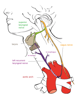
The recurrent laryngeal nerve (RLN) is a branch of the vagus nerve that supplies all the intrinsic muscles of the larynx, with the exception of the cricothyroid muscles. There are two recurrent laryngeal nerves, right and left. The right and left nerves are not symmetrical, with the left nerve looping under the aortic arch, and the right nerve looping under the right subclavian artery then traveling upwards. They both travel alongside of the trachea. Additionally, the nerves are one of few nerves that follow a recurrent course, moving in the opposite direction to the nerve they branch from, a fact from which they gain their name.

Rima glottidis Opening between the true vocal cords and the arytenoid cartilages of the larynx

The rima glottidis is the opening between the true vocal cords and the arytenoid cartilages of the larynx.

Laryngomalacia

Laryngomalacia is the most common cause of chronic stridor in infancy, in which the soft, immature cartilage of the upper larynx collapses inward during inhalation, causing airway obstruction. It can also be seen in older patients, especially those with neuromuscular conditions resulting in weakness of the muscles of the throat. However, the infantile form is much more common. Laryngomalacia is one of the most common laryngeal congenital disease in infancy and public education about the signs and symptoms of the disease is lacking.

Polyneuropathy in dogs and cats is a collection of peripheral nerve disorders that often are breed-related in these animals. Polyneuropathy indicates that multiple nerves are involved, unlike mononeuropathy. Polyneuropathy usually involves motor nerve dysfunction, also known as lower motor neuron disease. Symptoms include decreased or absent reflexes and muscle tone, weakness, or paralysis. It often occurs in the rear legs and is bilateral. Most are chronic problems with a slow onset of symptoms, but some occur suddenly.

Oropharyngeal dysphagia arises from abnormalities of muscles, nerves or structures of the oral cavity, pharynx, and upper esophageal sphincter.

Globus pharyngis

Globus pharyngis or globus sensation is the persistent but painless sensation of having a pill, food bolus, or some other sort of obstruction in the throat when there is none. Swallowing is typically performed normally, so it is not a true case of dysphagia, but it can become quite irritating. It is common, with 22–45% of people experiencing it at least once in their lifetime.

Vocal cord paresis, also known as recurrent laryngeal nerve paralysis or vocal fold paralysis, is an injury to one or both recurrent laryngeal nerves (RLNs), which control all muscles of the larynx except for the cricothyroid muscle. The RLN is important for speaking, breathing and swallowing.

Laryngeal saccules or laryngeal ventricles are soft tissue masses located between the vocal folds and the lateral wall of the larynx in canines. Their function is not well understood, but in brachycephalic breeds the saccules can become everted and protrude into the laryngeal opening, causing symptoms such as snoring, noisy breathing, coughing, nasal congestion, and shortness of breath in affected dogs.

Brachycephalic airway obstructive syndrome

Brachycephalic syndrome is a pathological condition affecting short nosed dogs and cats which can lead to severe respiratory distress. There are four different anatomical abnormalities that contribute to the disease, all of which occur more commonly in brachycephalic breeds:- an elongated soft palate, stenotic nares, a hypoplastic trachea, and everted laryngeal saccules. Because all of these components make it more difficult to breathe, in situations of exercise, stress, or heat, an animal with these abnormalities may be unable to take deep or fast enough breaths to blow off carbon dioxide. This leads to distress and further increases respiratory rate and heart rate, creating a vicious cycle that can quickly lead to a life-threatening situation.

Thyroplasty is a phonosurgical technique designed to improve the voice by altering the thyroid cartilage of the larynx, which houses the vocal cords in order to change the position or the length of the vocal cords.

Geriatric onset laryngeal paralysis polyneuropathy (GOLPP), previously described as idiopathic laryngeal paralysis (ILP), is a degenerative polyneuropathy that most commonly occurs in older medium-to-large breed dogs.

Arytenoid adduction is a surgical procedure used to treat vocal cord paralysis. A suture is used to emulate the action of the lateral cricoarytenoid muscle and position the paralyzed vocal cord closer to the midline. This allows the two vocal cords to meet and can improve speaking and swallowing ability for affected patients. Arytenoid adduction is often performed in conjunction with medialization thyroplasty.

Exercise-induced laryngeal obstruction (EILO) is a transient, reversible narrowing of the larynx that occurs during high intensity exercise. This acts to impair airflow and cause shortness of breath, stridor and often discomfort in the throat and upper chest. EILO is a very common cause of breathing difficulties in young athletic individuals but is often misdiagnosed as asthma or exercise-induced bronchoconstriction.

An elongated soft palate is a congenital hereditary disorder that negatively affect dogs and cats breathing and eating. A soft palate is considered elongated when it extends past the top of the epiglottis and/or past the middle of the tonsillar crypts. When the soft palate is elongated, it partially blocks the throat thereby creating breathing and feeding-related issues. An elongated soft palate is a symptom of Brachycephalic Obstructive Airway Syndrome (BOAS) and is common in brachycephalic dog breeds and has been reported in brachycephalic cat breeds as well. Some of the other BOAS related symptoms include stenotic nares, everted laryngeal saccules, and laryngeal collapse.

References

Stanley BJ, et al. Esophageal dysfunction in dogs with idiopathic laryngeal paralysis: A controlled cohort study. Veterinary Surgery 39(2), pg. 139–149, February 2010.

  1. Laryngeal paralysis in dogs and horses
  2. 1 2 Laryngeal Paralysis Archived 2007-11-26 at the Wayback Machine by Katharine Hillestad, DVM.