Madurella mycetomatis

Last updated

Contents

Madurella mycetomatis
Madurella mycetomatis UAMH7130.jpg
Scientific classification
Kingdom:
Phylum:
Class:
Order:
Family:
Genus:
Species:
M. mycetomatis
Binomial name
Madurella mycetomatis
Brumpt (1905) [1]
Synonyms
  • Streptothrix mycetomi Laveran (1902)
  • Glenospora khartoumensisChalmerrs & Archibald (1902)
  • Oospora tozeuri Nicolle & Pinoy (1908)
  • Madurella tozeuriPinoy (1919)
  • M. tabarkaeBlanc & Brun (1919)
  • M. AmericanaGammel (1926)
  • M. ikedaeGammel (1927)
  • M. LackawannaHanan & Zurett (1938)
  • M. virido brunneaRedaelli & Ciferri (1942)

Madurella mycetomatis is a fungus primarily reported in Central Africa as a cause of mycetoma in humans. It has been misclassified for many years, but with improvement of molecular techniques, its phylogenetic classification has been established. Many methods exist to identify M. mycetomatis, both in lesions and in culture. Histological examination is especially useful, as it has many unique morphological features. Strain-level differences in response to antifungal agents is informative for treatment and laboratory isolation of cultures.

History

Madurella mycetomatis underwent many name changes. In 1901, Brumpt described the first recorded case of mycosis caused by M. mycetomatis, identifying black granules in association with mycetoma. [2] In 1902, Laveran named the fungus Strepthothrix mycetomi, which he had identified from a mycetoma grain. In 1905, Brumpt corrected its genus to Madurella, in turn changing its name to Madurella mycetomi. [3] The fungus was grown in-vitro by Brault in 1912, so it could be studied in culture. [2] In 1977, the British Medical Research Council changed the name to the currently accepted name, Madurella mycetomatis. [3] With the binomial name determined, M. mycetomatis still remained incorrectly classified in the Pleosporales. This error was corrected in 2015 when M. mycetomatis was placed in the order Sordariales. [4] Maduramycosis or Madura foot, was first described from Madurai, South India, in 1842 by John Gill. [2]

Phylogeny

The genus Madurella contains only two well defined species: M. mycetomatis and M. grisea . Roughly a dozen other species of uncertain validity have been described as genus Madurella based on in vivo similarities and cultural sterility. Although similar, there were important physiological and morphological differences between the two well defined species, leading scientists to doubt their phylogeny. The development of ribosomal sequencing and other molecular techniques, led to the discovery that M. mycetomatis did not share a common ancestor with M. grisea and that M. mycetomatis belonged in the order Sordariales, [3] [5] further confirmed by genomic comparison against Chaetomium thermophilum , another member of the Sordariales. [6]

Genotypic variation can help explain geographical distribution of fungi and differences in host symptoms. Restriction endonuclease assay (REA) and random amplification of polymorphic DNA (RAPD) helped characterize the different genotypes of M. mycetomatis, namely 2 genotypic clusters in Africa, and 7 different genotypes from other continents. Further testing with amplified fragment length polymorphism (AFLP) found 3 clusters in Sudan, in contrast to the 2 clusters identified by RAPD, proving AFLP to be a more sensitive method. This showed that M. mycetomatis is not genetically homogenous, and explained the variability in host symptoms affected by the same etiological agent. [3] [7]

Physiology and ecology

Madurella mycetomatis has been identified in both soil and anthill samples, growing optimally at 37 ˚C, however can viably grow at up to 40 ˚C. [8] [9] This ability to grow at high temperatures is a feature that can be useful in identifying the fungus in culture. The fungus's ability, an inability, to break down various molecules can also be used to confirm its identity. Madurella mycetomatis is amylolytic yet is only weakly proteolytic, and has the ability to assimilate glucose, galactose, lactose and maltose, while unable to assimilate sucrose. [3] [8] Potassium nitrate, ammonium sulfate, asparagine and urea can also be used by the fungus. [9] Madurellamycetomatis produces 1,8-dihydroxynapthalene a precursor to melanin – a protein extracellularly attached to proteins. [9] Both molecules are responsible for the characteristic dark grain color. The melanin produced by the fungus has also been identified as a defense mechanism against processes such as hydrolytic enzymes, free radicals, redox buffering, antibodies and complement. [10] The fungus also produces pyomelanin, a brown diffusible pigment. [3]

Growth and morphology

Colony of Madurella mycetomatis (UAMH 9528) on potato dextrose agar incubated for 12 days at 37 degC Madurella mycetomatis UAMH9528.jpg
Colony of Madurella mycetomatis (UAMH 9528) on potato dextrose agar incubated for 12 days at 37 °C

The growth of M. mycetomatis is very slow and can be broken down into three stages. Initially the colony is dome shaped white-yellow or olivaceous brown in color. [9] The mycelium is covered in grey down, giving it a woolly texture. Following the initial stage, brownish aerial mycelia (1 to 5 μm) form and the colony starts producing a diffusible pigment called pyomelanin, and becomes smooth in texture. [3] Older colonies form masses of hyphae called sclerotia or grains. In nutritionally deficient or potato-carrot media, black grains (0.75 to 1 mm in diameter) with undifferentiated polygonal cells can be observed. [8] [9]

Grains of M. mycetomatis are hard and brittle, ranging between 0.5 and 1 mm (maximum being 2 mm), with masses from 2 to 4 mm. The grains are oval and often multi-lobed. They are reddish brown to black in color and texturally smooth or ridged. [9] [11] The grains are made up of an internal mass of hyphae, 2 to 5 μm in diameter, with terminal cells swelling from 12 – 15 μm (maximum being 30 μm) in diameter. [8] Overall two main types of grains are observed. The most common type is compact or filamentous, where a dark brown cement like amorphous, electron rich substance fills the voids surrounding the hyphal network. [9] The hyphal network differs in growth between the cortical and medullar region, with radial versus multidirectional growth respectively. When stained with hematoxylin and eosin it appears rust-brown in color. In contrast, the second type, vesicular, has a light colored medulla and a brown cortical region filled with hyphae and vesicles 6 to 14 μm in diameter. [12] Often it is difficult to determine the transition point from cortex to medulla. Lesions can have both the filamentous and vesicular type grains at the same time. [3] [8]

Although conidiation (conidium), a form of asexual reproduction, in M. mycetomatis is rare, two main types can be described in-vitro. In the first type oval to pyriform conidia, 3 to 5 μm can be observed. The conidia have truncated bases and are on the tips of simple or branched conidiophores. In-vitro, this type of conidiation can be observed in 50% of cultures on soil extract, hay infusion or water agar. [9] When grown on potato carrot agar or cornmeal agar the second type of conidiation is observed. This type is characterized by small spherical conidia (3 μm in diameter) on tapered tips of flask shaped phialides and collarettes. [8] [13] On SDA media M. mycetomatis is sterile. [3] [9] No sexual stage has been identified for M. mycetomatis. [6]

Mycosis

Madurella mycetomatis is the most common fungus with respect to causing mycetoma in humans, [14] a chronic localized inflammatory disease. [15] Madurellamycetomatis accounts for 70% of mycetoma cases in central Africa, especially common in Sudan. [3] Cases of mycetoma caused by this fungus have also been reported in West Africa, India, Venezuela, Curaçao, Brazil, Peru and Argentina. [11] The mode of entry for etiological agents of mycetoma, such as M. mycetomatis, is trauma, such as; snake bites, knives, splinters, thorns and insect bites. Thus having identified M. mycetomatis in soil and anthill samples substantiates its involvement in mycetoma. [9] Infected hosts from samples in Sudan show variability in clinical symptoms, this corroborates the heterogeneity of M. mycetomatis genotypes. [3] [7]

Detection

There are various methods available for the purpose of differentiating fungal species. Histological examination allows for the exploitation of unique morphological features of M. mycetomatis, including but not limited to the unique cement like substance and multi-lobed morphology of the grains. [3] [9] Molecular analysis allows for a more sensitive technique to discriminate between morphologically similar species. An internal transcribed spacer (ITS) is amplified by polymerase chain reaction (PCR). [4] Followed by restriction fragment length polymorphism (RFLP) digestion or gene sequencing to obtain the results. The highly variable nature of the ITS sequence between species not only allows for diagnosis, but also the identification of M. mycetomatis in soil samples. ITS has also been cited as the recommended method for isolation of M. mycetomatis. [16] Additionally, molecular analysis lead to the determination that M. mycetomatis did not share a common ancestor with M. grisea, and belonged in the Sordariales class. [3]

Anti-fungal susceptibility

Understanding how a fungus might react to various anti-fungal agents in-vitro can be beneficial when wanting to study or isolate particular organisms in culture. The Clinical & Laboratory Standards Institute (CLSI) M38A methods indicate that conidial suspensions are to be used when testing anti-fungal susceptibility of filamentous fungi. When following these methods for M. mycetomatis, alternative suspensions of hyphal fragments are required, as conidial forms are exceedingly rare. Table 1 summarizes the minimal inhibitory concentrations at 90% (MIC90) for various anti-fungal agents, with specific relation to M. mycetomatis. [3]

In vitro susceptibility of Madurellamycetomatisto various drugs
Anti-fungal drugMIC90 (μg/mL)MIC50 (μg/mL)MIC range (μg/mL)
Ketoconazole [17] 0.250.06≤0.031–1
Ravuconazole [18] 0.0160.004≤0.002–0.031
Posaconazole [17] 0.060.03≤0.03–0.125
Terbinafine [17] 1681–>16
Voriconazole [19] 0.1250.016–1
Isavuconazole [20] 0.0630.031≤0.016–0.125
Itraconazole [18] 0.250.063≤0.016–0.5

See also

Related Research Articles

<span class="mw-page-title-main">Hypha</span> Long, filamentous structure in fungi and Actinobacteria

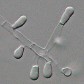
A hypha is a long, branching, filamentous structure of a fungus, oomycete, or actinobacterium. In most fungi, hyphae are the main mode of vegetative growth, and are collectively called a mycelium.

<span class="mw-page-title-main">Eumycetoma</span> Human and animal fungal infection

Eumycetoma, also known as Madura foot, is a persistent fungal infection of the skin and the tissues just under the skin, affecting most commonly the feet, although it can occur in hands and other body parts. It starts as a painless wet nodule, which may be present for years before ulceration, swelling, grainy discharge and weeping from sinuses and fistulae, followed by bone deformity.

<span class="mw-page-title-main">Sordariales</span> Order of fungi

The order Sordariales is one of the most diverse taxonomic groups within the Sordariomycetes.

<i>Cochliobolus lunatus</i> Fungal plant pathogen

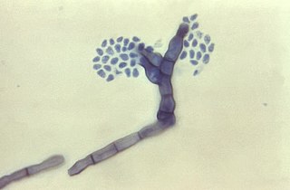
Cochliobolus lunatus is a fungal plant pathogen that can cause disease in humans and other animals. The anamorph of this fungus is known as Curvularia lunata, while C. lunatus denotes the teleomorph or sexual stage. They are, however, the same biological entity. C. lunatus is the most commonly reported species in clinical cases of reported Cochliobolus infection.

Exophiala jeanselmei is a saprotrophic fungus in the family Herpotrichiellaceae. Four varieties have been discovered: Exophiala jeanselmei var. heteromorpha, E. jeanselmei var. lecanii-corni, E. jeanselmei var. jeanselmei, and E. jeanselmei var. castellanii. Other species in the genus Exophiala such as E. dermatitidis and E. spinifera have been reported to have similar annellidic conidiogenesis and may therefore be difficult to differentiate.

<i>Madurella</i> Genus of fungi

Madurella is a fungal genus of uncertain position in the Sordariales, and sometimes classified as Mitosporic Ascomycota.

<i>Pseudallescheria boydii</i> Species of fungus

Pseudallescheria boydii is a species of fungus classified in the Ascomycota. It is associated with some forms of eumycetoma/maduromycosis and is the causative agent of pseudallescheriasis. Typically found in stagnant and polluted water, it has been implicated in the infection of immunocompromised and near-drowned pneumonia patients. Treatment of infections with P. boydii is complicated by resistance to many of the standard antifungal agents normally used to treat infections by filamentous fungi.

<i>Piedraia hortae</i> Species of fungus

Piedraia hortae is a superficial fungus that exists in the soils of tropical and subtropical environments and affects both sexes of all ages. The fungus grows very slowly, forming dark hyphae, which contain chlamydoconidia cells and black colonies when grown on agar. Piedraia hortae is a dermatophyte and causes a superficial fungal infection known as black piedra, which causes the formation of black nodules on the hair shaft and leads to progressive weakening of the hair. The infection usually infects hairs on the scalp and beard, but other varieties tend to grow on pubic hairs. The infection is usually treated with cutting or shaving of the hair and followed by the application of anti-fungal and topical agents. The fungus is used for cosmetic purposes to darken hair in some societies as a symbol of attractiveness.

<i>Trichophyton tonsurans</i> Species of fungus

Trichophyton tonsurans is a fungus in the family Arthrodermataceae that causes ringworm infection of the scalp. It was first recognized by David Gruby in 1844. Isolates are characterized as the "–" or negative mating type of the Arthroderma vanbreuseghemii complex. This species is thought to be conspecific with T. equinum, although the latter represents the "+" mating strain of the same biological species Despite their biological conspecificity, clones of the two mating types appear to have undergone evolutionary divergence with isolates of the T. tonsurans-type consistently associated with Tinea capitis whereas the T. equinum-type, as its name implies, is associated with horses as a regular host. Phylogenetic relationships were established in isolates from Northern Brazil, through fingerprinting polymorphic RAPD and M13 markers. There seems to be lower genomic variability in the T. tonsurans species due to allopatric divergence. Any phenotypic density is likely due to environmental factors, not genetic characteristics of the fungus.

Mycetoma is a chronic infection in the skin caused by either bacteria (actinomycetoma) or fungi (eumycetoma), typically resulting in a triad of painless firm skin lumps, the formation of weeping sinuses, and a discharge that contains grains. 80% occur in feet.

<span class="mw-page-title-main">Ectomycorrhiza</span> Non-penetrative symbiotic association between a fungus and the roots of a vascular plant

An ectomycorrhiza is a form of symbiotic relationship that occurs between a fungal symbiont, or mycobiont, and the roots of various plant species. The mycobiont is often from the phyla Basidiomycota and Ascomycota, and more rarely from the Zygomycota. Ectomycorrhizas form on the roots of around 2% of plant species, usually woody plants, including species from the birch, dipterocarp, myrtle, beech, willow, pine and rose families. Research on ectomycorrhizas is increasingly important in areas such as ecosystem management and restoration, forestry and agriculture.

<i>Aspergillus versicolor</i> Species of fungus

Aspergillus versicolor is a slow-growing species of filamentous fungus commonly found in damp indoor environments and on food products. It has a characteristic musty odor associated with moldy homes and is a major producer of the hepatotoxic and carcinogenic mycotoxin sterigmatocystin. Like other Aspergillus species, A. versicolor is an eye, nose, and throat irritant.

Coniochaeta hoffmannii, also known as Lecythophora hoffmannii, is an ascomycete fungus that grows commonly in soil. It has also been categorized as a soft-rot fungus capable of bringing the surface layer of timber into a state of decay, even when safeguarded with preservatives. Additionally, it has pathogenic properties, although it causes serious infection only in rare cases. A plant pathogen lacking a known sexual state, C. hoffmannii has been classified as a "dematiaceous fungus" despite its contradictory lack of pigmentation; both in vivo and in vitro, there is no correlation between its appearance and its classification.

Scedosporiosis is the general name for any mycosis – i.e., fungal infection – caused by a fungus from the genus Scedosporium. Current population-based studies suggest Scedosporium prolificans and Scedosporium apiospermum to be among the most common infecting agents from the genus, although infections caused by other members thereof are not unheard of. The latter is an asexual form (anamorph) of another fungus, Pseudallescheria boydii. The former is a "black yeast", currently not characterized as well, although both of them have been described as saprophytes.

<i>Fonsecaea compacta</i> Species of fungus

Fonsecaea compacta is a saprophytic fungal species found in the family Herpotrichiellaceae. It is a rare etiological agent of chromoblastomycosis, with low rates of correspondence observed from reports. The main active components of F. compacta are glycolipids, yet very little is known about its composition. F. compacta is widely regarded as a dysplastic variety of Fonsecaea pedrosoi, its morphological precursor. The genus Fonsecaea presently contains two species, F. pedrosoi and F. compacta. Over 100 strains of F. pedrosoi have been isolated but only two of F. compacta.

<i>Aspergillus clavatus</i> Species of fungus

Aspergillus clavatus is a species of fungus in the genus Aspergillus with conidia dimensions 3–4.5 x 2.5–4.5 μm. It is found in soil and animal manure. The fungus was first described scientifically in 1834 by the French mycologist John Baptiste Henri Joseph Desmazières.

<i>Phialophora verrucosa</i> Species of fungus

Phialophora verrucosa is a pathogenic, dematiaceous fungus that is a common cause of chromoblastomycosis. It has also been reported to cause subcutaneous phaeohyphomycosis and mycetoma in very rare cases. In the natural environment, it can be found in rotting wood, soil, wasp nests, and plant debris. P. verrucosa is sometimes referred to as Phialophora americana, a closely related environmental species which, along with P. verrucosa, is also categorized in the P. carrionii clade.

Edouard Drouhet was a physician, biologist, and medical mycologist who played a key role in understanding how anti-fungal agents such as ketoconazole and amphotericin-B can be used as therapeutic treatments in humans with superficial or deep-seated mycoses.

The Trematosphaeriaceae are a family of fungi in the order of Pleosporales. They are found world-wide with the greatest contributions found in Europe and Australia. It includes taxa that are found in the marine environment, such as Falciformispora lignatis which can be found in freshwater and marine habitats. Most are saprobic species.

Ahmed Hassan Fahal is a Sudanese Professor of Surgery at the University of Khartoum, who specialised in Mycetoma.

References

  1. Brumpt, E (1905). "Sur le mycétome à grains noirs, maladie produite par une Mucédinée dugenre Madurella n. g.". Comptes Rendus des Séances de la Société de Biologie et de ses Filiales. 58: 997–9.
  2. 1 2 3 McGinnis, Michael R (2012). Laboratory Handbook of Medical Mycology. London: Academic Press. ISBN   978-0123960146.
  3. 1 2 3 4 5 6 7 8 9 10 11 12 13 14 Liu, Dongyou (2011). Molecular Detection of Human Fungal Pathogens (1st ed.). Boca Raton, FL: CRC Press. ISBN   978-1439812402.
  4. 1 2 Samanta, Indranil (2015). Veterinary Mycology. New Delhi: Springer. ISBN   978-8132222798.
  5. Heitman, Joseph; Filler, Scott G.; Edwards Jr., John E.; Mitchell, Aaron P. (2006). Molecular Principles of Fungal Pathogens (1st ed.). Washington, DC: American Society Microbiology. ISBN   1555813682.
  6. 1 2 van de Sande, Wendy W. J. (1 January 2012). "Phylogenetic analysis of the complete mitochondrial genome of Madurella mycetomatis confirms its taxonomic position within the order Sordariales". PLOS ONE. 8 (3): e57774. doi: 10.1371/journal.pone.0057774 . PMC   3590280 . PMID   23483927.
  7. 1 2 Lopes, Maria M; Freitas, Graciete; Boiron, Patrick (2000). "Potential Utility of Random Amplified Polymorphic DNA (RAPD) and Restriction Endonuclease Assay (REA) as Typing Systems for Madurella mycetomatis". Microbiology. 40 (1): 1–5. doi:10.1007/s002849910001. PMID   10568795. S2CID   22344606.
  8. 1 2 3 4 5 6 Rippon, John Willard (1988). Medical mycology: the pathogenic fungi and the pathogenic actinomycetes (3rd ed.). Philadelphia, PA: Saunders. ISBN   0721624448.
  9. 1 2 3 4 5 6 7 8 9 10 11 Murray, Patrick R.; Baron, Ellen Jo (2003). Manual of clinical microbiology (8th ed.). Washington, DC: ASM Press. ISBN   1555817378.
  10. Ibrahim, Anahid Izzat; El Hassan, Ahmed Mohammed; Fahal, Ahmed; van de Sande, Wendy W (1 January 2013). "A histopathological exploration of the Madurella mycetomatis grain". PLOS ONE. 8 (3): e57774. Bibcode:2013PLoSO...857774I. doi: 10.1371/journal.pone.0057774 . PMC   3590280 . PMID   23483927.
  11. 1 2 Baker, Roger Denio (1971). The Pathologic Anatomy of Mycoses: Human Infection with Fungi, Actinomycetes and Algae (1st ed.). Springer Verlag. ISBN   3540051406.
  12. Dismukes, William E.; Pappas, Peter G.; Sobel, Jack D. (2003). Clinical Mycology (1st ed.). Oxford University Press. ISBN   0195148096.
  13. Gaspari, Anthony A.; Tyring, Stephen K.; Kaplan, Daniel H. (2016). Clinical and Basic Immunodermatology (2nd ed.). Springer. ISBN   978-3319297835.
  14. Howard, Dexter H. (2007). Pathogenic fungi in humans and animals (2nd ed.). New York, NY: Dekker. ISBN   978-0824706838.
  15. Kradin, Richard L. (2010). Diagnostic Pathology of Infectious Disease. Philadelphia, PA: Saunders Elsevier. ISBN   978-1416034292.
  16. "Madurella mycetomatis". Mycology Online. University of Adelaide.
  17. 1 2 3 Belkum, Alex van; Fahal, Ahmed H.; Sande, Wendy W. J. van de (1 April 2011). "In Vitro Susceptibility of Madurella mycetomatis to Posaconazole and Terbinafine". Antimicrobial Agents and Chemotherapy. 55 (4): 1771–3. doi:10.1128/AAC.01045-10. PMC   3067195 . PMID   21263050.
  18. 1 2 Ahmed, Sarah Abdalla; Kloezen, Wendy; Duncanson, Frederick; Zijlstra, Ed E.; Hoog, G. Sybren de; Fahal, Ahmed H.; Sande, Wendy W. J. van de (19 June 2014). "Madurella mycetomatis Is Highly Susceptible to Ravuconazole". PLOS Negl Trop Dis. 8 (6): e2942. doi: 10.1371/journal.pntd.0002942 . PMC   4063742 . PMID   24945848.
  19. van de Sande, Wendy W. J.; Luijendijk, Ad; Ahmed, Abdalla O. A.; Bakker-Woudenberg, Irma A. J. M.; van Belkum, Alex (1 April 2005). "Testing of the in vitro susceptibilities of Madurella mycetomatis to six antifungal agents by using the Sensititre system in comparison with a viability-based 2,3-bis(2-methoxy-4-nitro-5-sulfophenyl)-5- [(phenylamino)carbonyl]-2H-tetrazolium hydroxide (XTT) assay and a modified NCCLS method". Antimicrobial Agents and Chemotherapy. 49 (4): 1364–8. doi:10.1128/AAC.49.4.1364-1368.2005. PMC   1068587 . PMID   15793113.
  20. Kloezen, Wendy; Meis, Jacques F.; Curfs-Breuker, Ilse; Fahal, Ahmed H.; van de Sande, Wendy W. J. (1 November 2012). "In vitro antifungal activity of isavuconazole against Madurella mycetomatis". Antimicrobial Agents and Chemotherapy. 56 (11): 6054–6. doi:10.1128/AAC.01170-12. PMC   3486573 . PMID   22964246.