
Magnetic resonance imaging (MRI) is a medical imaging technique used in radiology to form pictures of the anatomy and the physiological processes of the body. MRI scanners use strong magnetic fields, magnetic field gradients, and radio waves to generate images of the organs in the body. MRI does not involve X-rays or the use of ionizing radiation, which distinguishes it from CT and PET scans. MRI is a medical application of nuclear magnetic resonance (NMR) which can also be used for imaging in other NMR applications, such as NMR spectroscopy.

Radiography is an imaging technique using X-rays, gamma rays, or similar ionizing radiation and non-ionizing radiation to view the internal form of an object. Applications of radiography include medical radiography and industrial radiography. Similar techniques are used in airport security. To create an image in conventional radiography, a beam of X-rays is produced by an X-ray generator and is projected toward the object. A certain amount of the X-rays or other radiation is absorbed by the object, dependent on the object's density and structural composition. The X-rays that pass through the object are captured behind the object by a detector. The generation of flat two dimensional images by this technique is called projectional radiography. In computed tomography an X-ray source and its associated detectors rotate around the subject which itself moves through the conical X-ray beam produced. Any given point within the subject is crossed from many directions by many different beams at different times. Information regarding attenuation of these beams is collated and subjected to computation to generate two dimensional images in three planes which can be further processed to produce a three dimensional image.

Radiology is the medical discipline that uses medical imaging to diagnose diseases and guide their treatment, within the bodies of humans and other animals. It began with radiography, but today it includes all imaging modalities, including those that use no electromagnetic radiation, as well as others that do, such as computed tomography (CT), fluoroscopy, and nuclear medicine including positron emission tomography (PET). Interventional radiology is the performance of usually minimally invasive medical procedures with the guidance of imaging technologies such as those mentioned above.
Medical physics deals with the application of the concepts and methods of physics to the prevention, diagnosis and treatment of human diseases with a specific goal of improving human health and well-being. Since 2008, medical physics has been included as a health profession according to International Standard Classification of Occupation of the International Labour Organization.

Medical imaging is the technique and process of imaging the interior of a body for clinical analysis and medical intervention, as well as visual representation of the function of some organs or tissues (physiology). Medical imaging seeks to reveal internal structures hidden by the skin and bones, as well as to diagnose and treat disease. Medical imaging also establishes a database of normal anatomy and physiology to make it possible to identify abnormalities. Although imaging of removed organs and tissues can be performed for medical reasons, such procedures are usually considered part of pathology instead of medical imaging.
A radiation therapist, therapeutic radiographer or radiotherapist is an allied health professional who works in the field of radiation oncology. Radiation therapists plan and administer radiation treatments to cancer patients in most Western countries including the United Kingdom, Australia, most European countries, and Canada, where the minimum education requirement is often a baccalaureate degree or postgraduate degrees in radiation therapy. Radiation therapists can also prescribe medications and radiation, interpret tests results, perform follow ups, reviews, and provide consultations to cancer patients in the United Kingdom and Ontario, Canada . In the United States, radiation therapists have a lower educational requirement and often require postgraduate education and certification in order to plan treatments.

Nuclear medicine or nucleology is a medical specialty involving the application of radioactive substances in the diagnosis and treatment of disease. Nuclear imaging, in a sense, is "radiology done inside out" because it records radiation emitting from within the body rather than radiation that is generated by external sources like X-rays. In addition, nuclear medicine scans differ from radiology, as the emphasis is not on imaging anatomy, but on the function. For such reason, it is called a physiological imaging modality. Single photon emission computed tomography (SPECT) and positron emission tomography (PET) scans are the two most common imaging modalities in nuclear medicine.
A medical physicist is a health professional with specialist education and training in the concepts and techniques of applying physics in medicine and competent to practice independently in one or more of the subfields (specialties) of medical physics. A medical physicist plays a fundamental role in applying physics to medicine, but particularly in the diagnosis and treatment of cancer. The scientific and technological progress in medical physics has led to a variety of skills that must be integrated into the role of a medical physicist in order for them to perform their job. The "medical services" provided to patients undergoing diagnostic and therapeutic treatments must, therefore, be the result of different but complementary skills. In general, the medical physicist is responsible for all scientific and technical aspects of imaging, radiation treatment, and radiation safety. It is their occupational role to ensure that medical modalities offered to patients are met with the utmost quality assurance. It is the medical physicist that manage and supervise the efforts of dosimetrists, therapists and technologists in that capacity.

A sonographer is an allied healthcare professional who specializes in the use of ultrasonic imaging devices to produce diagnostic images, scans, videos or three-dimensional volumes of anatomy and diagnostic data. The requirements for clinical practice vary greatly by country. Sonography requires specialized education and skills to acquire, analyze and optimize information in the image. Due to the high levels of decisional latitude and diagnostic input, sonographers have a high degree of responsibility in the diagnostic process. Many countries require medical sonographers to have professional certification. Sonographers have core knowledge in ultrasound physics, cross-sectional anatomy, physiology, and pathology.
The American College of Radiology (ACR), founded in 1923, is a professional medical society representing nearly 40,000 diagnostic radiologists, radiation oncologists, interventional radiologists, nuclear medicine physicians and medical physicists.

Radiographers, also known as radiologic technologists, diagnostic radiographers and medical radiation technologists are healthcare professionals who specialize in the imaging of human anatomy for the diagnosis and treatment of pathology. Radiographers are infrequently, and almost always erroneously, known as x-ray technicians. In countries that use the title radiologic technologist they are often informally referred to as techs in the clinical environment; this phrase has emerged in popular culture such as television programmes. The term radiographer can also refer to a therapeutic radiographer, also known as a radiation therapist.
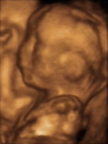
3D ultrasound is a medical ultrasound technique, often used in fetal, cardiac, trans-rectal and intra-vascular applications. 3D ultrasound refers specifically to the volume rendering of ultrasound data. When involving a series of 3D volumes collected over time, it can also be referred to as 4D ultrasound or real-time 3D ultrasound.
The American Society of Radiologic Technologists (ASRT) is a professional membership association that serves medical imaging technologists, radiation therapists, and radiologic science students. The organization, located in Albuquerque, New Mexico provides its members with ongoing education and professional development opportunities.

Cardiac magnetic resonance imaging, also known as cardiovascular MRI, is a magnetic resonance imaging (MRI) technology used for non-invasive assessment of the function and structure of the cardiovascular system. Conditions in which it is performed include congenital heart disease, cardiomyopathies and valvular heart disease, diseases of the aorta such as dissection, aneurysm and coarctation, coronary heart disease. It can also be used to look at pulmonary veins.
Nuclear medicine physicians, also called nuclear radiologists or simply nucleologists, are medical specialists that use tracers, usually radiopharmaceuticals, for diagnosis and therapy. Nuclear medicine procedures are the major clinical applications of molecular imaging and molecular therapy. In the United States, nuclear medicine physicians are certified by the American Board of Nuclear Medicine and the American Osteopathic Board of Nuclear Medicine.
Clinical physiology is both an academic discipline within the medical sciences and a clinical medical specialty for physicians in the health care systems of Sweden, Denmark and Finland. Clinical physiology is characterized as a branch of physiology that uses a functional approach to understand the pathophysiology of a disease.
Ferenc Andras Jolesz was a Hungarian-American physician and scientist best known for his research on image guided therapy, the process by which information derived from diagnostic imaging is used to improve the localization and targeting of diseased tissue to monitor and control treatment during surgical and interventional procedures. He pioneered the field of Magnetic Resonance Imaging-guided interventions and introduced of a variety of new medical procedures based on novel combinations of imaging and therapy delivery.
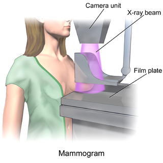
In medicine, breast imaging is a sub-speciality of diagnostic radiology that involves imaging of the breasts for screening or diagnostic purposes. There are various methods of breast imaging using a variety of technologies as described in detail below. Traditional screening and diagnostic mammography uses x-ray technology and has been the mainstay of breast imaging for many decades. Breast tomosynthesis is a relatively new digital x-ray mammography technique that produces multiple image slices of the breast similar to, but distinct from, computed tomography (CT). Xeromammography and galactography are somewhat outdated technologies that also use x-ray technology and are now used infrequently in the detection of breast cancer. Breast ultrasound is another technology employed in diagnosis and screening that can help differentiate between fluid filled and solid lesions, an important factor to determine if a lesion may be cancerous. Breast MRI is a technology typically reserved for high-risk patients and patients recently diagnosed with breast cancer. Lastly, scintimammography is used in a subgroup of patients who have abnormal mammograms or whose screening is not reliable on the basis of using traditional mammography or ultrasound.

Roderic Ivan Pettigrew is an American physicist, engineer, and physician who is CEO of EnHealth and Executive Dean for EnMed at Texas A&M University. From 2002-November 2017, he was the founding director of the National Institute of Biomedical Imaging and Bioengineering (NIBIB) at the National Institutes of Health (NIH). He is a pioneer and world expert in cardiovascular magnetic resonance imaging (MRI).
A clinical technologist, also known as a healthcare science practitioner, is a medical professional involved in the practical delivery of medical physics and clinical engineering services. In some locations there is considerable overlap in closely related terms, for example in many countries technologist and radiographer are synonyms, while in the United Kingdom they are considered separate professions. Clinical technologists can be found in nuclear medicine, radiotherapy, radiation protection, and rehabilitation engineering departments, and they are often described by their scope of practice.