Related Research Articles
Collagen is the main structural protein in the extracellular matrix found in the body's various connective tissues. As the main component of connective tissue, it is the most abundant protein in mammals, making up from 25% to 35% of the whole-body protein content. Collagen consists of amino acids bound together to form a triple helix of elongated fibril known as a collagen helix. It is mostly found in connective tissue such as cartilage, bones, tendons, ligaments, and skin.

A cell wall is a structural layer surrounding some types of cells, just outside the cell membrane. It can be tough, flexible, and sometimes rigid. It provides the cell with both structural support and protection, and also acts as a filtering mechanism. Cell walls are absent in many eukaryotes, including animals, but are present in some other ones like fungi, algae and plants, and in most prokaryotes. A major function is to act as pressure vessels, preventing over-expansion of the cell when water enters.

Cellulose is an organic compound with the formula (C
6H
10O
5)
n, a polysaccharide consisting of a linear chain of several hundred to many thousands of β(1→4) linked D-glucose units. Cellulose is an important structural component of the primary cell wall of green plants, many forms of algae and the oomycetes. Some species of bacteria secrete it to form biofilms. Cellulose is the most abundant organic polymer on Earth. The cellulose content of cotton fiber is 90%, that of wood is 40–50%, and that of dried hemp is approximately 57%.

In biology, the extracellular matrix (ECM), also called intercellular matrix, is a three-dimensional network consisting of extracellular macromolecules and minerals, such as collagen, enzymes, glycoproteins and hydroxyapatite that provide structural and biochemical support to surrounding cells. Because multicellularity evolved independently in different multicellular lineages, the composition of ECM varies between multicellular structures; however, cell adhesion, cell-to-cell communication and differentiation are common functions of the ECM.

Elastin is a protein that in humans is encoded by the ELN gene. Elastin is a key component of the extracellular matrix in gnathostomes. It is highly elastic and present in connective tissue allowing many tissues in the body to resume their shape after stretching or contracting. Elastin helps skin to return to its original position when it is poked or pinched. Elastin is also an important load-bearing tissue in the bodies of vertebrates and used in places where mechanical energy is required to be stored.
The basement membrane is a thin, pliable sheet-like type of extracellular matrix that provides cell and tissue support and acts as a platform for complex signalling. The basement membrane sits between epithelial tissues including mesothelium and endothelium, and the underlying connective tissue.
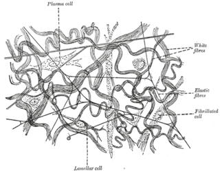
Elastic fibers are an essential component of the extracellular matrix composed of bundles of proteins (elastin) which are produced by a number of different cell types including fibroblasts, endothelial, smooth muscle, and airway epithelial cells. These fibers are able to stretch many times their length, and snap back to their original length when relaxed without loss of energy. Elastic fibers include elastin, elaunin and oxytalan.

Fibrillin is a glycoprotein, which is essential for the formation of elastic fibers found in connective tissue. Fibrillin is secreted into the extracellular matrix by fibroblasts and becomes incorporated into the insoluble microfibrils, which appear to provide a scaffold for deposition of elastin.

Fibrils are structural biological materials found in nearly all living organisms. Not to be confused with fibers or filaments, fibrils tend to have diameters ranging from 10-100 nanometers. Fibrils are not usually found alone but rather are parts of greater hierarchical structures commonly found in biological systems. Due to the prevalence of fibrils in biological systems, their study is of great importance in the fields of microbiology, biomechanics, and materials science.

Natural fibers or natural fibres are fibers that are produced by geological processes, or from the bodies of plants or animals. They can be used as a component of composite materials, where the orientation of fibers impacts the properties. Natural fibers can also be matted into sheets to make paper or felt.

The zonule of Zinn is a ring of fibrous strands forming a zonule that connects the ciliary body with the crystalline lens of the eye. These fibers are sometimes collectively referred to as the suspensory ligaments of the lens, as they act like suspensory ligaments.
Katanin is a microtubule-severing AAA protein. It is named after the Japanese sword called a katana. Katanin is a heterodimeric protein first discovered in sea urchins. It contains a 60 kDa ATPase subunit, encoded by KATNA1, which functions to sever microtubules. This subunit requires ATP and the presence of microtubules for activation. The second 80 kDA subunit, encoded by KATNB1, regulates the activity of the ATPase and localizes the protein to centrosomes. Electron microscopy shows that katanin forms 14–16 nm rings in its active oligomerized state on the walls of microtubules.

MASS syndrome is a medical disorder of the connective tissue similar to Marfan syndrome. MASS stands for: Mitral valve prolapse, Aortic root diameter at upper limits of normal for body size, Stretch marks of the skin, and Skeletal conditions similar to Marfan syndrome. It is caused by a mutation in the FBN1 gene, which encodes fibrillin-1. Fibrillin-1 is an extracellular matrix protein that is found in microfibrils; defects in the fibrillin-1 protein cause the malfunctioning of microfibrils, which results in improper stretching of ligaments, blood vessels, and skin.

The secondary cell wall is a structure found in many plant cells, located between the primary cell wall and the plasma membrane. The cell starts producing the secondary cell wall after the primary cell wall is complete and the cell has stopped expanding.

Fibrillin-1 is a protein that in humans is encoded by the FBN1 gene, located on chromosome 15. It is a large, extracellular matrix glycoprotein that serves as a structural component of 10-12 nm calcium-binding microfibrils. These microfibrils provide force bearing structural support in elastic and nonelastic connective tissue throughout the body. Mutations altering the protein can result in a variety of phenotypic effects differing widely in their severity, including fetal death, developmental problems, Marfan syndrome or in some cases Weill-Marchesani syndrome.

The UDP-forming form of cellulose synthase is the main enzyme that produces cellulose. Systematically, it is known as UDP-glucose:(1→4)-β-D-glucan 4-β-D-glucosyltransferase in enzymology. It catalyzes the chemical reaction:

Microfibrillar-associated protein 5 is a protein that in humans is encoded by the MFAP5 gene.

Bacterial cellulose is an organic compound with the formula (C
6H
10O
5)
n produced by certain types of bacteria. While cellulose is a basic structural material of most plants, it is also produced by bacteria, principally of the genera Acetobacter, Sarcina ventriculi and Agrobacterium. Bacterial, or microbial, cellulose has different properties from plant cellulose and is characterized by high purity, strength, moldability and increased water holding ability. In natural habitats, the majority of bacteria synthesize extracellular polysaccharides, such as cellulose, which form protective envelopes around the cells. While bacterial cellulose is produced in nature, many methods are currently being investigated to enhance cellulose growth from cultures in laboratories as a large-scale process. By controlling synthesis methods, the resulting microbial cellulose can be tailored to have specific desirable properties. For example, attention has been given to the bacteria Komagataeibacter xylinum due to its cellulose's unique mechanical properties and applications to biotechnology, microbiology, and materials science. Historically, bacterial cellulose has been limited to the manufacture of Nata de coco, a South-East Asian food product. With advances in the ability to synthesize and characterize bacterial cellulose, the material is being used for a wide variety of commercial applications including textiles, cosmetics, and food products, as well as medical applications. Many patents have been issued in microbial cellulose applications and several active areas of research are attempting to better characterize microbial cellulose and utilize it in new areas.

Peter Klock Hepler HonFRMS is the Constantine J. Gilgut and Ray Ethan Torrey Professor Emeritus in the Biology Department of the University of Massachusetts at Amherst who is notable for his work on elucidating the roles of calcium, membranes and the cytoskeleton in plant cell development and cell motility.
MFAP4 is an extracellular matrix protein encoded by the MFAP4 gene. It is part of the MFAP family of proteoglycans, which are involved in cell adhesion, intercellular interactions and the assembly and/or maintenance of elastic fibres.
References
- 1 2 3 Massam-Wu, Teresa; Chiu, Maybo; Choudhury, Rawshan; Chaudhry, Shazia S.; Baldwin, Andrew K.; McGovern, Amanda; Baldock, Claire; Shuttleworth, C. Adrian; Kielty, Cay M. (2010). "Assembly of fibrillin microfibrils governs extracellular deposition of latent TGFβ" (PDF). J Cell Sci. 123 (17): 3006–3018. doi:10.1242/jcs.073437. PMC 2923573 . PMID 20699357 – via JCS.
- ↑ Kielty, Cay M.; Baldock, Claire; Lee, David; Rock, Matthew J.; Ashworth, Jane L.; Shuttleworth, C. Adrian (28 February 2002). "Fibrillin: from microfibril assembly to biomechanical function". Philos Trans R Soc Lond B Biol Sci. 357 (1148): 207–217. doi:10.1098/rstb.2001.1029. PMC 1692929 . PMID 11911778.
- ↑ Reinhardt, Dieter P. (October–November 2014). "Microfibril-associated Disorders: Fibrillinopathies". Journal of Glaucoma. 23 (8 Suppl 1): S34-5. doi: 10.1097/IJG.0000000000000114 . ISSN 1057-0829. PMID 25275902. S2CID 31705597.
- ↑ Bonetti, Maria Ida (October 2009). "Microfibrils: a cornerstone of extracellular matrix and a key to understand Marfan syndrome". Italian Journal of Anatomy and Embryology = Archivio Italiano di Anatomia ed Embriologia. 114 (4): 201–224. ISSN 1122-6714. PMID 20578676.
| | This biochemistry article is a stub. You can help Wikipedia by expanding it. |