Orbitofrontal cortex

Last updated
Orbitofrontal cortex
MRI of orbitofrontal cortex.jpg
Approximate location of the OFC shown on a sagittal MRI
Gray729 orbital gyrus.png
Orbital surface of left frontal lobe.
Details
Part of Frontal lobe
Identifiers
Latin cortex orbitofrontalis
NeuroNames 91
NeuroLex ID birnlex_1049
FMA 242003
Anatomical terms of neuroanatomy

The orbitofrontal cortex (OFC) is a prefrontal cortex region in the frontal lobes of the brain which is involved in the cognitive process of decision-making. In non-human primates it consists of the association cortex areas Brodmann area 11, 12 and 13; in humans it consists of Brodmann area 10, 11 and 47. [1]

Contents

The OFC is functionally related to the ventromedial prefrontal cortex. [2] Therefore, the region is distinguished due to the distinct neural connections and the distinct functions it performs. [3] It is defined as the part of the prefrontal cortex that receives projections from the medial dorsal nucleus of the thalamus, and is thought to represent emotion, taste, smell and reward in decision making. [4] [5] [6] [7] [8] [9] [10] [11] It gets its name from its position immediately above the orbits in which the eyes are located. Considerable individual variability has been found in the OFC of humans. [12] A related area is found in rodents. [13]

Structure

The OFC is divided into multiple broad regions distinguished by cytoarchitecture, including brodmann area 47/12, brodmann area 11, brodmann area 14, brodmann area 13, and brodmann area 10. [14] Four gyri are split by a complex of sulci that most frequently resembles a "H" or a "K" pattern. Extending along the rostro-caudal axis, two sulci, the lateral and orbital sulci, are usually connected by the transverse orbital sulcus, which extends along a medial-lateral axis. Most medially, the medial orbital gyrus is separated from the gyrus rectus by the olfactory sulcus. [15] Anteriorly, both the gyrus rectus and the medial part of the medial orbital gyrus consist of area 11(m), and posteriorly, area 14. The posterior orbital gyrus consists mostly of area 13, and is bordered medially and laterally by the anterior limbs of the medial and lateral orbital sulci. Area 11 makes up a large part of the OFC involving both the lateral parts of the medial orbital gyrus as well as the anterior orbital gyrus. The lateral orbital gyrus consists mostly of area 47/12. [14] Most of the OFC is granular, although the caudal parts of area 13 and area 14 are agranular. [16] These caudal regions, which sometimes includes parts of the insular cortex, responds primarily to unprocessed sensory cues. [17]

Connections

The connectivity of the OFC varies somewhat along a rostral-caudal axis. The caudal OFC is more heavily interconnected with sensory regions, notably receiving direct input from the pyriform cortex. The caudal OFC is also the most heavily interconnected with the amygdala. [18] Rostrally, the OFC receives fewer direct sensory projections, and is less connected with the amygdala, but it is interconnected with the lateral prefrontal cortex and parahippocampus. [17] The connectivity of the OFC has also been conceptualized as being composed of two networks; an orbital network composed of most of the central parts of the OFC, including most of areas 47/12, 13, and 11; a medial network composed of the medial most and caudolateral regions of the OFC, as well as areas 24, 25 and 32 of the medial prefrontal cortex. [19] The medial and orbital networks are sometimes referred to as the "visceromotor network" and the "sensory network", respectively. [20]

Afferents

The OFC receives projections from multiple sensory modalities. The primary olfactory cortex, gustatory cortex, secondary somatosensory cortex, superior and inferior temporal gyrus(conveying visual information) all project to the OFC. [16] [21] [22] Evidence for auditory inputs is weak, although some neurons respond to auditory stimuli, indicating an indirect projection may exist. [19] The OFC also receives input from the medial dorsal nucleus, insular cortex, entorhinal cortex, perirhinal cortex, hypothalamus, and amygdala. [21] [23]

Efferents

The orbitofrontal cortex is reciprocally connected with the perirhinal and entorhinal cortices, [23] the amygdala, the hypothalamus, and parts of the medial temporal lobe. In addition to these outputs, the OFC also projects to the striatum, including the nucleus accumbens, caudate nucleus, and ventral putamen, as well as regions of the midbrain including the periaqueductal grey, and ventral tegmental area. [21] [24] OFC inputs to the amygdala synapse on multiple targets, including two robust pathways to the basolateral amygdala and intercalated cells of the amygdala, as well as a weaker direct projection to the central nucleus of the amygdala. [18]

Function

Multiple functions have been ascribed to the OFC including mediating context specific responding, [25] encoding contingencies in a flexible manner, encoding value, encoding inferred value, inhibiting responses, learning changes in contingency, emotional appraisal, [26] altering behavior through somatic markers, driving social behavior, and representing state spaces. [27] [28] While most of these theories explain certain aspects of electrophysiological observations and lesion related changes in behavior, they often fail to explain, or are contradicted by other findings. One proposal that explains the variety of OFC functions is that the OFC encodes state spaces, or the discrete configuration of internal and external characteristics associated with a situation and its contingencies [29] For example, the proposal that the OFC encodes economic value may be a reflection of the OFC encoding task state value. [25] The representation of task states could also explain the proposal that the OFC acts as a flexible map of contingencies, as a switch in task state would enable the encoding of new contingencies in one state, with the preservation of old contingencies in a separate state, enabling switching contingencies when the old task state becomes relevant again. [28] The representation of task states is supported by electrophysiological evidence demonstrating that the OFC responds to a diverse array of task features, and is capable of rapidly remapping during contingency shifts. [28] The representation of task states may influence behavior through multiple potential mechanisms. For example, the OFC is necessary for ventral tegmental area (VTA) neurons to produce a dopaminergic reward prediction error, and the OFC may encode expectations for computation of RPEs in the VTA. [25]

Specific functions have been ascribed to subregions of the OFC. The lateral OFC has been proposed to reflect potential choice value, enabling fictive(counterfactual) prediction errors to potentially mediate switching choices during reversal, extinction and devaluation. [30] Optogenetic activation of the lOFC enhances goal directed over habitual behavior, possibly reflecting increased sensitivity to potential choices and therefore increased switching. The mOFC, on the other hand, has been proposed to reflect relative subjective value. [26] In rodents, a similar function has been ascribed to the mOFC, encoding action value in a graded fashion, while the lOFC has been proposed to encode specific sensory features of outcomes. [31] The lOFC has also been proposed to encode stimulus outcome associations, which are then compared by value in the mOFC. [32] Meta analysis of neuroimaging studies in humans reveals that a medial-lateral valence gradient exists, with the medial OFC responding most often to reward, and the lateral OFC responding most often to punishment. A posterior-anterior abstractness gradient was also found, with the posterior OFC responding to more simple reward, and the anterior OFC responding more to abstract rewards. [33] Similar results were reported in a meta analysis of studies on primary versus secondary rewards. [34]

The OFC and basolateral amygdala (BLA) are highly interconnected, and their connectivity is necessary for devaluation tasks. Damage to either the BLA or the OFC before, but only the OFC after devaluation impairs performance. [35] While the BLA only responds to cues predicting salient outcomes in a graded fashion in accordance with value, the OFC responds to both value and the specific sensory attributes of cue-outcome associations. While OFC neurons that, early in learning, respond to outcome receipt normally transfer their response to the onset of cues that predict the outcome, damage to the BLA impairs this form of learning. [36]

The posterior orbitofrontal cortex (pOFC) is connected to the amygdala via multiple paths, that are capable of both upregulating and downregulating autonomic nervous system activity. [37] Tentative evidence suggests that the neuromodulator dopamine plays a role in mediating the balance between the inhibitory and excitatory pathways, with a high dopamine state driving autonomic activity. [38]

It has been suggested that the medial OFC is involved in making stimulus-reward associations and with the reinforcement of behavior, while the lateral OFC is involved in stimulus-outcome associations and the evaluation and possibly reversal of behavior. [39] Activity in the lateral OFC is found, for example, when subjects encode new expectations about punishment and social reprisal. [40] [41]

The mid-anterior OFC has been found to consistently track subjective pleasure in neuroimaging studies. A hedonic hotspot has been discovered in the anterior OFC, which is capable of enhancing liking response to sucrose. The OFC is also capable of biasing the affective responses induced by α-amino-3-hydroxy-5-methyl-4-isoxazolepropionic acid (AMPA) antagonism in the nucleus accumbens towards appetitive responses. [42]

The OFC is capable of modulating aggressive behavior via projections to interneurons in the amygdala that inhibit glutaminergic projections to the ventromedial hypothalamus. [43]

Electrophysiology

Neurons in the OFC respond both to primary reinforcers, as well as cues that predict rewards across multiple sensory domains. The evidence for responses to visual, gustatory, somatosensory, and olfactory stimuli is robust, but evidence for auditory responses are weaker. In a subset of OFC neurons, neural responses to rewards or reward cues are modulated by individual preference and by internal motivational states such as hunger. A fraction of neurons that respond to sensory cues predicting a reward are selective for reward, and exhibit reversal behavior when cue outcome relationships are swapped. Neurons in the OFC also exhibit responses to the absence of an expected reward, and punishment. Another population of neurons exhibits responses to novel stimuli and can “remember” familiar stimuli for up to a day. [44]

During cued reward or cued instrumental reward tasks, neurons in the OFC exhibit three general patterns of firing; firing in response to cues; firing before reward receipt; firing in response to reward receipt. In contrast to the medial prefrontal cortex and striatum, OFC neurons do not exhibit firing mediating by movement. Their reward-predictive responses are, however, shaped by attention: when shifting attention between two alternatives, the same OFC population will represent positively the value of a currently attended item, but negatively the value of the unattended item. [45] The encoding of reward magnitude is also flexible, and takes into account the relative values of present rewards. [46]

Humans

The human OFC is among the least-understood regions of the human brain. It has been proposed that the OFC is involved in sensory integration, in representing the affective value of reinforcers, and in decision-making and expectation. [1] In particular, the OFC seems to be important in signaling the expected rewards/punishments of an action given the particular details of a situation. [47] In doing this, the brain is capable of comparing the expected reward/punishment with the actual delivery of reward/punishment, thus, making the OFC critical for adaptive learning. This is supported by research in humans, non-human primates, and rodents.

Psychiatric disorders

The orbitofrontal cortex has been implicated in borderline personality disorder, [48] schizophrenia, major depressive disorder, bipolar disorder, obsessive-compulsive disorder, addiction, post-traumatic stress disorder, Autism, [49] and panic disorder. Although neuroimaging studies have provided evidence for dysfunction in a wide variety of psychiatric disorders, the enigmatic nature of the OFCs role in behavior complicates the understanding of its role in the pathophysiology of psychiatric disorders. [50] The function of the OFC is not known, but its anatomical connections with the ventral striatum, amygdala, hypothalamus, hippocampus, and periaqueductal grey support a role in mediating reward and fear related behaviors. [51]

Obsessive compulsive disorder

Meta analyses of neuroimaging studies in OCD report hyperactivity in areas generally considered to be part of the orbitofrontal segment of the cortico-basal ganglia-thalamo-cortical loop such as the caudate nucleus, thalamus and orbitofrontal cortex. OCD has been proposed to reflect a positive feedback loop due to mutual excitation of the OFC and subcortical structures. [52] While the OFC is usually overactive during symptom provocation tasks, cognitive tasks usually elicit hypoactivity of the OFC; [53] this may reflect a distinction between emotional and non emotional tasks, lateral and medial OFC, [54] or simply just inconsistent methodologies. [55]

Addiction

Animal models, and cell specific manipulations in relation to drug seeking behavior implicate dysfunction of the OFC in addiction. [56] Substance use disorders are associated with a variety of deficits related to flexible goal directed behavior and decision making. These deficits overlap with symptoms related to OFC lesions, and are also associated with reduced orbitofrontal grey matter, resting state hypometabolism, and blunted OFC activity during tasks involving decision making or goal directed behavior. In contrast to resting state and decision related activity, cues associated with drugs evoke robust OFC activity that correlates with craving. [57] Rodent studies also demonstrate that lOFC to BLA projections are necessary for cue induced reinstatement of self administration. These findings are all congruent with the role that the OFC plays in encoding the outcomes associated with certain stimuli. [58] [59] [60] The progression towards compulsive substance abuse may reflect a shift between model based decision making, where an internal model of future outcomes guides decisions, to model free learning, where decisions are based on reinforcement history. Model based learning involves the OFC and is flexible and goal directed, while model free learning is more rigid; as shift to more model free behavior due to dysfunction in the OFC, like that produced by drugs of misuse, could underlie drug seeking habits. [61]

Behavioral disorders

Conduct disorder is associated with both structural abnormalities, and functional abnormalities during affective tasks. [62] Abnormalities in OFC structure, activity, and functional connectivity have all been observed in association with aggression. [63]

Affective disorders

Neuroimaging studies have found abnormalities in the OFC in MDD and bipolar disorder. Consistent with the medial/reward and lateral/punishment gradient found in neuroimaging studies, some neuroimaging studies have observed elevated lateral OFC activity in depression, as well as reduced interconnectivity of the medial OFC, and enhanced interconnectivity in the lateral OFC. [64] Hypoactivity of the lateral OFC has been frequently observed in bipolar disorder, in particular during manic episodes. [64]

Research

Imaging

Using functional magnetic resonance imaging (fMRI) to image the human OFC is a challenge, because this brain region is in proximity to the air-filled sinuses. This means that artifact errors can occur in the signal processing, causing for example geometric distortions that are common when using echo-planar imaging (EPI) at higher magnetic field strengths. Extra care is therefore recommended for obtaining a good signal from the orbitofrontal cortex, and a number of strategies have been devised, such as automatic shimming at high static magnetic field strengths. [65]

Rodents

In rodents, the OFC is entirely agranular or dysgranular. [16] The OFC is divided into ventrolateral (VLO), lateral (LO), medial (MO) and dorsolateral (DLO) regions. [19] Using highly specific techniques to manipulate circuitry, such as optogenetics, the OFC has been implicated in OCD like behaviors, [66] and in the ability to use latent variables in decision making task. [67]

Clinical significance

Damage

Destruction of the OFC through acquired brain injury typically leads to a pattern of disinhibited behaviour. Examples include swearing excessively, hypersexuality, poor social interaction, compulsive gambling, drug use (including alcohol and tobacco), and poor empathising ability. Disinhibited behaviour by patients with some forms of frontotemporal dementia is thought to be caused by degeneration of the OFC. [68]

Disruption

When OFC connections are disrupted, a number of cognitive, behavioral, and emotional consequences may arise. Research supports that the main disorders associated with dysregulated OFC connectivity/circuitry center around decision-making, emotion regulation, impulsive control, and reward expectation. [69] [70] [71] [72] A recent multi-modal human neuroimaging study shows disrupted structural and functional connectivity of the OFC with the subcortical limbic structures (e.g., amygdala or hippocampus) and other frontal regions (e.g., dorsal prefrontal cortex or anterior cingulate cortex) correlates with abnormal OFC affect (e.g., fear) processing in clinically anxious adults. [73]

One clear extension of problems with decision-making is drug addiction/substance dependence, which can result from disruption of the striato-thalamo-orbitofrontal circuit. [72] [70] [74] Attention deficit hyperactivity disorder (ADHD) has also been implicated in dysfunction of neural reward circuitry controlling motivation, reward, and impulsivity, including OFC systems. [71] Other disorders of executive functioning and impulse control may be affected by OFC circuitry dysregulation, such as obsessive–compulsive disorder and trichotillomania [75] [76] [77]

Some dementias are also associated with OFC connectivity disruptions. The behavioral variant of frontotemporal dementia [78] is associated with neural atrophy patterns of white and gray matter projection fibers involved with OFC connectivity. [79] Finally, some research suggests that later stages of Alzheimer's disease be impacted by altered connectivity of OFC systems. [77]

Orbitofrontal epilepsy

Orbitofrontal epilepsy is rare, but does occur. The presentation of OFC epilepsy is fairly diverse, although common characteristics include sleep related symptoms, automatisms, and hypermotor symptoms. One review reported that auras were generally not common or nonspecific, while another reported that OFC epilepsy was associated auras involving somatosensory phenomenon and fear. [80] [81] [82]

Assessment

Visual discrimination test

The visual discrimination test has two components. In the first component, "reversal learning", participants are presented with one of two pictures, A and B. They learn that they will be rewarded if they press a button when picture A is displayed, but punished if they press the button when picture B is displayed. Once this rule has been established, the rule swaps. In other words, now it is correct to press the button for picture B, not picture A. Most healthy participants pick up on this rule reversal almost immediately, but patients with OFC damage continue to respond to the original pattern of reinforcement, although they are now being punished for persevering with it. Rolls et al. [83] noted that this pattern of behaviour is particularly unusual given that the patients reported that they understood the rule.

The second component of the test is "extinction". Again, participants learn to press the button for picture A but not picture B. However this time, instead of the rules reversing, the rule changes altogether. Now the participant will be punished for pressing the button in response to either picture. The correct response is not to press the button at all, but people with OFC dysfunction find it difficult to resist the temptation to press the button despite being punished for it.

Iowa Gambling Task

A simulation of real life decision-making, the Iowa gambling task is widely used in cognition and emotion research. [84] Participants are presented with four virtual decks of cards on a computer screen. They are told that each time they choose a card they stand to win some game money. They are told that the aim of the game is to win as much money as possible. Every so often, however, when they choose a card they will lose some money. The task is meant to be opaque, that is, participants are not meant to consciously work out the rule, and they are supposed to choose cards based on their "gut reaction." Two of the decks are "bad decks", which means that, over a long enough time, they will make a net loss; the other two decks are "good decks" and will make a net gain over time.

Most healthy participants sample cards from each deck, and after about 40 or 50 selections are fairly good at sticking to the good decks. Patients with OFC dysfunction, however, continue to perseverate with the bad decks, sometimes even though they know that they are losing money overall. Concurrent measurement of galvanic skin response shows that healthy participants show a "stress" reaction to hovering over the bad decks after only 10 trials, long before conscious sensation that the decks are bad. By contrast, patients with OFC dysfunction never develop this physiological reaction to impending punishment. Bechara and his colleagues explain this in terms of the somatic marker hypothesis. The Iowa gambling task is currently being used by a number of research groups using fMRI to investigate which brain regions are activated by the task in healthy volunteers as well as clinical groups with conditions such as schizophrenia and obsessive compulsive disorder.

The faux pas test is a series of vignettes recounting a social occasion during which someone said something that should not have been said, or an awkward occurrence. The participant's task is to identify what was said that was awkward, why it was awkward, how people would have felt in reaction to the faux pas and to a factual control question. Although first designed for use in people on the autism spectrum, [85] the test is also sensitive to patients with OFC dysfunction, who cannot judge when something socially awkward has happened despite appearing to understand the story perfectly well.

See also

Additional images

Related Research Articles

<span class="mw-page-title-main">Amygdala</span> Each of two small structures deep within the temporal lobe of complex vertebrates

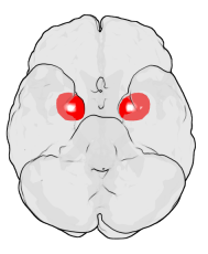
The amygdala is one of two almond-shaped clusters of nuclei located deep and medially within the temporal lobes of the brain's cerebrum in complex vertebrates, including humans. Shown to perform a primary role in the processing of memory, decision making, and emotional responses, the amygdalae are considered part of the limbic system. The term "amygdala" was first introduced by Karl Friedrich Burdach in 1822.

The mesolimbic pathway, sometimes referred to as the reward pathway, is a dopaminergic pathway in the brain. The pathway connects the ventral tegmental area in the midbrain to the ventral striatum of the basal ganglia in the forebrain. The ventral striatum includes the nucleus accumbens and the olfactory tubercle.

<span class="mw-page-title-main">Cingulate cortex</span> Part of the brain within the cerebral cortex

The cingulate cortex is a part of the brain situated in the medial aspect of the cerebral cortex. The cingulate cortex includes the entire cingulate gyrus, which lies immediately above the corpus callosum, and the continuation of this in the cingulate sulcus. The cingulate cortex is usually considered part of the limbic lobe.

<span class="mw-page-title-main">Caudate nucleus</span> Structure of the striatum in the basal ganglia of the brain

The caudate nucleus is one of the structures that make up the corpus striatum, which is a component of the basal ganglia in the human brain. While the caudate nucleus has long been associated with motor processes due to its role in Parkinson's disease, it plays important roles in various other nonmotor functions as well, including procedural learning, associative learning and inhibitory control of action, among other functions. The caudate is also one of the brain structures which compose the reward system and functions as part of the cortico–basal ganglia–thalamic loop.

<span class="mw-page-title-main">Olfactory bulb</span> Neural structure

The olfactory bulb is a neural structure of the vertebrate forebrain involved in olfaction, the sense of smell. It sends olfactory information to be further processed in the amygdala, the orbitofrontal cortex (OFC) and the hippocampus where it plays a role in emotion, memory and learning. The bulb is divided into two distinct structures: the main olfactory bulb and the accessory olfactory bulb. The main olfactory bulb connects to the amygdala via the piriform cortex of the primary olfactory cortex and directly projects from the main olfactory bulb to specific amygdala areas. The accessory olfactory bulb resides on the dorsal-posterior region of the main olfactory bulb and forms a parallel pathway. Destruction of the olfactory bulb results in ipsilateral anosmia, while irritative lesions of the uncus can result in olfactory and gustatory hallucinations.

<span class="mw-page-title-main">Frontal lobe</span> Part of the brain

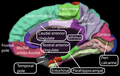
The frontal lobe is the largest of the four major lobes of the brain in mammals, and is located at the front of each cerebral hemisphere. It is parted from the parietal lobe by a groove between tissues called the central sulcus and from the temporal lobe by a deeper groove called the lateral sulcus. The most anterior rounded part of the frontal lobe is known as the frontal pole, one of the three poles of the cerebrum.

<span class="mw-page-title-main">Brodmann area 10</span> Brain area

Brodmann area 10 is the anterior-most portion of the prefrontal cortex in the human brain. BA10 was originally defined broadly in terms of its cytoarchitectonic traits as they were observed in the brains of cadavers, but because modern functional imaging cannot precisely identify these boundaries, the terms anterior prefrontal cortex, rostral prefrontal cortex and frontopolar prefrontal cortex are used to refer to the area in the most anterior part of the frontal cortex that approximately covers BA10—simply to emphasize the fact that BA10 does not include all parts of the prefrontal cortex.

<span class="mw-page-title-main">Brodmann area 11</span> Brain area

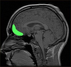
Brodmann area 11 is one of Brodmann's cytologically defined regions of the brain. It is in the orbitofrontal cortex which is above the eye sockets (orbitae). It is involved in decision making, processing rewards, and encoding new information into long-term memory..

<span class="mw-page-title-main">Dopaminergic pathways</span> Projection neurons in the brain that synthesize and release dopamine

Dopaminergic pathways in the human brain are involved in both physiological and behavioral processes including movement, cognition, executive functions, reward, motivation, and neuroendocrine control. Each pathway is a set of projection neurons, consisting of individual dopaminergic neurons.

<span class="mw-page-title-main">Ventral tegmental area</span> Group of neurons on the floor of the midbrain

The ventral tegmental area (VTA), also known as the ventral tegmental area of Tsai, or simply ventral tegmentum, is a group of neurons located close to the midline on the floor of the midbrain. The VTA is the origin of the dopaminergic cell bodies of the mesocorticolimbic dopamine system and other dopamine pathways; it is widely implicated in the drug and natural reward circuitry of the brain. The VTA plays an important role in a number of processes, including reward cognition and orgasm, among others, as well as several psychiatric disorders. Neurons in the VTA project to numerous areas of the brain, ranging from the prefrontal cortex to the caudal brainstem and several regions in between.

<span class="mw-page-title-main">Prefrontal cortex</span> Part of the brain responsible for personality, decision-making, and social behavior

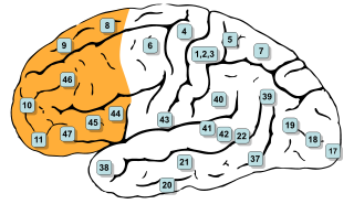
In mammalian brain anatomy, the prefrontal cortex (PFC) covers the front part of the frontal lobe of the cerebral cortex. The PFC contains the Brodmann areas BA8, BA9, BA10, BA11, BA12, BA13, BA14, BA24, BA25, BA32, BA44, BA45, BA46, and BA47.

<span class="mw-page-title-main">Affective neuroscience</span> Study of the neural mechanisms of emotion

Affective neuroscience is the study of how the brain processes emotions. This field combines neuroscience with the psychological study of personality, emotion, and mood. The basis of emotions and what emotions are remains an issue of debate within the field of affective neuroscience.

Frontostriatal circuits are neural pathways that connect frontal lobe regions with the basal ganglia (striatum) that mediate motor, cognitive, and behavioural functions within the brain. They receive inputs from dopaminergic, serotonergic, noradrenergic, and cholinergic cell groups that modulate information processing. Frontostriatal circuits are part of the executive functions. Executive functions include the following: selection and perception of important information, manipulation of information in working memory, planning and organization, behavioral control, adaptation to changes, and decision making. These circuits are involved in neurodegenerative disorders such as Alzheimer's disease and Parkinson's disease as well as neuropsychiatric disorders including schizophrenia, depression, obsessive compulsive disorder (OCD), and in neurodevelopmental disorder such as attention-deficit hyperactivity disorder (ADHD).

<span class="mw-page-title-main">Reward system</span> Group of neural structures responsible for motivation and desire

The reward system is a group of neural structures responsible for incentive salience, associative learning, and positively-valenced emotions, particularly ones involving pleasure as a core component. Reward is the attractive and motivational property of a stimulus that induces appetitive behavior, also known as approach behavior, and consummatory behavior. A rewarding stimulus has been described as "any stimulus, object, event, activity, or situation that has the potential to make us approach and consume it is by definition a reward". In operant conditioning, rewarding stimuli function as positive reinforcers; however, the converse statement also holds true: positive reinforcers are rewarding.

<span class="mw-page-title-main">Ventromedial prefrontal cortex</span> Body part

The ventromedial prefrontal cortex (vmPFC) is a part of the prefrontal cortex in the mammalian brain. The ventral medial prefrontal is located in the frontal lobe at the bottom of the cerebral hemispheres and is implicated in the processing of risk and fear, as it is critical in the regulation of amygdala activity in humans. It also plays a role in the inhibition of emotional responses, and in the process of decision-making and self-control. It is also involved in the cognitive evaluation of morality.

Scientific studies have found that different brain areas show altered activity in humans with major depressive disorder (MDD), and this has encouraged advocates of various theories that seek to identify a biochemical origin of the disease, as opposed to theories that emphasize psychological or situational causes. Factors spanning these causative groups include nutritional deficiencies in magnesium, vitamin D, and tryptophan with situational origin but biological impact. Several theories concerning the biologically based cause of depression have been suggested over the years, including theories revolving around monoamine neurotransmitters, neuroplasticity, neurogenesis, inflammation and the circadian rhythm. Physical illnesses, including hypothyroidism and mitochondrial disease, can also trigger depressive symptoms.

The biology of obsessive–compulsive disorder (OCD) refers biologically based theories about the mechanism of OCD. Cognitive models generally fall into the category of executive dysfunction or modulatory control. Neuroanatomically, functional and structural neuroimaging studies implicate the prefrontal cortex (PFC), basal ganglia (BG), insula, and posterior cingulate cortex (PCC). Genetic and neurochemical studies implicate glutamate and monoamine neurotransmitters, especially serotonin and dopamine.

Neuromorality is an emerging field of neuroscience that studies the connection between morality and neuronal function. Scientists use fMRI and psychological assessment together to investigate the neural basis of moral cognition and behavior. Evidence shows that the central hub of morality is the prefrontal cortex guiding activity to other nodes of the neuromoral network. A spectrum of functional characteristics within this network to give rise to both altruistic and psychopathological behavior. Evidence from the investigation of neuromorality has applications in both clinical neuropsychiatry and forensic neuropsychiatry.

<span class="mw-page-title-main">Biology of bipolar disorder</span> Biological Study Of Bipolar Disorder

Bipolar disorder is an affective disorder characterized by periods of elevated and depressed mood. The cause and mechanism of bipolar disorder is not yet known, and the study of its biological origins is ongoing. Although no single gene causes the disorder, a number of genes are linked to increase risk of the disorder, and various gene environment interactions may play a role in predisposing individuals to developing bipolar disorder. Neuroimaging and postmortem studies have found abnormalities in a variety of brain regions, and most commonly implicated regions include the ventral prefrontal cortex and amygdala. Dysfunction in emotional circuits located in these regions have been hypothesized as a mechanism for bipolar disorder. A number of lines of evidence suggests abnormalities in neurotransmission, intracellular signalling, and cellular functioning as possibly playing a role in bipolar disorder.

Social cognitive neuroscience is the scientific study of the biological processes underpinning social cognition. Specifically, it uses the tools of neuroscience to study "the mental mechanisms that create, frame, regulate, and respond to our experience of the social world". Social cognitive neuroscience uses the epistemological foundations of cognitive neuroscience, and is closely related to social neuroscience. Social cognitive neuroscience employs human neuroimaging, typically using functional magnetic resonance imaging (fMRI). Human brain stimulation techniques such as transcranial magnetic stimulation and transcranial direct-current stimulation are also used. In nonhuman animals, direct electrophysiological recordings and electrical stimulation of single cells and neuronal populations are utilized for investigating lower-level social cognitive processes.

References

  1. 1 2 Kringelbach M. L. (2005). "The orbitofrontal cortex: linking reward to hedonic experience". Nature Reviews Neuroscience. 6 (9): 691–702. doi:10.1038/nrn1747. PMID   16136173. S2CID   205500365.
  2. Phillips, LH., MacPherson, SE. & Della Sala, S. (2002). 'Age, cognition and emotion: the role of anatomical segregation in the frontal lobes: the role of anatomical segregation in the frontal lobes'. in J Grafman (ed.), Handbook of Neuropsychology: the frontal lobes. Elsevier Science, Amsterdam, pp. 73-98.
  3. Barbas H, Ghashghaei H, Rempel-Clower N, Xiao D (2002) Anatomic basis of functional specialization in prefrontal cortices in primates. In: Handbook of Neuropsychology (Grafman J, ed), pp 1-27. Amsterdam: Elsevier Science B.V.
  4. Gottfried JA, Zald DH. On the scent of human olfactory orbitofrontal cortex: meta-analysis and comparison to non-human primates. Brain Res Rev 2005;50:287–304.
  5. Rolls ET. The functions of the orbitofrontal cortex. Brain Cogn 2004;55:11–29.
  6. Kringelbach ML. The human orbitofrontal cortex: linking reward to hedonic experience. Nat Rev Neurosci 2005;6:691–702
  7. Rushworth M, Behrens T, Rudebeck P, Walton M. Contrasting roles for cingulate and orbitofrontal cortex in decisions and social behaviour. Trends Cogn Sci 2007;11: 168–176.
  8. Dixon ML, Thiruchselvam R, Todd RM, ChristoffK. Emotion and the prefrontal cortex: an integrative review. Psychol Bull 2017;143:1033–1081.
  9. Kringelbach ML, Rolls ET. The functional neuroanatomy of the human orbitofrontal cortex: evidence from neuroimaging and neuropsychology. Prog Neurobiol 2004;72: 341–372.
  10. Damasio AR. Descartes’Error: Emotion, Reason and the Human Brain. New York: Putnam; 1994.
  11. Fuster, J.M. The Prefrontal Cortex, (Raven Press, New York, 1997).
  12. Isamah N, Faison W, Payne ME, MacFall J, Steffens DC, Beyer JL, Krishnan R, Taylor WD (2010). "Variability in Frontotemporal Brain Structure: The Importance of Recruitment of African Americans in Neuroscience Research". PLOS ONE. 5 (10): e13642. Bibcode:2010PLoSO...513642I. doi: 10.1371/journal.pone.0013642 . PMC   2964318 . PMID   21049028.
  13. Uylings HB, Groenewegen HJ, Kolb B (2003). "Do rats have a prefrontal cortex?". Behav Brain Res. 146 (1–2): 3–17. doi:10.1016/j.bbr.2003.09.028. PMID   14643455. S2CID   32136463.
  14. 1 2 Mackey, Sott; Petrides, Michael (2006). "Chapter 2: The orbitofrontal cortex: sulcal and gyral morphology and architecture". In Zald, David H.; Rauch, Scott (eds.). The Orbitofrontal Cortex. New York: Oxford University Press. p. 34. ISBN   9780198565741.
  15. Mackey, Sott; Petrides, Michael (2006). "Chapter 2: The orbitofrontal cortex: sulcal and gyral morphology and architecture". In Zald, David H.; Rauch, Scott (eds.). The Orbitofrontal Cortex. New York: Oxford University Press. p. 24. ISBN   9780198565741.
  16. 1 2 3 Passingham, Richard E.; Wise, Steven P. (1012). "Chapter 4 Orbital prefrontal cortex: choosing objects based on outcomes". The Neurobiology of the Prefrontal Cortex: Anatomy, Evolution and Origin of Insight. Great Clarendon Street, Oxford: Oxford University Press. p. 97. ISBN   9780199552917.
  17. 1 2 Haber, SN; Behrens, TE (3 September 2014). "The neural network underlying incentive-based learning: implications for interpreting circuit disruptions in psychiatric disorders". Neuron. 83 (5): 1019–39. doi:10.1016/j.neuron.2014.08.031. PMC   4255982 . PMID   25189208.
  18. 1 2 Barbas, Helen; Zikopoulos, Basilis (2006). "Chapter 4: Sequential and parallel circuits for emotional processing in the primate orbitofrontal cortex". In Rauch, Scott L.; Zald, David H. (eds.). The Orbitofrontal Cortex. New York: Oxford University Press. p. 67.
  19. 1 2 3 Price, Joseph L. (2006). "Chapter 3: Connections of the orbital cortex". In Rauch, Scott L.; Zald, David H. (eds.). The Orbitofrontal Cortex. New York: Oxford University Press. p. 42.
  20. Rudebeck, PH; Murray, EA (December 2011). "Balkanizing the primate orbitofrontal cortex: distinct subregions for comparing and contrasting values". Annals of the New York Academy of Sciences. 1239 (1): 1–13. Bibcode:2011NYASA1239....1R. doi:10.1111/j.1749-6632.2011.06267.x. PMC   3951748 . PMID   22145870.
  21. 1 2 3 Rolls, ET (March 2000). "The orbitofrontal cortex and reward". Cerebral Cortex. 10 (3): 284–94. doi: 10.1093/cercor/10.3.284 . PMID   10731223.
  22. Rolls, ET (November 2004). "Convergence of sensory systems in the orbitofrontal cortex in primates and brain design for emotion". The Anatomical Record Part A: Discoveries in Molecular, Cellular, and Evolutionary Biology. 281 (1): 1212–25. doi: 10.1002/ar.a.20126 . PMID   15470678.
  23. 1 2 Rempel-Clower, NL (December 2007). "Role of orbitofrontal cortex connections in emotion". Annals of the New York Academy of Sciences. 1121 (1): 72–86. Bibcode:2007NYASA1121...72R. doi:10.1196/annals.1401.026. PMID   17846152. S2CID   21317263.
  24. Price, Joseph L. (2006). "Chapter 3: Connections of the orbital cortex". In Rauch, Scott L.; Zald, David H. (eds.). The Orbitofrontal Cortex. New York: Oxford University Press. p. 45.
  25. 1 2 3 Wikenheiser, AM; Schoenbaum, G (August 2016). "Over the river, through the woods: cognitive maps in the hippocampus and orbitofrontal cortex". Nature Reviews. Neuroscience. 17 (8): 513–23. doi:10.1038/nrn.2016.56. PMC   5541258 . PMID   27256552.
  26. 1 2 Fettes, P; Schulze, L; Downar, J (2017). "Cortico-Striatal-Thalamic Loop Circuits of the Orbitofrontal Cortex: Promising Therapeutic Targets in Psychiatric Illness". Frontiers in Systems Neuroscience. 11: 25. doi: 10.3389/fnsys.2017.00025 . PMC   5406748 . PMID   28496402.
  27. Wilson, Robert C.; Takahashi, Yuji K.; Schoenbaum, Geoffrey; Niv, Yael (January 2014). "Orbitofrontal Cortex as a Cognitive Map of Task Space". Neuron. 81 (2): 267–279. doi:10.1016/j.neuron.2013.11.005. ISSN   0896-6273. PMC   4001869 . PMID   24462094.
  28. 1 2 3 Sadacca, BF; Wikenheiser, AM; Schoenbaum, G (14 March 2017). "Toward a theoretical role for tonic norepinephrine in the orbitofrontal cortex in facilitating flexible learning". Neuroscience. 345: 124–129. doi:10.1016/j.neuroscience.2016.04.017. PMC   5461826 . PMID   27102419.
  29. Stalnaker, TA; Cooch, NK; Schoenbaum, G (May 2015). "What the orbitofrontal cortex does not do". Nature Neuroscience. 18 (5): 620–7. doi:10.1038/nn.3982. PMC   5541252 . PMID   25919962.
  30. Tobia, M. J.; Guo, R.; Schwarze, U.; Boehmer, W.; Gläscher, J.; Finckh, B.; Marschner, A.; Büchel, C.; Obermayer, K.; Sommer, T. (2014-04-01). "Neural systems for choice and valuation with counterfactual learning signals". NeuroImage. 89: 57–69. doi:10.1016/j.neuroimage.2013.11.051. ISSN   1053-8119. PMID   24321554. S2CID   35280557.
  31. Izquierdo, A (1 November 2017). "Functional Heterogeneity within Rat Orbitofrontal Cortex in Reward Learning and Decision Making". The Journal of Neuroscience. 37 (44): 10529–10540. doi:10.1523/JNEUROSCI.1678-17.2017. PMC   6596524 . PMID   29093055.
  32. Rudebeck, PH; Murray, EA (December 2011). "Balkanizing the primate orbitofrontal cortex: distinct subregions for comparing and contrasting values". Annals of the New York Academy of Sciences. 1239 (1): 1–13. Bibcode:2011NYASA1239....1R. doi:10.1111/j.1749-6632.2011.06267.x. PMC   3951748 . PMID   22145870.
  33. Kringelbach, ML; Rolls, ET (April 2004). "The functional neuroanatomy of the human orbitofrontal cortex: evidence from neuroimaging and neuropsychology". Progress in Neurobiology. 72 (5): 341–72. doi:10.1016/j.pneurobio.2004.03.006. PMID   15157726. S2CID   13624163.
  34. Sescousse, G; Caldú, X; Segura, B; Dreher, JC (May 2013). "Processing of primary and secondary rewards: a quantitative meta-analysis and review of human functional neuroimaging studies". Neuroscience and Biobehavioral Reviews. 37 (4): 681–96. doi:10.1016/j.neubiorev.2013.02.002. hdl: 2066/117487 . PMID   23415703. S2CID   8335094.
  35. Padoa-Schioppa, C; Conen, KE (15 November 2017). "Orbitofrontal Cortex: A Neural Circuit for Economic Decisions". Neuron. 96 (4): 736–754. doi:10.1016/j.neuron.2017.09.031. PMC   5726577 . PMID   29144973.
  36. Sharpe, MJ; Schoenbaum, G (May 2016). "Back to basics: Making predictions in the orbitofrontal-amygdala circuit". Neurobiology of Learning and Memory. 131: 201–6. doi:10.1016/j.nlm.2016.04.009. PMC   5541254 . PMID   27112314.
  37. Barbas, H (August 2007). "Flow of information for emotions through temporal and orbitofrontal pathways". Journal of Anatomy. 211 (2): 237–49. doi:10.1111/j.1469-7580.2007.00777.x. PMC   2375774 . PMID   17635630. The posterior orbitofrontal cortex targets dual systems in the amygdala which have opposite effects on central autonomic structures. Both pathways originate in posterior orbitofrontal cortex, but one targets heavily the inhibitory intercalated masses, whose activation can ultimately disinhibit central autonomic structures during emotional arousal.
  38. Zikopoulos, B; Höistad, M; John, Y; Barbas, H (17 May 2017). "Posterior Orbitofrontal and Anterior Cingulate Pathways to the Amygdala Target Inhibitory and Excitatory Systems with Opposite Functions". The Journal of Neuroscience. 37 (20): 5051–5064. doi:10.1523/JNEUROSCI.3940-16.2017. PMC   5444191 . PMID   28411274. The specific innervation of inhibitory systems in the amygdala found here, along with the differential impact that dopamine has on them, makes it possible to hypothesize how distinct autonomic states may be achieved. A strong pOFC influence on IM that activates DARPP-32+ and CB+ neurons may help modulate autonomic function by downregulating CeM and thereby facilitate social interactions in primates....On the other hand, in a panic condition, when survival is perceived to be threatened, dopamine levels markedly increase. DARPP-32+ neurons in IM may thus be primarily inhibited, rendering the pOFC pathway ineffective.
  39. Walton M. E.; Behrens T. E.; Buckley M. J.; Rudebeck P. H.; Rushworth M. F. (2010). "Separable learning systems in the macaque brain and the role of orbitofrontal cortex in contingent learning". Neuron. 65 (6): 927–939. doi:10.1016/j.neuron.2010.02.027. PMC   3566584 . PMID   20346766.
  40. Campbell-Meiklejohn D. K.; Kanai R.; Bahrami B.; Bach D. R.; Dolan R. J.; Roepstorff A.; Frith C. D. (2012). "Structure of orbitofrontal cortex predicts social influence". Current Biology. 22 (4): R123–R124. doi:10.1016/j.cub.2012.01.012. PMC   3315000 . PMID   22361146.
  41. Tanferna A.; López-Jiménez L.; Blas J.; Hiraldo F.; Sergio F. (2012). "How Expert Advice Influences Decision Making". PLOS ONE. 7 (11): e49748. Bibcode:2012PLoSO...749748M. doi: 10.1371/journal.pone.0049748 . PMC   3504100 . PMID   23185425.
  42. Berridge, KC; Kringelbach, ML (6 May 2015). "Pleasure systems in the brain". Neuron. 86 (3): 646–64. doi:10.1016/j.neuron.2015.02.018. PMC   4425246 . PMID   25950633.
  43. Numan, Michael (2015). Neurobiology of Social Behavior: Toward an Understanding of the Prosocial and Antisocial Brain. Londong: Elsevier Science. p. 85.
  44. Rolls, Edmund T. (2006). "Chapter 5 The Neurophysiology and Functions of the Orbitofrontal Cortex". In Zald, David H.; Rauch, Scott L. (eds.). The Orbitofrontal Cortex. New York: Oxford University Press.
  45. Hunt LT; Malalasekera WMN; de Berker AO; Miranda B; Farmer S; Behrens TEJ; Kennerley SW (26 September 2018). "Triple dissociation of attention and decision computations across prefrontal cortex". Nature Neuroscience. 21 (9): 1471–1481. doi:10.1038/s41593-018-0239-5. PMC   6331040 . PMID   30258238.
  46. Schultz, Wolfram; Tremblay, Leon (2006). "Chapter 7: Involvement of primate orbitofrontal neurons in reward, uncertainty, and learning 173 Wolfram Schultz and Leon Tremblay". In Zald, David H.; Rauch, Scott :L. (eds.). The Orbitofrontal Cortex. New York: Oxford University Press.
  47. Schoenbaum G, Takahashi Y, Liu T, McDannald M (2011). "Does the orbitofrontal cortex signal value?". Annals of the New York Academy of Sciences. 1239 (1): 87–99. Bibcode:2011NYASA1239...87S. doi:10.1111/j.1749-6632.2011.06210.x. PMC   3530400 . PMID   22145878.
  48. Berlin, HA; Rolls, ET; Iversen, SD (October 2005). "Borderline Personality Disorder, Impulsivity, and the Orbitofrontal Cortex". Archives of Clinical Neuropsychology. 20 (7): 862–863.
  49. Ha, Sungji; Sohn, In-Jung; Kim, Namwook; Sim, Hyeon Jeong; Cheon, Keun-Ah (December 2015). "Characteristics of Brains in Autism Spectrum Disorder: Structure, Function and Connectivity across the Lifespan". Experimental Neurobiology. 24 (4): 273–284. doi:10.5607/en.2015.24.4.273. ISSN   1226-2560. PMC   4688328 . PMID   26713076.
  50. Jackowski, AP; Araújo Filho, GM; Almeida, AG; Araújo, CM; Reis, M; Nery, F; Batista, IR; Silva, I; Lacerda, AL (June 2012). "The involvement of the orbitofrontal cortex in psychiatric disorders: an update of neuroimaging findings". Revista Brasileira de Psiquiatria. 34 (2): 207–12. doi: 10.1590/S1516-44462012000200014 . PMID   22729418.
  51. Milad, MR; Rauch, SL (December 2007). "The role of the orbitofrontal cortex in anxiety disorders". Annals of the New York Academy of Sciences. 1121 (1): 546–61. Bibcode:2007NYASA1121..546M. doi:10.1196/annals.1401.006. PMID   17698998. S2CID   34467365.
  52. Nakao, T; Okada, K; Kanba, S (August 2014). "Neurobiological model of obsessive-compulsive disorder: evidence from recent neuropsychological and neuroimaging findings". Psychiatry and Clinical Neurosciences. 68 (8): 587–605. doi: 10.1111/pcn.12195 . PMID   24762196. S2CID   5528241.
  53. Fineberg, NA; Potenza, MN; Chamberlain, SR; Berlin, HA; Menzies, L; Bechara, A; Sahakian, BJ; Robbins, TW; Bullmore, ET; Hollander, E (February 2010). "Probing compulsive and impulsive behaviors, from animal models to endophenotypes: a narrative review". Neuropsychopharmacology. 35 (3): 591–604. doi:10.1038/npp.2009.185. PMC   3055606 . PMID   19940844.
  54. Milad, MR; Rauch, SL (January 2012). "Obsessive-compulsive disorder: beyond segregated cortico-striatal pathways". Trends in Cognitive Sciences. 16 (1): 43–51. doi:10.1016/j.tics.2011.11.003. PMC   4955838 . PMID   22138231.
  55. Vaghi, M; Robbins, T. "TASK-BASED FUNCTIONAL NEUROIMAGING STUDIES OF OBSESSIVE-COMPULSIVE DISORDER: A HYPOTHESIS-DRIVEN REVIEW". In Pittenger, Christopher (ed.). Obsessive Compulsive Disorder, Phenomenology, Pathophysiology and Treatment. Oxford University Press. pp. 239–240.
  56. Schoenbaum, G; Chang, CY; Lucantonio, F; Takahashi, YK (December 2016). "Thinking Outside the Box: Orbitofrontal Cortex, Imagination, and How We Can Treat Addiction". Neuropsychopharmacology. 41 (13): 2966–2976. doi:10.1038/npp.2016.147. PMC   5101562 . PMID   27510424.
  57. Koob, GF; Volkow, ND (January 2010). "Neurocircuitry of addiction". Neuropsychopharmacology. 35 (1): 217–38. doi:10.1038/npp.2009.110. PMC   2805560 . PMID   19710631.
  58. Moorman, DE (2 February 2018). "The role of the orbitofrontal cortex in alcohol use, abuse, and dependence". Progress in Neuro-psychopharmacology & Biological Psychiatry. 87 (Pt A): 85–107. doi:10.1016/j.pnpbp.2018.01.010. PMC   6072631 . PMID   29355587.
  59. Gowin, JL; Mackey, S; Paulus, MP (1 September 2013). "Altered risk-related processing in substance users: imbalance of pain and gain". Drug and Alcohol Dependence. 132 (1–2): 13–21. doi:10.1016/j.drugalcdep.2013.03.019. PMC   3748224 . PMID   23623507. Individuals with SUDs show several processing abnormalities during risk-taking decision-making, which include altered valuation of options (VMPFC) and outcomes (OFC and striatum), poor estimation of uncertainty (ACC and insular cortex), diminished executive control (DLPFC), and an attenuated influence of emotional salience (amygdala), and reduced responsiveness to somatic markers (somatosensory cortex). These neural processing differences during risk-taking among individuals with SUDs have been linked to poorer behavioral performance on risk-taking tasks and a more extensive history of substance use
  60. Chase, HW; Eickhoff, SB; Laird, AR; Hogarth, L (15 October 2011). "The neural basis of drug stimulus processing and craving: an activation likelihood estimation meta-analysis". Biological Psychiatry. 70 (8): 785–93. doi:10.1016/j.biopsych.2011.05.025. PMC   4827617 . PMID   21757184. A medial region of the OFC showed greater activation by drug cues compared with control cues and was consistently activated in the nontreatment-seeking subgroup. There is substantial evidence that this region plays a role in appetitive behavior and decision making (86,87), in particular with regard to expectations of reward (88) predicted by conditioned stimuli (89–94), which can control instrumental action selectio
  61. Lucantonio, F; Caprioli, D; Schoenbaum, G (January 2014). "Transition from 'model-based' to 'model-free' behavioral control in addiction: Involvement of the orbitofrontal cortex and dorsolateral striatum". Neuropharmacology. 76 Pt B: 407–15. doi:10.1016/j.neuropharm.2013.05.033. PMC   3809026 . PMID   23752095.
  62. Rubia, K (15 June 2011). ""Cool" inferior frontostriatal dysfunction in attention-deficit/hyperactivity disorder versus "hot" ventromedial orbitofrontal-limbic dysfunction in conduct disorder: a review". Biological Psychiatry. 69 (12): e69–87. doi:10.1016/j.biopsych.2010.09.023. PMID   21094938. S2CID   14987165.
  63. Rosell, DR; Siever, LJ (June 2015). "The neurobiology of aggression and violence". CNS Spectrums. 20 (3): 254–79. doi: 10.1017/S109285291500019X . PMID   25936249.
  64. 1 2 Rolls, ET (September 2016). "A non-reward attractor theory of depression" (PDF). Neuroscience and Biobehavioral Reviews. 68: 47–58. doi:10.1016/j.neubiorev.2016.05.007. PMID   27181908. S2CID   8145667.
  65. J. Wilson; M. Jenkinson; I. E. T. de Araujo; Morten L. Kringelbach; E. T. Rolls & Peter Jezzard (October 2002). "Fast, fully automated global and local magnetic field optimization for fMRI of the human brain". NeuroImage . 17 (2): 967–976. doi:10.1016/S1053-8119(02)91172-9. PMID   12377170.
  66. Ahmari, SE; Dougherty, DD (August 2015). "Dissecting Ocd Circuits: From Animal Models to Targeted Treatments". Depression and Anxiety. 32 (8): 550–62. doi:10.1002/da.22367. PMC   4515165 . PMID   25952989.
  67. Vertechi, Pietro; Lottem, Eran; Sarra, Dario; Godinho, Beatriz; Treves, Isaac; Quendera, Tiago; Lohuis, Matthijs Nicolai Oude; Mainen, Zachary F. (2020-04-08). "Inference-Based Decisions in a Hidden State Foraging Task: Differential Contributions of Prefrontal Cortical Areas". Neuron. 106 (1): 166–176.e6. doi: 10.1016/j.neuron.2020.01.017 . ISSN   0896-6273. PMC   7146546 . PMID   32048995.
  68. Snowden J. S.; Bathgate D.; Varma A.; Blackshaw A.; Gibbons Z. C.; Neary D. (2001). "Distinct behavioural profiles in frontotemporal dementia and semantic dementia". J Neurol Neurosurg Psychiatry. 70 (3): 323–332. doi:10.1136/jnnp.70.3.323. PMC   1737271 . PMID   11181853.
  69. Xiao, Xiong; Deng, Hanfei; Wei, Lei; Huang, Yanwang; Wang, Zuoren (September 2016). "Neural activity of orbitofrontal cortex contributes to control of waiting". The European Journal of Neuroscience. 44 (6): 2300–2313. doi:10.1111/ejn.13320. ISSN   1460-9568. PMID   27336203. S2CID   205105682.
  70. 1 2 Paulus M. P.; Hozack N. E.; Zauscher B. E.; Frank L.; Brown G. G.; Braff D. L.; Schuckit M. A. (2002). "Behavioral and Functional Neuroimaging Evidence for Prefrontal Dysfunction in Methamphetamine-Dependent Subjects". Neuropsychopharmacology. 26 (1): 53–63. doi: 10.1016/s0893-133x(01)00334-7 . PMID   11751032.
  71. 1 2 Toplak M. E.; Jain U.; Tannock R. (2005). "Executive and motivational processes in adolescents with Attention-Deficit-Hyperactivity Disorder (ADHD)". Behavioral and Brain Functions. 1 (1): 8–20. doi: 10.1186/1744-9081-1-8 . PMC   1183187 . PMID   15982413.
  72. 1 2 Verdejo-Garcia A.; Bechara A.; Recknor E. C.; Perez-Garcia M. (2006). "Executive dysfunction in substance dependent individuals during drug use and abstinence: An examination of the behavioral, cognitive and emotional correlates of addiction". Journal of the International Neuropsychological Society. 12 (3): 405–415. doi:10.1017/s1355617706060486. PMID   16903133. S2CID   15939155.
  73. Cha, Jiook; Greenberg, Tsafrir; Carlson, Joshua M.; DeDora, Daniel J.; Hajcak, Greg; Mujica-Parodi, Lilianne R. (2014-03-12). "Circuit-Wide Structural and Functional Measures Predict Ventromedial Prefrontal Cortex Fear Generalization: Implications for Generalized Anxiety Disorder". The Journal of Neuroscience. 34 (11): 4043–4053. doi:10.1523/JNEUROSCI.3372-13.2014. ISSN   0270-6474. PMC   6705282 . PMID   24623781.
  74. Volkow N.D.; Fowler J.S. (2000). "Addiction a disease of compulsion and drive: involvement of the orbitofrontal cortex". Cerebral Cortex. 10 (3): 318–325. doi: 10.1093/cercor/10.3.318 . PMID   10731226.
  75. Chamberlain S. R.; Odlaug B. L.; Boulougouris V.; Fineberg N. A.; Grant J. E. (2009). "Trichotillomania: Neurobiology and treatment". Neuroscience and Biobehavioral Reviews. 33 (6): 831–842. doi:10.1016/j.neubiorev.2009.02.002. PMID   19428495. S2CID   6956143.
  76. Menzies L. (2008). "Integrating evidence from neuroimaging and neuropsychological studies of obsessive-compulsive disorder: The orbitofronto-striatal model revisited". Neuroscience and Biobehavioral Reviews. 32 (3): 525–549. doi:10.1016/j.neubiorev.2007.09.005. PMC   2889493 . PMID   18061263.
  77. 1 2 Tekin S.; Cummings J. L. (2002). "Frontal-subcortical neuronal circuits and clinical neuropsychiatry: An update". Journal of Psychosomatic Research. 53 (2): 647–654. doi:10.1016/s0022-3999(02)00428-2. PMID   12169339.
  78. Rahman S.; Sahakian B. J.; Hodges J. R.; Rogers R. D.; Robbins T. W. (1999). "Specific cognitive deficits in early behavioural variant frontotemporal dementia". Brain. 122 (8): 1469–1493. doi: 10.1093/brain/122.8.1469 . PMID   10430832.
  79. Seeley W. W.; Crawford R.; Rascovsky K.; Kramer J. H.; Weiner M.; Miller B. L.; Gorno-Tempini L. (2008). "Frontal paralimbic network atrophy in very mild behavioral variant frontotemporal dementia". Archives of Neurology. 65 (2): 249–255. doi:10.1001/archneurol.2007.38. PMC   2544627 . PMID   18268196.
  80. Chibane, IS; Boucher, O; Dubeau, F; Tran, TPY; Mohamed, I; McLachlan, R; Sadler, RM; Desbiens, R; Carmant, L; Nguyen, DK (November 2017). "Orbitofrontal epilepsy: Case series and review of literature". Epilepsy & Behavior. 76: 32–38. doi:10.1016/j.yebeh.2017.08.038. PMID   28928072. S2CID   13656956.
  81. Gold, JA; Sher, Y; Maldonado, JR (2016). "Frontal Lobe Epilepsy: A Primer for Psychiatrists and a Systematic Review of Psychiatric Manifestations". Psychosomatics. 57 (5): 445–64. doi:10.1016/j.psym.2016.05.005. PMID   27494984.
  82. Smith, JR; Sillay, K; Winkler, P; King, DW; Loring, DW (2004). "Orbitofrontal epilepsy: electroclinical analysis of surgical cases and literature review". Stereotactic and Functional Neurosurgery. 82 (1): 20–5. doi:10.1159/000076656. PMID   15007215. S2CID   18811550.
  83. Rolls E. T.; Hornak J.; Wade D.; McGrath J. (1994). "Emotion-related learning in patients with social and emotional changes associated with frontal lobe damage". J Neurol Neurosurg Psychiatry. 57 (12): 1518–1524. doi:10.1136/jnnp.57.12.1518. PMC   1073235 . PMID   7798983.
  84. Bechara A.; Damasio A. R.; Damasio H.; Anderson S.W. (1994). "Insensitivity to future consequences following damage to human prefrontal cortex". Cognition. 50 (1–3): 7–15. doi:10.1016/0010-0277(94)90018-3. PMID   8039375. S2CID   204981454.
  85. Stone V.E.; Baron-Cohen S.; Knight R. T. (1998a). "Frontal Lobe Contributions to Theory of Mind". Journal of Medical Investigation. 10 (5): 640–656. CiteSeerX   10.1.1.330.1488 . doi:10.1162/089892998562942. PMID   9802997. S2CID   207724498.