
Skin is the layer of usually soft, flexible outer tissue covering the body of a vertebrate animal, with three main functions: protection, regulation, and sensation.
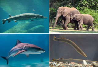
Vertebrates are deuterostomal animals with bony or cartilaginous axial endoskeleton — known as the vertebral column, spine or backbone — around and along the spinal cord, including all fish, amphibians, reptiles, birds and mammals. The vertebrates consist of all the taxa within the subphylum Vertebrata and represent the overwhelming majority of the phylum Chordata, with currently about 69,963 species described.

In zoology, a scale is a small rigid plate that grows out of an animal's skin to provide protection. In lepidopterans, scales are plates on the surface of the insect wing, and provide coloration. Scales are quite common and have evolved multiple times through convergent evolution, with varying structure and function.
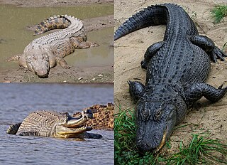
Crocodilia is an order of mostly large, predatory, semiaquatic reptiles known as crocodilians. They first appeared 94 million years ago in the Late Cretaceous period and are the closest living relatives of birds, as the two groups are the only known survivors of the Archosauria. Members of the order's total group, the clade Pseudosuchia, appeared about 250 million years ago in the Early Triassic period, and diversified during the Mesozoic era. The order Crocodilia includes the true crocodiles, the alligators and caimans, and the gharial and false gharial. Although the term crocodiles is sometimes used to refer to all of these, crocodilians is a less ambiguous vernacular term for members of this group.

Euteleostomi is a successful clade that includes more than 90% of the living species of vertebrates. Both its major subgroups are successful today: Actinopterygii includes most extant bony fish species, and Sarcopterygii includes the tetrapods.
The quadratojugal is a skull bone present in many vertebrates, including some living reptiles and amphibians.

A scute or scutum is a bony external plate or scale overlaid with horn, as on the shell of a turtle, the skin of crocodilians, and the feet of birds. The term is also used to describe the anterior portion of the mesonotum in insects as well as some arachnids.
A dermal bone or investing bone or membrane bone is a bony structure derived from intramembranous ossification forming components of the vertebrate skeleton, including much of the skull, jaws, gill covers, shoulder girdle, fin rays (lepidotrichia), and the shells of turtles and armadillos. In contrast to endochondral bone, dermal bone does not form from cartilage that then calcifies, and it is often ornamented. Dermal bone is formed within the dermis and grows by accretion only – the outer portion of the bone is deposited by osteoblasts.

Mymoorapelta is a nodosaurid ankylosaur from the Late Jurassic Morrison Formation of western Colorado and central Utah, USA. The animal is known from a single species, Mymoorapelta maysi, and few specimens are known. The most complete specimen is the holotype individual from the Mygatt-Moore Quarry, which includes osteoderms, a partial skull, vertebrae, and other bones. It was initially described by James Kirkland and Kenneth Carpenter in 1994. Along with Gargoyleosaurus, it is one of the earliest known nodosaurids.

Proterochampsa is an extinct genus of proterochampsid archosauriform from the Late Triassic of South America. The genus is the namesake of the family Proterochampsidae, and the broader clade Proterochampsia. Like other proterochampsids, Proterochampsa are quadruped tetrapods superficially similar in appearance to modern crocodiles, although the two groups are not closely related. Proterochampsids can be distinguished from other related archosauriformes by characters such as a dorsoventrally flattened, triangular skull with a long, narrow snout at the anterior end and that expands transversally at the posterior end, asymmetric feet, and a lack of postfrontal bones in the skull, with the nares located near the midline. Proterochampsa is additionally defined by characters of dermal sculpturing consisting of nodular protuberances on the skull, antorbital fenestrae facing dorsally, and a restricted antorbital fossa on the maxilla. The genus comprises two known species: Proterochampsa barrionuevoi and Proterochampsa nodosa. P. barrionuevoi specimens have been discovered in the Ischigualasto Formation in northwestern Argentina, while P. nodosa specimens have been found in the Santa Maria supersequence in southeastern Brazil. The two species are distinct in several characters, including that P. nodosa has larger, more well-developed nodular protuberances, a more gradually narrowing snout, and a higher occiput than P. barrionuevoi. Of the two, P. nodosa is thought to have less derived features than P. barrionuevoi.

The turtle shell is a shield for the ventral and dorsal parts of turtles, completely enclosing all the vital organs of the turtle and in some cases even the head. It is constructed of modified bony elements such as the ribs, parts of the pelvis and other bones found in most reptiles. The bone of the shell consists of both skeletal and dermal bone, showing that the complete enclosure of the shell likely evolved by including dermal armor into the rib cage.

Eutretauranosuchus is an extinct genus of goniopholidid crocodyliform. E. delfsi is the only known species within the genus.

Armadillosuchus is an extinct genus of sphagesaurid crocodylomorph. It was described in February 2009 from the late Campanian to early Maastrichtian Adamantina Formation of the Bauru Basin in Brazil, dating to approximately 70 Ma. Armadillosuchus was among the larger and more robust sphagesaurids, with a total length of approximately 2 metres (6.6 ft).

The skull roof or the roofing bones of the skull are a set of bones covering the brain, eyes and nostrils in bony fishes and all land-living vertebrates. The bones are derived from dermal bone and are part of the dermatocranium.
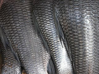
A fish scale is a small rigid plate that grows out of the skin of a fish. The skin of most jawed fishes is covered with these protective scales, which can also provide effective camouflage through the use of reflection and colouration, as well as possible hydrodynamic advantages. The term scale derives from the Old French escale, meaning a shell pod or husk.
Enameloid, also known as durodentine or vitrodentine, is an enamel-like tissue found in many fish. It is the primary outer component of shark odontodes. Although the origin of enameloid is debated, it is probably homologous to dentine rather than true enamel, despite its enamel-like strength and development. The term covers any hyper-mineralized tissue with an organic "scaffold" consisting of ectodermal and ectomesenchymal proteins.

Most bony fishes have two sets of jaws made mainly of bone. The primary oral jaws open and close the mouth, and a second set of pharyngeal jaws are positioned at the back of the throat. The oral jaws are used to capture and manipulate prey by biting and crushing. The pharyngeal jaws, so-called because they are positioned within the pharynx, are used to further process the food and move it from the mouth to the stomach.
This glossary explains technical terms commonly employed in the description of dinosaur body fossils. Besides dinosaur-specific terms, it covers terms with wider usage, when these are of central importance in the study of dinosaurs or when their discussion in the context of dinosaurs is beneficial. The glossary does not cover ichnological and bone histological terms, nor does it cover measurements.
Osteoderms are dermal bone structures that support the upper layer of skin and serve as protection against the elements in a large variety of extinct and extant organisms, especially reptiles. This structure is commonly called "dermal armor" and serves to protect the organism, while also helping with temperature regulation. Osteoderms represent hard tissue components of the integument, making them easy to identify in fossil examination. This dermal armor is found prominently in many lizards. Some early amphibians have this armor, but it is lost in modern species with the exception a ventral plate, called the gastralia.
Postparietals are cranial bones present in fish and many tetrapods. Although initially a pair of bones, many lineages possess postparietals which were fused into a single bone. The postparietals were dermal bones situated along the midline of the skull, behind the parietal bones. They formed part of the rear edge of the skull roof, and the lateral edge of each postparietal often contacts the tabular and supratemporal bones. In fish, the postparietals are elongated, typically the largest components of the skull roof. Tetrapods possessed shorter postparietals, which were reduced further and shifted towards the braincase in amniotes. At several points in synapsid evolution, the postparietals fused to each other and the tabulars during embryological development. This fusion produces the interparietal bone, which is inherited by mammals. Postparietals are common in extinct amphibians and early reptiles. However, most living amphibians and living reptiles lack postparietal bones, with a few exceptions.

