Parabiosis

Last updated

Parabiosis is a laboratory technique used in physiological research, derived from the Greek word meaning "living beside." The technique involves the surgical joining of two living organisms in such a way that they develop a single, shared physiological system. Through this unique approach, researchers can study the exchange of blood, hormones, and other substances between the two organisms, allowing for the examination of a wide range of physiological phenomena and interactions. Parabiosis has been employed in various fields of study, including stem cell research, endocrinology, aging research, and immunology.

Contents

Physiology

Parabiotic experiments

Left: A headless Cecropia Moth joined with a pupa of the Polyphemus silkworm. Right: The abdomen of a Cecropia moth joined with a Cecropia pupa The Biological bulletin (19755290053).jpg
Left: A headless Cecropia Moth joined with a pupa of the Polyphemus silkworm. Right: The abdomen of a Cecropia moth joined with a Cecropia pupa

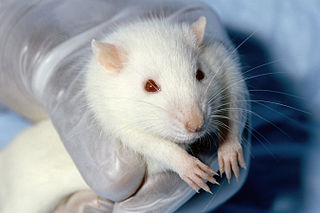
Parabiosis combines two living organisms which are joined surgically and develop single, shared physiological systems. [1] [2] Researchers can prove that the feedback system in one animal is circulated and affects the second animal via blood and plasma exchange.

Parabiotic experiments were pioneered by Paul Bert in the mid-1800s. He postulated that surgically connected animals could share a circulatory system. Bert was awarded the Prize of Experimental Physiology of the French Academy of Science in 1866 for his discoveries. [3]

One limitation of the experiments is that outbred rats cannot be used because it can lead to a significant loss of pairs due to intoxication of the blood supply from a dissimilar rat. [4]

Discovery of leptin and the role of the hypothalamus in obesity

Many of the parabiotic experiments since 1950 involve research regarding metabolism. One of these experiments was published in 1959 by G. R. Hervey in the Journal of Physiology . This experiment supported the theory that damage to the hypothalamus, particularly the ventromedial hypothalamus, leads to obesity caused by the overconsumption of food. The study's rats were from the same litter, which had been a closed colony for multiple years. The two rats in each pair had no more than a 3% difference in weight. Rats were paired at four weeks old. Unpaired rats were used as controls. The rats were conjoined in three ways. In early experiments, the peritoneal cavities were opened and connected between the two rats. In later experiments, to avoid the risk of tangling the two rats’ intestines together, smaller cuts were made. After further refinement of the experimental procedure, the abdominal cavities were not opened, and the rats were conjoined at the hip bone with minimal cutting. To prove that the two animals were sharing blood, researchers injected dye into one rat's veins, and the pigment would show up in the conjoined rat.

In each pair, one rat became obese and exhibited hyperphagia. The weight of the rat with the surgical lesion rose rapidly for a few months, then reached a plateau as a direct result of the surgical procedure. After the procedure, the rat with the impaired hypothalamus ate voraciously while the paired rat's appetite decreased. The paired rat became obviously thin throughout the experiment, even rejecting food when it was offered. [5] [6]

Later studies identified this satiety factor as the adipose-derived hormone leptin. Many hormones and metabolites were proven not to be the satiety factor that caused one rat to starve in the experiments. Leptin seemed like a viable candidate. Starting in 1977, Ruth B.S. Harris, a graduate student under Hervey, repeated previous studies about parabiosis in rats and mice. Due to the discovery of leptin, she analyzed leptin concentrations of the mice in the parabiotic experiments. After injecting leptin into each pair's obese mouse, she found that leptin circulated between the conjoined animals, but the circulation of leptin took some time to reach equilibrium. As a result of the injections, the almost immediate weight loss resulted in the parabiotic pairs due to increased inhibition. Approximately 50–70% of fat was lost in pairs. The obese mouse lost only fat. The lean mouse lost muscle mass and fat. Harris concluded that leptin levels are increased in obese animals, but other factors could also affect them. Also, leptin was determined to decrease fat storage in both obese and thin animals. [4]

Early parabiotic experiments also included cancer research. One study, published in 1966 by Friedell, studied radiation's effects with X-rays on ovarian tumors. To study the tumors, two adult female rats were conjoined. The left rat was shielded, and the right rat was exposed to high levels of radiation. The rats were given a controlled amount of food and water. 149 of 328 pairs showed possible ovarian tumors in the irradiated animals, but not in their partners. This result matched previous studies of single rats. [7]

Aging research

Chronic diseases of age are studied by conjoining an older animal with a younger animal. Known as heterochronic parabiosis, this process has been used in studies to investigate the age-related and disease-related changes in the composition of the blood, especially plasma proteome. [8] This process could be used to research cardiovascular disease, diabetes, osteoarthritis, and Alzheimer's disease. As animals age, their oligodendrocytes reduce in efficiency, resulting in decreased myelination, causing negative effects on the central nervous system (CNS). Julia Ruckh and fellow researchers have used parabiosis to study remyelination from adult stem cells to see if conjoining young with older mice could reverse or delay this process. The two mice were conjoined in the experiment, and demyelination was induced via injection into the older mice. The experiment determined that the younger mice's factors reversed CNS demyelination in older mice by revitalizing the oligodendrocytes. The monocytes from the younger mice also enhanced the older mice's ability to clear myelin debris because the young monocytes can clear lipids from myelin sheaths more effectively than older monocytes. The conjoining of the two animals reversed the effects of age on the myelination cells. The ability of the young mouse's cells was unaffected. Enhanced immunity from the younger mouse also promoted the general health of the older mouse in each pair. The results of this experiment could lead to therapy processes for people with demyelinating diseases like multiple sclerosis. [9] [3]

Natural examples

The term is also applicable to spontaneously occurring conditions such as in conjoined twins. [10]

Obligate parasitic reproduction of Anglerfish of the family Ceratiidae, in which the circulatory systems of the males and females unite completely. Without the attachment of males to females, the endocrine functions cannot mature; the individuals fail to develop properly and die young and without reproducing. [11]

Plants growing closely together roots or stems in intimate contact sometimes form natural grafts. In parasitic plants such as mistletoe and dodder the haustoria unite the circulatory systems of the host and the parasite so intimately that parasitic twiners such as Cassytha may act as vectors carrying disease organisms from one host plant to another. [12]

Ecology

Social organisms sharing nests

Crematogaster modiglianii and Camponotus rufifemur ants sharing a nest Shattuck 53813, Crematogaster modiglianii and Camponotus rufifemur parabiosis, Danum Valley, Sabah-web (5042353719) (2).jpg
Crematogaster modiglianii and Camponotus rufifemur ants sharing a nest

Ant colonies can share their nests with essentially unrelated species of ants. They did not obviously share anything beyond the nests' upkeep, even segregating their brood, so these were very surprising observations; most ants are radically intolerant of intruders, usually including even intruders of their own species.

In the early 20th century Auguste-Henri Forel coined the term "parabiosis" for such associations, and it was adopted by the likes of William Morton Wheeler. [13] [14] Furthermore, there is evidence for the partitioning of functions of work between the two species in the nest. [15] Early reports that parabiotic ant colonies forage and feed together peacefully also have been qualified by observations that revealed ants of one species in such an association aggressively displacing members of the other species from artificially provided food, while also profiting by following their recruitment trails to new food sources. [14] Benefits from shared nest defence and maintenance even when there is neither direct cooperation nor interaction between the two associated populations in a nest. [16]

Etymology

Parabiosis derives most directly from Neo-Latin, [10] but the Latin in turn derives from two classical Greek roots. The first is παρά (para) for "beside" or "next to". In modern etymology, this root appears in various senses, such as "close to", "outside of", and "different".

The second classical Greek root from which the Latin derives is βίος (bios), meaning "life."

See also

Related Research Articles

<span class="mw-page-title-main">Eating</span> Ingestion of food

Eating is the ingestion of food. In biology, this is typically done to provide a heterotrophic organism with energy and nutrients and to allow for growth. Animals and other heterotrophs must eat in order to survive — carnivores eat other animals, herbivores eat plants, omnivores consume a mixture of both plant and animal matter, and detritivores eat detritus. Fungi digest organic matter outside their bodies as opposed to animals that digest their food inside their bodies.

<span class="mw-page-title-main">Leptin</span> Hormone that inhibits hunger

Leptin also obese protein is a protein hormone predominantly made by adipocytes. Its primary role is likely to regulate long-term energy balance.

<span class="mw-page-title-main">Appetite</span> Desire to eat food

Appetite is the desire to eat food items, usually due to hunger. Appealing foods can stimulate appetite even when hunger is absent, although appetite can be greatly reduced by satiety. Appetite exists in all higher life-forms, and serves to regulate adequate energy intake to maintain metabolic needs. It is regulated by a close interplay between the digestive tract, adipose tissue and the brain. Appetite has a relationship with every individual's behavior. Appetitive behaviour also known as approach behaviour, and consummatory behaviour, are the only processes that involve energy intake, whereas all other behaviours affect the release of energy. When stressed, appetite levels may increase and result in an increase of food intake. Decreased desire to eat is termed anorexia, while polyphagia is increased eating. Dysregulation of appetite contributes to anorexia nervosa, bulimia nervosa, cachexia, overeating, and binge eating disorder.

<span class="mw-page-title-main">Adipose tissue</span> Loose connective tissue composed mostly by adipocytes

Adipose tissue is a loose connective tissue composed mostly of adipocytes. It also contains the stromal vascular fraction (SVF) of cells including preadipocytes, fibroblasts, vascular endothelial cells and a variety of immune cells such as adipose tissue macrophages. Its main role is to store energy in the form of lipids, although it also cushions and insulates the body.

<span class="mw-page-title-main">Ghrelin</span> Peptide hormone involved in appetite regulation

Ghrelin is a hormone primarily produced by enteroendocrine cells of the gastrointestinal tract, especially the stomach, and is often called a "hunger hormone" because it increases the drive to eat. Blood levels of ghrelin are highest before meals when hungry, returning to lower levels after mealtimes. Ghrelin may help prepare for food intake by increasing gastric motility and stimulating the secretion of gastric acid.

<span class="mw-page-title-main">Laboratory mouse</span> Mouse used for scientific research

The laboratory mouse or lab mouse is a small mammal of the order Rodentia which is bred and used for scientific research or feeders for certain pets. Laboratory mice are usually of the species Mus musculus. They are the most commonly used mammalian research model and are used for research in genetics, physiology, psychology, medicine and other scientific disciplines. Mice belong to the Euarchontoglires clade, which includes humans. This close relationship, the associated high homology with humans, their ease of maintenance and handling, and their high reproduction rate, make mice particularly suitable models for human-oriented research. The laboratory mouse genome has been sequenced and many mouse genes have human homologues. Lab mice are sold at pet stores for snake food and can also be kept as pets.

<span class="mw-page-title-main">Neuropeptide Y</span> Mammalian protein found in Homo sapiens

Neuropeptide Y (NPY) is a 36 amino-acid neuropeptide that is involved in various physiological and homeostatic processes in both the central and peripheral nervous systems. It is secreted alongside other neurotransmitters such as GABA and glutamate. 

<span class="mw-page-title-main">Laboratory rat</span> Rat used for scientific research

Laboratory rats or lab rats are strains of the rat subspecies Rattus norvegicus domestica which are bred and kept for scientific research. While less commonly used for research than laboratory mice, rats have served as an important animal model for research in psychology and biomedical science.

<span class="mw-page-title-main">Ventromedial nucleus of the hypothalamus</span> Nucleus of the hypothalamus

The ventromedial nucleus of the hypothalamus is a nucleus of the hypothalamus. In 2007, Kurrasch et al. found that the ventromedial hypothalamus is a distinct morphological nucleus involved in terminating hunger, fear, thermoregulation, and sexual activity. This nuclear region is involved in the recognition of the feeling of fullness.

<span class="mw-page-title-main">Jeffrey M. Friedman</span>

Jeffrey M. Friedman is a molecular geneticist at New York City's Rockefeller University and an Investigator of the Howard Hughes Medical Institute. His discovery of the hormone leptin and its role in regulating body weight has had a major role in the area of human obesity. Friedman is a physician scientist studying the genetic mechanisms that regulate body weight. His research on various aspects of obesity received national attention in late 1994, when it was announced that he and his colleagues had isolated the mouse ob gene and its human homologue. They subsequently found that injections of the encoded protein, leptin, decreases body weight of mice by reducing food intake and increasing energy expenditure. Current research is aimed at understanding the genetic basis of obesity in human and the mechanisms by which leptin transmits its weight-reducing signal.

<span class="mw-page-title-main">Animal testing on rodents</span> Overview article

Rodents are commonly used in animal testing, particularly mice and rats, but also guinea pigs, hamsters, gerbils and others. Mice are the most commonly used vertebrate species, due to their availability, size, low cost, ease of handling, and fast reproduction rate.

<span class="mw-page-title-main">Leptin receptor</span> Type I cytokine receptor

Leptin receptor, also known as LEP-R or OB-R, is a type I cytokine receptor, a protein that in humans is encoded by the LEPR gene. LEP-R functions as a receptor for the fat cell-specific hormone leptin. LEP-R has also been designated as CD295. Its location is the cell membrane, and it has extracellular, trans-membrane and intracellular sections.

<span class="mw-page-title-main">Free fatty acid receptor 3</span> Protein-coding gene in the species Homo sapiens

Free fatty acid receptor 3 protein is a G protein coupled receptor that in humans is encoded by the FFAR3 gene. GPRs reside on cell surfaces, bind specific signaling molecules, and thereby are activated to trigger certain functional responses in their parent cells. FFAR3 is a member of the free fatty acid receptor group of GPRs that includes FFAR1, FFAR2, and FFAR4. All of these FFARs are activated by fatty acids. FFAR3 and FFAR2 are activated by certain short-chain fatty acids (SC-FAs), i.e., fatty acids consisting of 2 to 6 carbon atoms whereas FFFAR1 and FFAR4 are activated by certain fatty acids that are 6 to more than 21 carbon atoms long. Hydroxycarboxylic acid receptor 2 is also activated by a SC-FA that activate FFAR3, i.e., butyric acid.

The diet-induced obesity model is an animal model used to study obesity using animals that have obesity caused by being fed high-fat or high-density diets. It is intended to mimic the most common cause of obesity in humans. Typically mice, rats, dogs, or non-human primates are used in these models. These animals can then be used to study in vivo obesity, obesity's comorbidities, and other related diseases. Users of such models must take into account the duration and type of diet as well as the environmental conditions and age of the animals, as each may promote different bodyweights, fat percentages, or behaviors.

Douglas L. Coleman was a scientist and professor emeritus at the Jackson Laboratory, in Bar Harbor, Maine. His work predicted that there exists a hormone that can cause mice to feel full, and that a mutation in the gene encoding this hormone can lead to obesity. The gene and corresponding hormone were discovered about 20 years later by Jeffrey M. Friedman, Rudolph Leibel, and their research teams at Rockefeller University, which Friedman named leptin.

A knockout mouse, or knock-out mouse, is a genetically modified mouse in which researchers have inactivated, or "knocked out", an existing gene by replacing it or disrupting it with an artificial piece of DNA. They are important animal models for studying the role of genes which have been sequenced but whose functions have not been determined. By causing a specific gene to be inactive in the mouse, and observing any differences from normal behaviour or physiology, researchers can infer its probable function.

Ingestive behaviors encompass all eating and drinking behaviors. These actions are influenced by physiological regulatory mechanisms; these mechanisms exist to control and establish homeostasis within the human body. Disruptions in these ingestive regulatory mechanisms can result in eating disorders such as obesity, anorexia, and bulimia.

<span class="mw-page-title-main">Rudolph Leibel</span>

Rudolph Leibel is the Christopher J. Murphy Professor of Diabetes Research, Professor of Pediatrics and Medicine at Columbia University Medical Center, and Director of the Division of Molecular Genetics in the Department of Pediatrics. He is also co-director of the Naomi Berrie Diabetes Center and executive director of the Russell and Angelica Berrie Program in Cellular Therapy, Co-director of the New York Obesity Research Center and the Columbia University Diabetes and Endocrinology Research Center.

<span class="mw-page-title-main">Pathophysiology of obesity</span> Physiological processes in obese people

Pathophysiology of obesity is the study of disordered physiological processes that cause, result from, or are otherwise associated with obesity. A number of possible pathophysiological mechanisms have been identified which may contribute in the development and maintenance of obesity.

The food-entrainable oscillator (FEO) is a circadian clock that can be entrained by varying the time of food presentation. It was discovered when a rhythm was found in rat activity. This was called food anticipatory activity (FAA), and this is when the wheel-running activity of mice decreases after feeding, and then rapidly increases in the hours leading up to feeding. FAA appears to be present in non-mammals (pigeons/fish), but research heavily focuses on its presence in mammals. This rhythmic activity does not require the suprachiasmatic nucleus (SCN), the central circadian oscillator in mammals, implying the existence of an oscillator, the FEO, outside of the SCN, but the mechanism and location of the FEO is not yet known. There is ongoing research to investigate if the FEO is the only non-light entrainable oscillator in the body.

References

  1. Biju Parekkadan & Martin L. Yarmush (eds). Stem Cell Bioengineering. Chapter 10: "Parabiosis in aging research and regenerative medicine" Artech House 2009 ISBN   978-1596934023
  2. Zarrow, M. X. Experimental Endocrinology: A Sourcebook of Basic Techniques Academic Press 1964 ISBN   978-0124143609
  3. 1 2 Eggel, A.; Wyss-Coray, T. (2014). "Parabiosis for the study of age-related chronic disease". Swiss Medical Weekly. 144: 13914. doi:10.4414/smw.2014.13914. PMC   4082987 . PMID   24496774.
  4. 1 2 Harris, R. B. S. (2013). "Is Leptin the Parabiotic "Satiety" Factor ? Past and Present Interpretations". Appetite. 61 (1): 111–118. doi:10.1016/j.appet.2012.08.006. PMC   3749919 . PMID   22889986.
  5. Hervey, G. R. (1959). "The effects of lesions in the hypothalamus in parabiotic rats". The Journal of Physiology. 145 (2): 336–352. doi:10.1113/jphysiol.1959.sp006145. PMC   1356830 . PMID   13642304.
  6. Coleman, D (2010). "A historical perspective on leptin". Nature Medicine. 16 (10): 1097–1099. doi:10.1038/nm1010-1097. PMID   20930752. S2CID   21890417.
  7. Friedell, G. H.; Sommers, S. C.; Chute, R. N.; Warren, S. (1966). "Ovarian tumorigenesis in irradiated parabiotic rats". Cancer Research. 3 (3): 427–434. PMID   5930688.
  8. Pluvinage, John V.; Wyss-Coray, Tony (February 2020). "Systemic factors as mediators of brain homeostasis, ageing and neurodegeneration". Nature Reviews Neuroscience. 21 (2): 93–102. doi:10.1038/s41583-019-0255-9. ISSN   1471-003X. PMID   31913356. S2CID   210044841.
  9. Ruckh, Julia M.; Zhao, Jing-Wei; Shadrach, Jennifer L.; Peter; Nageswara Rao, Tata; Wagers, Amy J.; Franklin, Robin J.M. (2012). "Rejuvenation of Regeneration in the Aging Central Nervous System". Cell Stem Cell. 10 (1): 96–103. doi:10.1016/j.stem.2011.11.019. PMC   3714794 . PMID   22226359.
  10. 1 2 "Parabiosis Definition & Meaning - Merriam-Webster".
  11. Rohde, Klaus. Marine Parasitology. CSIRO Publishing 2005. ISBN   978-0643090255
  12. Haynes, Alan R., Coile, Nancy C., Schubert. Timothy S.; "Comparison of Two Parasitic Vines: Dodder (Cuscuta) and Woe Vine (Cassytha)." Botany Circular No. 30. Fla. Dept Agric. & Consumer Services January/February 1996. Division of Plant Industry
  13. Wheeler, William Morton (1921). "A New Case of Parabiosis and the "Ant Gardens" of British Guiana". Ecology. 2 (2): 89–103. doi:10.2307/1928921. JSTOR   1928921.
  14. 1 2 Swain, R. B. (1980). "Trophic competition among parabiotic ants". Insectes Sociaux. 27 (4): 377–390. doi:10.1007/BF02223730. S2CID   39194355.
  15. Menzel, Florian; Linsenmair, Karl Eduard; Blüthgen, Nico (2008). "Selective interspecific tolerance in tropical Crematogaster–Camponotus associations". Animal Behaviour. 75 (3): 837–846. doi:10.1016/j.anbehav.2007.07.005. S2CID   140210373.
  16. Menzel, F.; Blüthgen, N. (2010). "Parabiotic associations between tropical ants: equal partnership or parasitic exploitation?". Journal of Animal Ecology. 79 (1): 71–81. doi: 10.1111/j.1365-2656.2009.01628.x . PMID   19891712.