Peduncular hallucinosis

Last updated
Peduncular hallucinosis
Other namesLhermitte's peduncular hallucinosis
Specialty Psychiatry, Neurology
Causesvascular and infectious midbrain, pontine and thalamic lesions, local subarachnoid hemorrhage, compression by tumors, basilar migraine, basilar vascular hypoplasia, and following regional surgical or angiographic interventions. [1]

Peduncular hallucinosis (PH) is a rare neurological phenomenon that causes vivid visual hallucinations that typically occur in dark environments and last for several minutes. Unlike some other kinds of hallucinations, the hallucinations that patients with PH experience are very realistic, and often involve people and environments that are familiar to the affected individuals. Because the content of the hallucinations is never exceptionally bizarre, patients can rarely distinguish between the hallucinations and reality. [2]

Contents

In 1922, the French neurologist Jean Lhermitte documented the case of a patient who was experiencing visual hallucinations that were suggestive of localized damage to the midbrain and pons. After other similar case studies were published, this syndrome was labeled "peduncular hallucinosis."

The accumulation of additional cases by Lhermitte and by others influenced academic medical debate about hallucinations and about behavioral neurology. Lhermitte provided a full account of his work in this area in his book "Les hallucinations: clinique et physiopathologie," which was published in Paris in 1951 by Doin publishing. Contemporary researchers, with access to new technologies in medical brain imaging, have confirmed the brain localization of these unusual hallucinations.

Signs and symptoms

The hallucinations are normally colorful, vivid images that occur during wakefulness, predominantly at night. [3] Lilliputian hallucinations (also called Alice in Wonderland syndrome), hallucinations in which people or animals appear smaller than they would be in real life, are common in cases of peduncular hallucinosis. [1] Most patients exhibit abnormal sleep patterns characterized by insomnia and daytime drowsiness. [4] Peduncular hallucinosis has been described as a “release phenomenon” due to damage to the ascending reticular activating system, which is supported by the sleep disturbance characteristic of this syndrome. [5] In most cases, people are aware that the hallucinations are not real. However, some people experience agitation and delusion and mistake their hallucinations for reality. [3]

Cause

Peduncular hallucinosis is attributed to a range of various pathologies such as vascular and infectious midbrain, pontine and thalamic lesions, local subarachnoid hemorrhage, compression by tumors, basilar migraine, basilar vascular hypoplasia, and following regional surgical or angiographic interventions. [1] These pathologies are mainly near the base of the brain and the hallucinations have gone away in patients that had their pathology corrected such as the removal of a tumor. [1] The most commonly reported hallucinations are animals, people of any age, scary or deformed faces and heads, landscapes, or people walking in a line. [1]

Lesions

The lesions that disturb brainstem reticular formation or thalamic targets seem to be the source behind peduncular hallucinosis. [1] For example, lesions affecting the dorsal raphe system can lead to hallucinations by preventing ascending inhibition to the dorsal lateral geniculate nucleus (LGN). [1] This inhibition may hyper-excite the LGN, inducing visual hallucinations. Lesions of the retina and areas proximal to it can remove or alter the inputs coming in from the external environment to the visual system. [1] Peduncular hallucinosis therefore might emanate from disturbances in distal portions of the visual system. Lesions in the frontal and temporal lobes can also lead to complex visual hallucinations because the lobes connect to the visual system via the lateral geniculate nucleus and medial pulvinar. [1] In addition, visual processing and salience can be disrupted by thalamic lesions which affect important structures such as the pulvinar. [1]

The effect lesions on the brainstem have on the ascending reticular activating system (ARAS) has also been hypothesized. It was proposed that since the ARAS plays a role in consciousness and waking, the lesions of the brainstem common to peduncular hallucinosis may “disrupt ARAS impulses from the brainstem reticular formation” and, as a consequence, lead to the sleep disturbances characteristic of peduncular hallucinosis. [6] The use of drugs such as Olanzapine may help treat sleep disturbances as it has been found to “improve sleep continuity, sleep quality, and [to] increase slow wave sleep.” [6]

Correlation between other diseases

People diagnosed with Parkinson's disease, narcolepsy-cataplexy syndrome, delirium tremens, Lewy body dementia, and temporal lobe epilepsy are more prone to complex visual hallucinations such as peduncular hallucinosis. [1] Peduncular hallucinosis is more common in patients with a long duration of Parkinson's disease and also with a long treatment history, depression, and cognitive impairment. [4] Paranoid delusions are common in these patients even though the hallucinations can occur during clear sensorium. [4]

Differences from other visual hallucinations

Other visual hallucinations tend to stem from psychological disorders. Whereas a person with a psychological disorder thinks their hallucinations are real, people with peduncular hallucinosis normally know that the visual hallucinations they see are not real. Peduncular hallucinations are independent of seizures, unlike some other visual hallucinations.

Diagnosis

Diagnosis can be made by multimodal approach such as by detailed history regarding sleep pattern, vividness of images, intact reality testing, and association of any brain pathology particularly brain stem areas, pons, and mid brain,[ clarification needed ] evidence of any tumor, parkinsonism, Lewy body dementia.[ citation needed ]

Treatment

Treatment of any kind of complex visual hallucination requires an understanding of the different pathologies in order to correctly diagnose and treat. If a person is taking a pro-hallucinogenic medication, the first step is to stop taking it. Sometimes improvement will occur spontaneously and pharmacotherapy is not necessary. While there is not a lot of evidence of effective pharmacological treatment, antipsychotics and anticonvulsants have been used in some cases to control hallucinations. [1] Since peduncular hallucinosis occurs due to an excess of serotonin, modern antipsychotics are used to block both dopamine and serotonin receptors, preventing the overstimulation of the lateral geniculate nucleus (LGN). [6] Carbamazepine increases GABA, which prevents the LGN from firing, thereby increasing the inhibition of the LGN. [6] Regular antipsychotics as well as antidepressants can also be helpful in reducing or eliminating peduncular hallucinosis.

More invasive treatments include corrective surgery such as cataract surgery, laser photocoagulation of the retina, and use of optical correcting devices. [1] Tumor removal can also help to relieve compression in the brain, which can decrease or eliminate peduncular hallucinosis. Some hallucinations may be due to underlying cardiovascular disease, so in these cases the appropriate treatment includes control of hypertension and diabetes. [1] As described, the type of treatment varies widely depending on the causation behind the complex visual hallucinations.

History

The first documented case of peduncular hallucinosis was by French neurologist and neuropsychiatrist Jean Lhermitte, which described a 72-year-old woman’s visual hallucinations. [1] The hallucinations occurred during normal conscious state and the patient’s neurological signs were associated with those characteristic of an infarct to the midbrain and pons. [1] Von Bogaert, Lhermitte’s colleague, named this type of hallucination “peduncular,” in reference to the cerebral peduncles, as well as to the midbrain and its surroundings. [1] In 1925, Von Bogaert was the first to describe the pathophysiology of peduncular hallucinosis through an autopsy of a patient. [5] His autopsy revealed the infarction of many areas of the brain including the inferolateral red nucleus, superior colliculus, periaqueductal gray, third nerve nucleus, superior cerebellar peduncle, substantia nigra, and pulvinar. [5] Later in 1932, Lhermitte, Levy, and Trelles discovered an association between peduncular hallucinosis and “pigmentary degeneration of the periaqueductal gray and the degeneration of the occulomotor nucleus.” [3] Posterior thalamic lesions were also found to be linked to peduncular hallucinosis by De Morsier. [3] More recently, magnetic resonance imaging (MRI) has been used to localize lesions in the brain characteristic of peduncular hallucinosis. In 1987, the first case of peduncular hallucinosis was reported in which MRI was used to locate a midbrain lesion. [7]

Related Research Articles

<span class="mw-page-title-main">Thalamus</span> Structure within the brain

The thalamus is a large mass of gray matter located in the dorsal part of the diencephalon. Nerve fibers project out of the thalamus to the cerebral cortex in all directions, known as the thalamocortical radiations, allowing hub-like exchanges of information. It has several functions, such as the relaying of sensory signals, including motor signals to the cerebral cortex and the regulation of consciousness, sleep, and alertness.

Blindsight is the ability of people who are cortically blind to respond to visual stimuli that they do not consciously see due to lesions in the primary visual cortex, also known as the striate cortex or Brodmann Area 17. The term was coined by Lawrence Weiskrantz and his colleagues in a paper published in a 1974 issue of Brain. A previous paper studying the discriminatory capacity of a cortically blind patient was published in Nature in 1973. The assumed existence of blindsight is controversial, with some arguing that it is merely degraded conscious vision.

<span class="mw-page-title-main">Brainstem</span> Posterior part of the brain, adjoining and structurally continuous

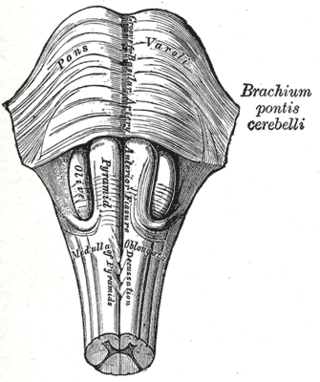
The brainstem is the stalk-like part of the brain that interconnects the cerebrum and diencephalon with the spinal cord. In the human brain, the brainstem is composed of the midbrain, the pons, and the medulla oblongata. The midbrain is continuous with the thalamus of the diencephalon through the tentorial notch.

<span class="mw-page-title-main">Trochlear nerve</span> Cranial nerve IV, for eye movements

The trochlear nerve, also known as the fourth cranial nerve, cranial nerve IV, or CN IV, is a cranial nerve that innervates a single muscle - the superior oblique muscle of the eye. Unlike most other cranial nerves, the trochlear nerve is exclusively a motor nerve.

<span class="mw-page-title-main">Lateral geniculate nucleus</span> Component of the visual system in the brains thalamus

In neuroanatomy, the lateral geniculate nucleus is a structure in the thalamus and a key component of the mammalian visual pathway. It is a small, ovoid, ventral projection of the thalamus where the thalamus connects with the optic nerve. There are two LGNs, one on the left and another on the right side of the thalamus. In humans, both LGNs have six layers of neurons alternating with optic fibers.

Visual release hallucinations, also known as Charles Bonnet syndrome or CBS, are a type of psychophysical visual disturbance in which a person with partial or severe blindness experiences visual hallucinations.

<span class="mw-page-title-main">Pulvinar nuclei</span>

The pulvinar nuclei or nuclei of the pulvinar are the nuclei located in the thalamus. As a group they make up the collection called the pulvinar of the thalamus, usually just called the pulvinar.

In the anatomy of the brain, the centromedian nucleus, also known as the centrum medianum, is a part of the intralaminar thalamic nuclei (ITN) in the thalamus. There are two centromedian nuclei arranged bilaterally.

<span class="mw-page-title-main">Pretectal area</span> Structure in the midbrain which mediates responses to ambient light

In neuroanatomy, the pretectal area, or pretectum, is a midbrain structure composed of seven nuclei and comprises part of the subcortical visual system. Through reciprocal bilateral projections from the retina, it is involved primarily in mediating behavioral responses to acute changes in ambient light such as the pupillary light reflex, the optokinetic reflex, and temporary changes to the circadian rhythm. In addition to the pretectum's role in the visual system, the anterior pretectal nucleus has been found to mediate somatosensory and nociceptive information.

<span class="mw-page-title-main">Reticular formation</span> Spinal trigeminal nucleus

The reticular formation is a set of interconnected nuclei that are located throughout the brainstem. It is not anatomically well defined, because it includes neurons located in different parts of the brain. The neurons of the reticular formation make up a complex set of networks in the core of the brainstem that extend from the upper part of the midbrain to the lower part of the medulla oblongata. The reticular formation includes ascending pathways to the cortex in the ascending reticular activating system (ARAS) and descending pathways to the spinal cord via the reticulospinal tracts.

<span class="mw-page-title-main">Central neurogenic hyperventilation</span> Abnormal pattern of breathing

Central neurogenic hyperventilation (CNH) is an abnormal pattern of breathing characterized by deep and rapid breaths at a rate of at least 25 breaths per minute. Increasing irregularity of this respiratory rate generally is a sign that the patient will enter into coma. CNH is unrelated to other forms of hyperventilation, like Kussmaul's respirations. CNH is the human body's response to reduced carbon dioxide levels in the blood. This reduction in carbon dioxide is caused by contraction of cranial arteries from damage caused by lesions in the brain stem. However, the mechanism by which CNH arises as a result from these lesions is still very poorly understood. Current research has yet to provide an effective means of treatment for the rare number of patients who are diagnosed with this condition.

<span class="mw-page-title-main">Thalamocortical radiations</span> Neural pathways between the thalamus and cerebral cortex

In neuroanatomy, thalamocortical radiations also known as thalamocortical fibres, are the efferent fibres that project from the thalamus to distinct areas of the cerebral cortex. They form fibre bundles that emerge from the lateral surface of the thalamus.

<span class="mw-page-title-main">Posterior cerebral artery</span> Artery which supplies blood to the occipital lobe of the brain

The posterior cerebral artery (PCA) is one of a pair of cerebral arteries that supply oxygenated blood to the occipital lobe, part of the back of the human brain. The two arteries originate from the distal end of the basilar artery, where it bifurcates into the left and right posterior cerebral arteries. These anastomose with the middle cerebral arteries and internal carotid arteries via the posterior communicating arteries.

<span class="mw-page-title-main">Parinaud's syndrome</span> Inability to move the eyes up and down

Parinaud's syndrome is a constellation of neurological signs indicating injury to the dorsal midbrain. More specifically, compression of the vertical gaze center at the rostral interstitial nucleus of medial longitudinal fasciculus (riMLF).

The zona incerta (ZI) is a horizontally elongated region of gray matter in the subthalamus below the thalamus. Its connections project extensively over the brain from the cerebral cortex down into the spinal cord.

Intention tremor is a dyskinetic disorder characterized by a broad, coarse, and low-frequency tremor evident during deliberate and visually-guided movement. An intention tremor is usually perpendicular to the direction of movement. When experiencing an intention tremor, one often overshoots or undershoots one's target, a condition known as dysmetria. Intention tremor is the result of dysfunction of the cerebellum, particularly on the same side as the tremor in the lateral zone, which controls visually guided movements. Depending on the location of cerebellar damage, these tremors can be either unilateral or bilateral.

Utilization behavior (UB) is a type of neurobehavioral phenomena that involves someone grabbing objects in view and starting the 'appropriate' behavior associated with it at an 'inappropriate' time. Patients exhibiting utilization behavior have difficulty resisting the impulse to operate or manipulate objects which are in their visual field and within reach. Characteristics of UB include unintentional, unconscious actions triggered by the immediate environment. The unpreventable excessive behavior has been linked to lesions in the frontal lobe. UB has also been referred to as "bilateral magnetic apraxia" and "hypermetamorphosis".

Conjugate gaze palsies are neurological disorders affecting the ability to move both eyes in the same direction. These palsies can affect gaze in a horizontal, upward, or downward direction. These entities overlap with ophthalmoparesis and ophthalmoplegia.

Prosopometamorphopsia is a visual disorder characterized by altered perceptions of faces. Facial features are distorted in a variety of ways including drooping, swelling, discoloration, and shifts of position. Prosopometamorphopsia is distinct from prosopagnosia which is characterised by the inability to recognise faces. About 75 cases of prosopometamorphopsia have been reported in the scientific literature. In about half of the reported cases, features on both sides of the face appear distorted. In the other half of cases, distortions are restricted to one side of the face and this condition is called hemi-prosopometamorphopsia.

<span class="mw-page-title-main">Visual pathway lesions</span> Overview about the lesions of visual pathways

The visual pathway consists of structures that carry visual information from the retina to the brain. Lesions in that pathway cause a variety of visual field defects. In the visual system of human eye, the visual information processed by retinal photoreceptor cells travel in the following way:
Retina→Optic nerve→Optic chiasma →Optic tract→Lateral geniculate body→Optic radiation→Primary visual cortex

References

  1. 1 2 3 4 5 6 7 8 9 10 11 12 13 14 15 16 17 Mocellin, Ramon; Walterfang, Mark; Velakoulis, Dennis (September 2006). "Neuropsychiatry of complex visual hallucinations". The Australian and New Zealand Journal of Psychiatry. 40 (9): 742–751. doi:10.1080/j.1440-1614.2006.01878.x. ISSN   0004-8674. PMID   16911748. S2CID   12151224.
  2. Benke, Thomas (2006). "Peduncular hallucinosis". Journal of Neurology. 253 (12): 1561–1571. doi:10.1007/s00415-0060-0254-4. PMID   17006630. S2CID   25990312.
  3. 1 2 3 4 Kumar, R., Wahi, J., Banerji, D., & Sharma, K. (1999). Peduncular Hallucinosis: An Unusual Sequel to Surgical Intervention in the Suprasellar Region. British Journal of Neurosurgery, 13(5), 500-503.
  4. 1 2 3 Benke, T. (2006). Peduncular Hallucinosis - a Syndrome of Impaired Reality Monitoring. Journal of Neurology, 253(12), 1561-1571.
  5. 1 2 3 FEINBERG W, M., & RAPCSAK S, Z. (1989). Peduncular Hallucinosis Following Paramedian Thalamic Infarction. Neurology, 39(11), 1535-1536.
  6. 1 2 3 4 Spiegel, D., Eastern Virginia Medical School Dept. of Psychiatry, interviewed by C. Byars, Oct. 12, 2009.
  7. Howlett, D., Downie, A., Banerjee, A., Tonge, K., & Oakeley, H. (1994). MRI of an Unusual Case of Peduncular Hallucinosis (Lhermitte's Syndrome). Neuroradiology, 36(2), 121-122.