Perfluorodecanoic acid

Last updated
Perfluorodecanoic acid
Perfluorodecanoic acid.svg
Names
Preferred IUPAC name
Nonadecafluorodecanoic acid
Other names
PFDA
C10 PFCA
Identifiers
3D model (JSmol)
ChEMBL
ChemSpider
ECHA InfoCard 100.005.819 OOjs UI icon edit-ltr-progressive.svg
EC Number
  • 206-400-3
35659
PubChem CID
UNII
  • InChI=1S/C10HF19O2/c11-2(12,1(30)31)3(13,14)4(15,16)5(17,18)6(19,20)7(21,22)8(23,24)9(25,26)10(27,28)29/h(H,30,31)
    Key: PCIUEQPBYFRTEM-UHFFFAOYSA-N
  • C(=O)(C(C(C(C(C(C(C(C(C(F)(F)F)(F)F)(F)F)(F)F)(F)F)(F)F)(F)F)(F)F)(F)F)O
Properties
C10HF19O2
Molar mass 514.086 g·mol−1
Melting point 77–81 °C (171–178 °F; 350–354 K) [1]
Boiling point 218 °C (424 °F; 491 K) [1]
Except where otherwise noted, data are given for materials in their standard state (at 25 °C [77 °F], 100 kPa).

Perfluorodecanoic acid (PFDA) is a fluorosurfactant and has been used in industry, [2] with applications as wetting agent and flame retardant. [3]

Contents

It was recently linked to health concerns, [4] like other fluorosurfactants, leading to proposed restrictions on its use. [5] In 2020, a California bill banned its use as an intentionally added ingredient in cosmetics. [6]

It has been proposed as a chemical probe to study peroxisome proliferation. [7] [8]

Perfluorodecanoic acid (PFDA) is a member of the group of polyfluoroalkyl substances (PFAS), more specific is it also a perfluoroalkyl acid (PFAA). PFAS, like PFDA, are man-made and are not naturally occurring in nature. Over the last decades they have been used in consumer products and industrial applications. It is a fluorosurfactant with a unique hydrophobicity and oleophobicity. [9] PFDA is well resistant to heat, oil, stains, grease and water, therefore it has been used in stain and greaseproof coating for furniture, packaging and carpet. Next to that, PFDA has also been found in nano-and impregnation-sprays, outdoor textiles, gloves, ski wax, leather, cosmetics, medical equipment and paper-based food containers. [9] [10] PFDA has a relatively high toxicity and can promote tumor growth. [11]

Mechanism of action

Via contaminated water or soil, plants can take up PFDA. This may lead to exposure and accumulation of PFDA in humans and other organisms. [11] In addition, exposure is possible via inhalation of indoor and outdoor air and ingestion of drinking water and food. [9] Direct dermal contact with PFDA-containing products is the main route of exposure. [11]

PFDA has been shown to increase the expression of two cytochrome P450 enzymes, namely Cyp2B10 and 4A14 in mouse liver. [12] In addition, it has been shown to activate the peroxisome proliferator-activated receptor alpha (PPARα). This receptor regulates lipid metabolism. [12]

A study looked at the harmful effects of PFDA on the antioxidative defense system in erythrocytes (red blood cells). Their results indicated that PFDA could influence the contents and activity of the biomolecules: GSH, MDA, SOD, CAT and GPx. This can lead to lipid peroxidation and oxidative injury of erythrocytes. The carbon chain length plays an important role, exposure to PFDA resulted in more obvious alterations of these biomolecules than shorter carbon chains of PFAA's. [13]

Further, has merging evidence showed that PFDA exposure can be associated with higher plasma triglyceride concentration in humans. [14] It is however unknown how PFDA might affect adipogenesis. HepG2 cells and 3T3-L1 differentiation model were used to detect the effects and mechanism of PFDA on lipid metabolism. PFDA showed to promote cellular triglyceride accumulation and triglyceride content in a concentration dependent manner. It also activated the NLP3 inflammasome. The inflammasome is crucial for induction of lipogenic genes expression in fatty acid synthase (FAS), hydroxymethyl glutaryl coenzyme A synthase (HMGCS) and stearoyl-CoA desaturase 1 (SCD1). [14] Besides, a suggestion can be made that PFDA may promote adipogenesis via an NLRP3 inflammasome-mediates SREBP1 pathway. Also, the expression of SREBP1, which is an important regulator of lipid metabolism, and its target genes were increased after PFDA treatment. The PFDA-induced SREBP1 enhanced expression can be terminated by caspase-1 inhibitor and by siNLRP3. [14]

Metabolism

PFDA is resistant to hydrolysis, photolysis and biodegradation, this causes persistence of the compound in the environment. [15] With its long carbon chain and carboxylate group, PFDA has some similar structure to amino acids. But it does not biodegrade according to the route of fatty acid metabolism. [16]

Structure and reactivity

Perfluorodecanoic acid is a compound with a carbon chain of 10. At 9 of the carbons the hydrogens are replaced by all fluor atoms, the last carbon is the carboxylate group. The length of the PFDA carbon chain is greater than that of PFOAs (perfluorooctanoic acids) and PFOS (perfluorooctane sulfonic acid) indicating that it is possibly more toxic. [17]

Perfluorodecanoic acid is a chemically inert due to relatively high organic bond strength and fluorine's electron negativity, which makes it resistant to advanced oxidation processes. [18] It is also resistant to hydrolysis and has thermal and photochemical stability unless certain reaction conditions are introduced, e.g., PFDA can be decomposed in hot water in the presence of S2O82-. [19] Photochemical decomposition with Na2S is another way of breaking up PFDA molecules. [18]

Synthesis and reactions

The first main road of perfluorochemicals (to which PFDA belongs) is electrochemical fluorination (ECF). This reaction occurs during an electrochemical hydrolysis of hydrofluoric acid (anhydrous) at a cell potential of 4.5 to 7 V. Several compounds can be used as starting material, for example, carboxylic acids (RCOOH), acyl chlorides (RCOCl) or sulfonic acid chlorides (RSO2Cl). The second step of the reaction is hydrolysis (addition of NaOH) to obtain the final products: [20]

RCOOH + nHF → RFCOOH + nH2O + by-products
RFCOOH + NaOH → RFCOONa + H2O
RCOCl + nHF → RFCO + nHCl + by-products
RFCO + NaOH→ RFCOONa+ H2O
RSO2Cl + nHF → RFSO2F + nHCl + by-products
RFSO2F + 2NaOH→ RFSO3Na+ NaF+ H2O

The second main synthesis road for perfluorodecanoic acid used commercially is telomerization. [21] Since PFDA has an even number of carbons, the staring material should be pentafluoroethyl iodide. [20] The process follows the general scheme: [21]

Telomerization             Tetrafluoroethylene                     ↓            Perfluoroalkyl iodide                     ↓         Fluorotelomer alcohols (FTOH)                     ↓         Perfluorochemicals (e.g. PFDA)

Availability

Studies have shown that, fortunately, there is a decreasing trend in the concentration of PFDA in Danish pregnant women from years of 2008- 2013. Germany and Denmark also follow this trend, while the concentration of this chemical is growing in Japan, Korea, Greenland, and Northern Norway. [22] Under normal conditions, the amount of PFDA (perfluorodecanoic acid) in European individuals was 0.8 ng/mL in 2013. [23]

Efficacy and side effects

Efficacy

Perfluorodecanoic Acid (PFDA) is a widely used industrial chemical that is persistent in the environment and can accumulate in the body. It has been detected in the blood of people and animals worldwide. The EPA has issued a lifetime health advisory for PFDA in drinking water of 70 parts per trillion (ppt). PFDA is not a medication and doesn't have any approved medical uses. It's a man-made chemical used in industrial applications and numerous studies point to its negative health effects. [24] [25]

Side effects

Perfluorodecanoic acid (PFDA) is a chemical of significant concern due to its classification as a Persistent, Bioaccumulative, and Toxic (PBT) substance. It has been identified as a potential carcinogen, with an acute oral toxicity estimate indicating a lethal dose for 50% of rats at 57 mg/kg. The substance is known to cause a range of acute symptoms as side effects such as a burning sensation, coughing, wheezing, and difficulty in breathing, which require immediate medical attention. [26]

There is little research done on the influence of PFDA on humans, most studies are done on animals. The human Reference Dose of PFDA can be calculated from mouse studies. In order to do that the NOAEL (no observed adverse effect level) should be divided by 10000. [23]

Toxicity

Research has shown that PFDA inhibits peroxisomal β-oxidation, a crucial metabolic process for energy production from fatty acids and is associated with DNA damage. Even at low levels, can damage this DNA in ovarian cells. This damage comes in the form of double-strand breaks, which are like tears in the DNA. Our bodies have a built-in repair system to fix these tears, however, PFDA disrupts this repair system and induces genotoxicity. This unrepaired DNA damage can lead to mutations. The mistakes get copied during cell division, potentially leading to genomic instability. Over time, these mutations can accumulate, potentially leading to the uncontrolled growth characteristic of cancer. Since ovaries are involved in reproduction, damage to ovarian cells caused by PFDA might be a factor in reproductive health problems. [27] It is also a potent and long-lasting toxin that may contribute to tumor formation. Studies have linked PFDA exposure to exacerbated adiposity and hepatic lipid accumulation, especially when combined with a high-fat diet, indicating a risk factor for liver problems. [28]

One important antioxidant molecule in the liver that helps protect cells from damage caused by free radicals is glutathione (GSH). PFDA exposure increases the total amount of GSH in the liver. This impact suggests an attempt by the body to counteract oxidative stress caused by the chemical. [29] Furthermore, exposure to PFDA has been associated with miscarriage, [30] liver damage, inflammation, and various negative effects on the heart, thyroid, and reproductive systems. It disrupts hormonal and immune system functions and can cause cellular damage through the activation of the PPARα receptor, affecting liver function and triggering oxidative stress, and the NLRP3 inflammasome pathway in both human cells and mouse tissues. [31]

PFDA interferes with the NF-κB pathway that induces the production of proinflammatory cytokines. [32] In gastric cells, PFDA has been found to increase the production of pro-inflammatory molecules IL-1β and IL-18, suggesting a role in stomach inflammation. Essentially, NLRP3 acts like a switch that turns on the production of mature IL-1β and IL-18. The active IL-1β and IL-18 are then released from the cell. They travel to nearby cells and trigger an inflammatory response. [33] It may also promote the proliferation of gastric epithelial cells, potentially preventing them from entering a state of senescence, which is a natural process to prevent uncontrolled cell growth. This increased growth could be a concern since uncontrolled cell growth is a hallmark of cancer. [34]

The toxicokinetic profile of PFDA reveals a long half-life in the body (52-66 days in rats), regardless of sex. Yet, female rats showed higher levels of PFDA accumulation in their bloodstream than males after exposure. Their bodies also showed slower clearance rates. [35] Similar to rats, PFDA in humans likely accumulates in the liver and triggers peroxisomal beta-oxidation. Data based on biomonitoring in Korea states that the margin of exposure of PFDA in human males was about two times higher than that of females (100.5, male; 27.7, female), indicating a lower risk for males. [36]

It's important to remember that most of the research on PFDA's health effects has been conducted on animals. While these studies’ findings raise concerns about the potential health risks of PFDA in humans, further investigation is needed to confirm these findings and fully understand the long-term health effects of PFDA exposure in humans.

Effects on animals

A study on rats has shown that the administration of PFDA can cause certain changes in the thyroid gland function. While the concentration of hormone T3 (triiodothyronine) in blood remained the same, gland weight and levels of thyroxine have decreased. Interestingly, this has led to only an 8% reduction in the metabolic rate of these rats. [37]

A single dose of 5mg/kg of PFDA is enough to cause these symptoms in rodents, as well as harm to the rough endoplasmic reticulum in the hepatic cells as well as abnormal mitochondria. Except for its effect on the liver and thyroid gland, one study on pigs has shown that this chemical may prevent fertilization by causing oocyte death. [38]

Another study on fish has found that PFDA alters the production of sex hormones and thus leads to endocrine disturbance. This is done through increased levels of 17β-estradiol (E2) that lead to upregulation of the cyp19b and cyp19a gene. These genes are necessary for the production of aromatase and regulate the production of estrogen from androgen. [39]

Related Research Articles

<span class="mw-page-title-main">Safrole</span> Chemical compound

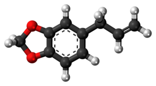
Safrole is an organic compound with the formula CH2O2C6H3CH2CH=CH2. It is a colorless oily liquid, although impure samples can appear yellow. A member of the phenylpropanoid family of natural products, it is found in sassafras plants, among others. Small amounts are found in a wide variety of plants, where it functions as a natural antifeedant. Ocotea pretiosa, which grows in Brazil, and Sassafras albidum, which grows in eastern North America, are the main natural sources of safrole. It has a characteristic "sweet-shop" aroma.

Genotoxicity is the property of chemical agents that damage the genetic information within a cell causing mutations, which may lead to cancer. While genotoxicity is often confused with mutagenicity, all mutagens are genotoxic, but some genotoxic substances are not mutagenic. The alteration can have direct or indirect effects on the DNA: the induction of mutations, mistimed event activation, and direct DNA damage leading to mutations. The permanent, heritable changes can affect either somatic cells of the organism or germ cells to be passed on to future generations. Cells prevent expression of the genotoxic mutation by either DNA repair or apoptosis; however, the damage may not always be fixed leading to mutagenesis.

<span class="mw-page-title-main">CYP2E1</span> Protein-coding gene in the species Homo sapiens

Cytochrome P450 2E1 is a member of the cytochrome P450 mixed-function oxidase system, which is involved in the metabolism of xenobiotics in the body. This class of enzymes is divided up into a number of subcategories, including CYP1, CYP2, and CYP3, which as a group are largely responsible for the breakdown of foreign compounds in mammals.

In biochemistry and metabolism, beta oxidation (also β-oxidation) is the catabolic process by which fatty acid molecules are broken down in the cytosol in prokaryotes and in the mitochondria in eukaryotes to generate acetyl-CoA. Acetyl-CoA enters the citric acid cycle, generating NADH and FADH2, which are electron carriers used in the electron transport chain. It is named as such because the beta carbon of the fatty acid chain undergoes oxidation and is converted to a carbonyl group to start the cycle all over again. Beta-oxidation is primarily facilitated by the mitochondrial trifunctional protein, an enzyme complex associated with the inner mitochondrial membrane, although very long chain fatty acids are oxidized in peroxisomes.

<span class="mw-page-title-main">Arecoline</span> Stimulant alkaloid

Arecoline is a nicotinic acid-based mild parasympathomimetic stimulant alkaloid found in the areca nut, the fruit of the areca palm. It is an odourless oily liquid. It can bring a sense of enhanced alertness and energy along with mild feelings of euphoria and relaxation. The psychoactive effects are comparable to that of nicotine.

Phytol is an acyclic hydrogenated diterpene alcohol that is used as a precursor for the manufacture of synthetic forms of vitamin E and vitamin K1, as well as in the fragrance industry. Its other commercial uses include cosmetics, shampoos, toilet soaps, and detergents, as well as in some cannabis distillates as a diluent or for flavoring. Its worldwide use has been estimated to be approximately 0.1–1.0 metric tons per year.

<span class="mw-page-title-main">Citrinin</span> Chemical compound

Citrinin is a mycotoxin which is often found in food. It is a secondary metabolite produced by fungi that contaminates long-stored food and it causes different toxic effects, like nephrotoxic, hepatotoxic and cytotoxic effects. Citrinin is mainly found in stored grains, but sometimes also in fruits and other plant products.

<span class="mw-page-title-main">Diisobutyl phthalate</span> Chemical compound

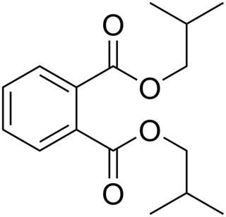
Diisobutyl phthalate (DIBP) is a phthalate ester having the structural formula C6H4(COOCH2CH 2)2. It is formed by the esterification of isobutanol and phthalic anhydride. This and other phthalates are used as plasticizers due to their flexibility and durability. They are found in many industrial and personal products, such as lacquers, nail polish and cosmetics. DIBP can be absorbed via oral ingestion and dermal exposure. When it comes to excretion, DIBP is first converted into the hydrolytic monoester monoisobutyl phthalate (MIBP). The primary excretory route is urine, with biliary excretion being noted in minor amounts. DIBP has lower density and freezing point than the related compound dibutyl phthalate (DBP).

<span class="mw-page-title-main">Dimethyl phthalate</span> Chemical compound

Dimethyl phthalate (DMP) is an organic compound and phthalate ester. it is a colourless and oily liquid that is soluble in organic solvents, but which is only poorly soluble in water.

<span class="mw-page-title-main">NLRP3</span> Human protein and coding gene

NLR family pyrin domain containing 3 (NLRP3), is a protein that in humans is encoded by the NLRP3 gene located on the long arm of chromosome 1.

<span class="mw-page-title-main">Ursolic acid</span> Pentacyclic chemical compound found in fruits

Ursolic acid, is a pentacyclic triterpenoid identified in the epicuticular waxes of apples as early as 1920 and widely found in the peels of fruits, as well as in herbs and spices like rosemary and thyme.

Pyroptosis is a highly inflammatory form of lytic programmed cell death that occurs most frequently upon infection with intracellular pathogens and is likely to form part of the antimicrobial response. This process promotes the rapid clearance of various bacterial, viral, fungal and protozoan infections by removing intracellular replication niches and enhancing the host's defensive responses. Pyroptosis can take place in immune cells and is also reported to occur in keratinocytes and some epithelial cells.

<span class="mw-page-title-main">Nodularin</span> Chemical compound

Nodularins are potent toxins produced by the cyanobacterium Nodularia spumigena, among others. This aquatic, photosynthetic cyanobacterium forms visible colonies that present as algal blooms in brackish water bodies throughout the world. The late summer blooms of Nodularia spumigena are among the largest cyanobacterial mass occurrences in the world. Cyanobacteria are composed of many toxic substances, most notably of microcystins and nodularins: the two are not easily differentiated. A significant homology of structure and function exists between the two, and microcystins have been studied in greater detail. Because of this, facts from microcystins are often extended to nodularins.

<i>Cornus officinalis</i> Species of flowering plant

Cornus officinalis, the Japanese cornel or Japanese cornelian cherry, is a species of flowering plant in the dogwood family Cornaceae. Despite its name, it is native to China and Korea as well as Japan. It is not to be confused with C. mas, which is also known as the Cornelian cherry. It is not closely related to the true cherries of the genus Prunus.

Inflammasomes are cytosolic multiprotein complexs of the innate immune system responsible for the activation of inflammatory responses and cell death. They are formed as a result of specific cytosolic pattern recognition receptors (PRRs) sensing microbe-derived pathogen-associated molecular patterns (PAMPs), damage-associated molecular patterns (DAMPs) from the host cell, or homeostatic disruptions. Activation and assembly of the inflammasome promotes the activation of caspase-1, which then proteolytically cleaves pro-inflammatory cytokines, interleukin 1β (IL-1β) and interleukin 18 (IL-18), as well as the pore-forming molecule gasdermin D (GSDMD). The N-terminal GSDMD fragment resulting from this cleavage induces a pro-inflammatory form of programmed cell death distinct from apoptosis, referred to as pyroptosis, which is responsible for the release of mature cytokines. Additionally, inflammasomes can act as integral components of larger cell death-inducing complexes called PANoptosomes, which drive another distinct form of pro-inflammatory cell death called PANoptosis.

<span class="mw-page-title-main">Ampelopsin</span> Chemical compound

Ampelopsin, also known as dihydromyricetin and DHM, when purported as an effective ingredient in supplements and other tonics, is a flavanonol, a type of flavonoid. It is extracted from the Japanese raisin tree and found in Ampelopsis species japonica, megalophylla, and grossedentata; Cercidiphyllum japonicum; Hovenia dulcis; Rhododendron cinnabarinum; some Pinus species; and some Cedrus species, as well as in Salix sachalinensis.

<span class="mw-page-title-main">Riddelliine</span> Chemical compound

Riddelliine is a chemical compound classified as a pyrrolizidine alkaloid. It was first isolated from Senecio riddellii and is also found in a variety of plants including Jacobaea vulgaris, Senecio vulgaris, and others plants in the genus Senecio.

<span class="mw-page-title-main">Chrysophanol</span> Chemical compound

Chrysophanol, also known as chrysophanic acid, is a fungal isolate and a natural anthraquinone. It is a C-3 methyl substituted chrysazin of the trihydroxyanthraquinone family.

Gaseous signaling molecules are gaseous molecules that are either synthesized internally (endogenously) in the organism, tissue or cell or are received by the organism, tissue or cell from outside and that are used to transmit chemical signals which induce certain physiological or biochemical changes in the organism, tissue or cell. The term is applied to, for example, oxygen, carbon dioxide, sulfur dioxide, nitrous oxide, hydrogen cyanide, ammonia, methane, hydrogen, ethylene, etc.

<span class="mw-page-title-main">Monocrotaline</span> Chemical compound

Monocrotaline (MCT) is a pyrrolizidine alkaloid that is present in plants of the Crotalaria genus. These species can synthesise MCT out of amino acids and can cause liver, lung and kidney damage in various organisms. Initial stress factors are released intracellular upon binding of MCT to BMPR2 receptors and elevated MAPK phosphorylation levels are induced, which can cause cancer in Homo sapiens. MCT can be detoxified in rats via oxidation, followed by glutathione-conjugation and hydrolysis.

References

  1. 1 2 "Perfluorodecanoic acid 98%". Sigma-Aldrich.
  2. Reich, Ieva L.; Reich, Hans J.; Menahan, Lawrence A.; Peterson, Richard E. (October 1987). "Synthesis of 14C-labeled perfluorooctanoic and perfluorodecanoic acids; purification of perfluorodecanoic acid". Journal of Labelled Compounds and Radiopharmaceuticals. 24 (10): 1235–1244. doi:10.1002/jlcr.2580241011.
  3. Harris, M (April 1989). "Developmental toxicity of perfluorodecanoic acid in C57BL/6N mice". Fundamental and Applied Toxicology. 12 (3): 442–448. doi:10.1016/0272-0590(89)90018-3. PMID   2731659.
  4. "Danish study links perfluorinated chemicals to miscarriage". Chemical Watch. Retrieved 25 March 2019.
  5. "Germany and Sweden propose restrictions on six PFASs". Chemical Watch. 14 May 2015. Retrieved 25 March 2019.
  6. "Assembly Bill No. 2762". State of California. September 30, 2020. Retrieved 10 October 2020.
  7. Vanden Heuvel, John P. (October 1996). "Perfluorodecanoic acid as a useful pharmacologic tool for the study of peroxisome proliferation". General Pharmacology: The Vascular System. 27 (7): 1123–1129. doi:10.1016/0306-3623(95)00126-3. PMID   8981056.
  8. Chen, Li-Chuan; Tatum, Vickie; Glauert, Howard P.; Chow, Ching K. (2001). "Peroxisome proliferator perfluorodecanoic acid alters glutathione and related enzymes". Journal of Biochemical and Molecular Toxicology. 15 (2): 107–113. doi:10.1002/jbt.6. ISSN   1095-6670. PMID   11284052. S2CID   27570192.
  9. 1 2 3 Iris assessments. Perfluorodecanoic Acid (PFDA). Available via: https://cfpub.epa.gov/ncea/iris_drafts/recordisplay.cfm?deid=354408#:~:text=PFDA%20has%20been%20used%20in,%2C%20ski%20wax%2C%20and%20leather.
  10. Shu-Zi Deng, Chang-Long Xu, Zhong-Feng Xu, Li-Ying Zhou, Shu-Juan Xie, Kang-Na Wei, Yuan-Chang Jin, Zhao-Cheng Zeng, Xiang-Jun Yang, Shu-Hua Tan, Hai-Long Wang, Perfluorodecanoic acid induces meiotic defects and deterioration of mice oocytes in vitro, Toxicology, Volume 460, 2021, 152884, ISSN 0300-483X, https://doi.org/10.1016/j.tox.2021.152884.
  11. 1 2 3 Noah Peter Christian, Chemical toxicity of per- and poly-fluorinated alkyl substances (PFAS), Editor(s): Philip Wexler, Encyclopedia of Toxicology (Fourth Edition), Academic Press, 2024, Pages 747-756, ISBN 9780323854344, https://doi.org/10.1016/B978-0-12-824315-2.01052-6
  12. 1 2 Mengchen Xu, Tong Zhang, Chao Lv, Qigui Niu, Wansong Zong, Jingchun Tang, Rutao Liu, Perfluorodecanoic acid-induced oxidative stress and DNA damage investigated at the cellular and molecular levels, Ecotoxicology and Environmental Safety, Volume 185, 2019, 109699, ISSN 0147-6513, https://doi.org/10.1016/j.ecoenv.2019.109699
  13. J. Agric. Food Chem. 2018, 66, 25, 6414–6420, Publication Date:June 4, 2018, https://doi.org/10.1021/acs.jafc.8b02197.
  14. 1 2 3 Taotao Wang, Hong Xu, Yu Guo, Zhanming Li, Hua Ye, Liang Wu, Yuanxin Guo, Dongxu Wang, Perfluorodecanoic acid promotes adipogenesis via NLRP3 inflammasome-mediated pathway in HepG2 and 3T3-L1 cells, Food and Chemical Toxicology, Volume 171, 2023, 113520, ISSN 0278-6915, https://doi.org/10.1016/j.fct.2022.113520
  15. National Center for Biotechnology Information (2024). PubChem Compound Summary for CID 9555, Perfluorodecanoic acid. Retrieved February 26, 2024 from https://pubchem.ncbi.nlm.nih.gov/compound/Perfluorodecanoic-acid
  16. Efficient Decomposition of Perfluorocarboxylic Acids and Alternative Fluorochemical Surfactants in Hot Water. Hisao Hori, Yumiko Nagaoka, Misako Murayama, and Shuzo Kutsuna. Environmental Science & Technology 2008 42 (19), 7438-7443 DOI: 10.1021/es800832p
  17. Gao, Sichen, and Rutao Liu. “Comprehensive Insights into the Interaction Mechanism between Perfluorodecanoic Acid and Human Serum Albumin.” New Journal of Chemistry, vol. 42, no. 11, 2018, pp. 9065–9073, https://doi.org/10.1039/c8nj00124c
  18. 1 2 B.B. Wang, M.H. Cao, Z.J. Tan, L.L. Wang, S.H. Yuan, J. Chen, Photochemical decomposition of perfluorodecanoic acid in aqueous solution with VUV light irradiation, Journal of Hazardous Materials, Volume 181, Issues 1–3, 2010, Pages 187-192, ISSN 0304-3894, https://doi.org/10.1016/j.jhazmat.2010.04.115
  19. Efficient Decomposition of Perfluorocarboxylic Acids and Alternative Fluorochemical Surfactants in Hot Water. Hisao Hori, Yumiko Nagaoka, Misako Murayama, and Shuzo Kutsuna. Environmental Science & Technology 2008 42 (19), 7438-7443 DOI: 10.1021/es800832p
  20. 1 2 Järnberg, U., Holmström, K., van Bavel, B., & Kärrman, A. (2007). Perfluoroalkylated acids and related compounds (PFAS) in the Swedish environment. Retrieved from Stockholms universitet, institutionen för tillämpad miljövetenskap (ITM) website: https://urn.kb.se/resolve?urn=urn:nbn:se:naturvardsverket:diva-415
  21. 1 2 Melissa M. Schultz, Douglas F. Barofsky, and Jennifer A. Field. Fluorinated Alkyl Surfactants. Environmental Engineering Science.Sep 2003.487-501. http://doi.org/10.1089/109287503768335959 Published in Volume: 20 Issue 5: July 6, 2004
  22. Bjerregaard-Olesen C, Bach CC, Long M, Ghisari M, Bossi R, Bech BH, Nohr EA, Henriksen TB, Olsen J, Bonefeld-Jørgensen EC. Time trends of perfluorinated alkyl acids in serum from Danish pregnant women 2008-2013. Environ Int. 2016 May;91:14-21. doi: 10.1016/j.envint.2016.02.010. Epub 2016 Feb 16. PMID: 26891270
  23. 1 2 Luo M, Tan Z, Dai M, Song D, Lin J, Xie M, Yang J, Sun L, Wei D, Zhao J, Gonzalez FJ, Liu A. Dual action of peroxisome proliferator-activated receptor alpha in perfluorodecanoic acid-induced hepatotoxicity. Arch Toxicol. 2017 Feb;91(2):897-907. doi: 10.1007/s00204-016-1779-7. Epub 2016 Jun 25. PMID: 27344344; PMCID: PMC6350782
  24. Agency for Toxic Substances and Disease Registry. (2024). ATSDR website: https://www.atsdr.cdc.gov/
  25. U.S. Environmental Protection Agency. (2024). Integrated Risk Information System [IRIS]. https://iris.epa.gov/ChemicalLanding/&substance_nmbr=702
  26. “Perfluorodecanoic Acid Safety Data Sheet” (2023) Created pursuant to the requirements of Regulation (EC) No. 1907/2006 and Regulation (EC) No. 1272/2008. Revision date: 22-Sep-2023. Available at: https://eur-lex.europa.eu/eli/reg/2008/1272/oj
  27. Qin, Y., Yuan, X., Cui, Z., Chen, W., Xu, S., Chen, K., Wang, F., Zheng, F., Ni, H., Shen, H. M., Wu, Y., & Xia, D. (2023). Low dose PFDA induces DNA damage and DNA repair inhibition by promoting nuclear cGAS accumulation in ovarian epithelial cells. Ecotoxicology and environmental safety, 265, 115503. https://doi.org/10.1016/j.ecoenv.2023.115503
  28. ] Du, Y., Paglicawan, L., Soomro, S., Abunofal, O., Baig, S., Vanarsa, K., Hicks, J., & Mohan, C. (2021). Epigallocatechin-3-Gallate Dampens Non-Alcoholic Fatty Liver by Modulating Liver Function, Lipid Profile and Macrophage Polarization. Nutrients, 13(2), 599. https://doi.org/10.3390/nu13020599
  29. Chen, L.-C., Tatum, V., Glauert, H.P. and Chow, C.K. (2001), Peroxisome proliferator perfluorodecanoic acid alters glutathione and related enzymes. J. Biochem. Mol. Toxicol., 15: 107-113. https://doi.org/10.1002/jbt.6
  30. Xuping Gao, Wanze Ni, Sui Zhu, Yanxin Wu, Yunfeng Cui, Junrong Ma, Yanhua Liu, Jinlong Qiao, Yanbin Ye, Pan Yang, Chaoqun Liu, Fangfang Zeng,Per- and polyfluoroalkyl substances exposure during pregnancy and adverse pregnancy and birth outcomes: A systematic review and meta-analysis, Environmental Research, Volume 201, 2021, 111632, ISSN 0013-9351, https://doi.org/10.1016/j.envres.2021.111632
  31. Dongxu Wang, Qiang Gao, Taotao Wang, Zhipeng Kan, Xin Li, Lizhen Hu, Chuan-yi Peng, Frank Qian, Yijun Wang, Daniel Granato, Green tea polyphenols and epigallocatechin-3-gallate protect against perfluorodecanoic acid induced liver damage and inflammation in mice by inhibiting NLRP3 inflammasome activation, Food Research International, Volume 127, 2020, 108628, ISSN 0963-9969, https://doi.org/10.1016/j.foodres.2019.108628
  32. Rachel P. Frawley, Matthew Smith, Mark F. Cesta, Schantel Hayes-Bouknight, Chad Blystone, Grace E. Kissling, Shawn Harris & Dori Germolec (2018) Immunotoxic and hepatotoxic effects of perfluoro-n-decanoic acid (PFDA) on female Harlan Sprague–Dawley rats and B6C3F1/N mice when administered by oral gavage for 28 days, Journal of Immunotoxicology, 15:1, 41-52, DOI: 10.1080/1547691X.2018.1445145
  33. Zhou, X., Dong, T., Fan, Z. et al. Perfluorodecanoic acid stimulates NLRP3 inflammasome assembly in gastric cells. Sci Rep 7, 45468 (2017). https://doi.org/10.1038/srep45468
  34. Dong T., Peng Y., Zhong N., Liu F., Zhang H., Xu M., Liu R., Han M., Tian X., Jia J., Chang L., Guo L., Liu S. et al Perfluorodecanoic acid (PFDA) promotes gastric cell proliferation via sPLA2-IIA. Oncotarget. 2017; 8: 50911-50920. https://doi.org/10.18632/oncotarget.17284
  35. Dzierlenga AL, Robinson VG, Waidyanatha S, DeVito MJ, Eifrid MA, Gibbs ST, Granville CA, Blystone CR. Toxicokinetics of perfluorohexanoic acid (PFHxA), perfluorooctanoic acid (PFOA) and perfluorodecanoic acid (PFDA) in male and female Hsd:Sprague dawley SD rats following intravenous or gavage administration. Xenobiotica. 2020 Jun;50(6):722-732. DOI: 10.1080/00498254.2019.1683776
  36. Kim, SJ., Choi, EJ., Choi, GW. et al. Exploring sex differences in human health risk assessment for PFNA and PFDA using a PBPK model. Arch Toxicol 93, 311–330 (2019). https://doi.org/10.1007/s00204-018-2365-y
  37. Marc J. Van Rafelghem, Stanley L. Inhorn, Richard E. Peterson, Effects of perfluorodecanoic acid on thyroid status in rats, Toxicology and Applied Pharmacology, Volume 87, Issue 3, 1987, Pages 430-439, ISSN 0041-008X, https://doi.org/10.1016/0041-008X(87)90248-1
  38. Domínguez, Z. Salazar, M. Betancourt, Y. Ducolomb, E. Casas, F. Fernández, I. Bahena, A. Salomón, M. Teteltitla, R. Martínez, A. Chaparro, P. Cuapio, C. Salazar-López, E. Bonilla, Effect of perfluorodecanoic acid on pig oocyte viability, intracellular calcium levels and gap junction intercellular communication during oocyte maturation in vitro, Toxicology in Vitro, Volume 58, 2019, Pages 224-229, ISSN 0887-2333, https://doi.org/10.1016/j.tiv.2019.03.041
  39. Areum Jo, Kyunghee Ji, Kyungho Choi, Endocrine disruption effects of long-term exposure to perfluorodecanoic acid (PFDA) and perfluorotridecanoic acid (PFTrDA) in zebrafish (Danio rerio) and related mechanisms, Chemosphere, Volume 108, 2014, Pages 360-366, ISSN 0045-6535, https://doi.org/10.1016/j.chemosphere.2014.01.080