Phantom eye syndrome

Last updated
Phantom eye syndrome
EyeMuscles.gif
Anatomy of the eye. The external eye muscles are shown in red.
Specialty Psychiatry, Neurology
Duration11-15 days
Frequency5%

Phantom eye syndrome (PES) is a phantom pain in the eye and visual hallucinations after the removal of an eye (enucleation, evisceration).

Contents

Symptoms

Many patients experience one or more phantom phenomena after the removal of the eye:

Pathogenesis

Causes

Triggers of Phantom Eye Syndrome encompass a range of factors that can initiate or intensify phantom sensations and pain following eye removal. These triggers commonly include fatigue, stress, and fluctuations in lighting conditions. Some cases suggest a correlation between the duration of pain prior to eye removal and the presence of preoperative conditions, such as headache or eye pain, with the likelihood of experiencing subsequent phantom sensations. [3]

Phantom pain and non-painful phantom sensations

Phantom sensations in Phantom Eye Syndrome (PES) encompass various tactile perceptions such as paresthesia, dysesthesia, and hyperpathia, excluding pain. These sensations can manifest in different forms, including kinetic, kinesthetic, or exteroceptive perceptions, and are commonly experienced by almost all PES patients. [4] Some cases have highlighted the prevalence of Phantom Eye Pain (PEP) in PES, with rates as high as 47% reported. PEP includes pain felt around the amputated eye (periocular pain), contributing to a higher prevalence compared to studies defining PEP solely as pain in the amputated eye. Frequency and characteristics of PEP vary, with paroxysmal episodes lasting for a few seconds or minutes being common, and weather conditions such as cold and humid weather serving as major triggering factors. Furthermore, approximately 38% of PES patients may experience neuropathic pain, indicating a need for targeted medication approaches. [5]

Phantom pain and non-painful phantom sensations result from changes in the central nervous system due to denervation of a body part. [6] [7] Phantom eye pain is considerably less common than phantom limb pain. The prevalence of phantom pain after limb amputation ranged from 50% to 78%. The prevalence of phantom eye pain, in contrast, is about 30%.

Post-amputation changes in the cortical representation of body parts adjacent to the amputated limb are believed to contribute to the development of phantom pain and non-painful phantom sensations. One reason for the smaller number of patients with phantom eye pain compared with those with phantom limb pain may be the smaller cortical somatosensory representation of the eye compared with the limbs.

In limb amputees, some, [8] but not all, studies have found a correlation between preoperative pain in the affected limb and postoperative phantom pain. There is a significant association between painful and non-painful phantom experiences, preoperative pain in the symptomatic eye and headache. [9] Based on the present data, it is difficult to determine if headaches or preoperative eye pain play a causal role in the development of phantom phenomena or if headache, preoperative eye pain, and postoperative phantom eye experiences are only epiphenomena of an underlying factor. However, a study in humans demonstrated that experimental pain leads to a rapid reorganization of the somatosensory cortex. [10] This study suggests that preoperative and postoperative pain may be an important co-factor for somatosensory reorganization and the development of phantom experiences.

Visual hallucinations

Individuals with Phantom Eye Syndrome (PES) may experience phantom visual sensations (PVS), which involve perceiving images that are not actually present in the removed eye. These hallucinations often resemble real-world objects, ranging from people and animals to various structures and scenes. Visual hallucinations in PES can be categorized into elementary, consisting of simple visual phenomena without distinct meaning, and complex, involving more detailed and formed perceptions that may relate to past experiences. [4] Rates of PVS range from 30% to 48% in eye amputated patients with the episodic nature of hallucinations persisting for at least 2 years post-surgery. [11]

Enucleation of an eye and, similarly, retinal damage, leads to a cascade of events in the cortical areas receiving visual input. Cortical GABAergic (GABA: Gamma-aminobutyric acid) inhibition decreases, and cortical glutamatergic excitation increases, followed by increased visual excitability or even spontaneous activity in the visual cortex. [12] It is believed that spontaneous activity in the denervated visual cortex is the neural correlate of visual hallucinations.

Quality of Life and Psychological Support

Anxiety often accompanies phantom eye symptoms (PES) in patients undergoing enucleation for uveal melanoma. While the exact nature of this relationship is complex, studies suggest that elevated anxiety levels may precede the onset of PES, particularly phantom eye pain (PEP). However, it's important to note that anxiety does not appear to influence the persistence of PES over time. Understanding and addressing anxiety in these patients may help alleviate distress associated with PES and improve overall well-being during the post-operative period. [11] Moreover, Eye removal can have more broad psychological impacts including insecurity, rejection, and fear of social marginalization. Ocular prosthesis can aid in restoring aesthetics and promoting psychological well-being. Psychological support, alongside medical intervention, is essential for managing anxiety and depression in PES patients. [4]

Treatment

Treatment on painful phantom eye syndrome is limited and does not point out a standard treatment protocol but possible treatment pathways include resting techniques, pharmacologic, non-pharmacologic, surgery, drug therapy, and psychological [4] [13] [5] . Individualized treatment strategies that combine various treatment pathways is potentially the treatment method. Triggers for phantom eye syndrome are generally related to stressors such as fatigue, extreme lighting conditions (dark or bright), opening and closing the eyes, and psychological stress [4] [5] . Resting techniques such as sleeping, using artificial tears, and closing eyes for extended periods of time have been proven to reduced negative symptoms associated with phantom eye syndrome such as phantom eye pain (PEP), phantom visions, and/or phantom sensations [5] . Pharmacologic techniques used for treating PEP depend on the pain level. Low pain can be treated through paracetamol and non-steroidal anti-inflammatory medications; medium level pain can be treated with tramadol or codeine; and high level pain can be treated with morphine [5] . Possible drug therapy treatments for phantom eye syndrome can incorporate antidepressants, anticonvulsants, sodium channel blockers, N-methyl-D-aspartate receptor antagonists, and opioids. Possible psychological treatment techniques include cognitive-behavioral therapy (trauma-focused or reprocessing), reassurance therapy (specifically in the case of hallucinations), and mind-body therapies (enhancing self-awareness and self-regulation of symptoms) [13] [14] [15] .

In the instance of introducing a ocular prosthesis (glass eye), it is important that the procedure prioritizes proper fitting of the ocular prosthesis, transparency with the patient, and meticulous monitoring of emotional state preceding and following insertion [2] . Ocular prothesis can mitigate symptoms of physiological distress related to phantom eye syndrome through simulating the functions of a normal eye socket [4] .

Overall, the literature promotes treatment that focuses on increasing quality of life through addressing personalized emotional and physical phantom eye syndrome stressors [4] .

See also

Related Research Articles

<span class="mw-page-title-main">Amputation</span> Medical procedure that removes a part of the body

Amputation is the removal of a limb by trauma, medical illness, or surgery. As a surgical measure, it is used to control pain or a disease process in the affected limb, such as malignancy or gangrene. In some cases, it is carried out on individuals as a preventive surgery for such problems. A special case is that of congenital amputation, a congenital disorder, where fetal limbs have been cut off by constrictive bands. In some countries, judicial amputation is currently used to punish people who commit crimes. Amputation has also been used as a tactic in war and acts of terrorism; it may also occur as a war injury. In some cultures and religions, minor amputations or mutilations are considered a ritual accomplishment. When done by a person, the person executing the amputation is an amputator. The oldest evidence of this practice comes from a skeleton found buried in Liang Tebo cave, East Kalimantan, Indonesian Borneo dating back to at least 31,000 years ago, where it was done when the amputee was a young child.

<span class="mw-page-title-main">Pain</span> Type of distressing feeling

Pain is a distressing feeling often caused by intense or damaging stimuli. The International Association for the Study of Pain defines pain as "an unpleasant sensory and emotional experience associated with, or resembling that associated with, actual or potential tissue damage."

<span class="mw-page-title-main">Hallucination</span> Perception that only seems real

A hallucination is a perception in the absence of an external stimulus that has the compelling sense of reality. Hallucinations are vivid, substantial, and are perceived to be located in external objective space. Hallucination is a combination of two conscious states of brain wakefulness and REM sleep. They are distinguishable from several related phenomena, such as dreaming, which does not involve wakefulness; pseudohallucination, which does not mimic real perception, and is accurately perceived as unreal; illusion, which involves distorted or misinterpreted real perception; and mental imagery, which does not mimic real perception, and is under voluntary control. Hallucinations also differ from "delusional perceptions", in which a correctly sensed and interpreted stimulus is given some additional significance.

<span class="mw-page-title-main">Alice in Wonderland syndrome</span> Neurological disorder that distorts perception of objects size and distance

Alice in Wonderland syndrome (AIWS), also known as Todd's syndrome or dysmetropsia, is a neurological disorder that distorts perception. People with this syndrome may experience distortions in their visual perception of objects, such as appearing smaller (micropsia) or larger (macropsia), or appearing to be closer (pelopsia) or farther (teleopsia) than they are. Distortion may also occur for senses other than vision.

<span class="mw-page-title-main">Phantom limb</span> Sensation that an amputated or missing limb is attached

A phantom limb is the sensation that an amputated or missing limb is still attached. It is a chronic condition which is often resistant to treatment. When the cut ends of sensory fibres are stimulated during thigh movements, the patient feels as if the sensation is arising from the non-existent limb. Sometimes the patient might feel pain in the non-existent limb. Approximately 80–100% of individuals with an amputation experience sensations in their amputated limb. However, only a small percentage will experience painful phantom limb sensation. These sensations are relatively common in amputees and usually resolve within two to three years without treatment. Research continues to explore the underlying mechanisms of phantom limb pain (PLP) and effective treatment options.

<span class="mw-page-title-main">V. S. Ramachandran</span> Indian-American neuroscientist

Vilayanur Subramanian Ramachandran is an Indian-American neuroscientist. He is known for his wide-ranging experiments and theories in behavioral neurology, including the invention of the mirror box. Ramachandran is a distinguished professor in UCSD's Department of Psychology, where he is the director of the Center for Brain and Cognition.

Visual release hallucinations, also known as Charles Bonnet syndrome or CBS, are a type of psychophysical visual disturbance in which a person with partial or severe blindness experiences visual hallucinations.

Cortical maps are collections (areas) of minicolumns in the brain cortex that have been identified as performing a specific information processing function.

<span class="mw-page-title-main">Mirror therapy</span> Treatment for some kinds of pain

Mirror therapy (MT) or mirror visual feedback (MVF) is a therapy for pain or disability that affects one side of the patient more than the other side. It was invented by Vilayanur S. Ramachandran to treat post-amputation patients who had phantom limb pain (PLP). Ramachandran created a visual illusion of two intact limbs by putting the patient's affected limb into a "mirror box," with a mirror down the center.

Phantom pain is a painful perception that an individual experiences relating to a limb or an organ that is not physically part of the body, either because it was removed or was never there in the first place.

Neuroplasticity, also known as neural plasticity or brain plasticity, is the ability of neural networks in the brain to change through growth and reorganization. It is when the brain is rewired to function in some way that differs from how it previously functioned. These changes range from individual neuron pathways making new connections, to systematic adjustments like cortical remapping or neural oscillation. Other forms of neuroplasticity include homologous area adaptation, cross modal reassignment, map expansion, and compensatory masquerade. Examples of neuroplasticity include circuit and network changes that result from learning a new ability, information acquisition, environmental influences, pregnancy, caloric intake, practice/training, and psychological stress.

<span class="mw-page-title-main">Aura (symptom)</span> Symptom of epilepsy and migraine

An aura is a perceptual disturbance experienced by some with epilepsy or migraine. An epileptic aura is a seizure.

Dysesthesia is an unpleasant, abnormal sense of touch. Its etymology comes from the Greek word "dys," meaning "bad," and "aesthesis," which means "sensation". It often presents as pain but may also present as an inappropriate, but not discomforting, sensation. It is caused by lesions of the nervous system, peripheral or central, and it involves sensations, whether spontaneous or evoked, such as burning, wetness, itching, electric shock, and pins and needles. Dysesthesia can include sensations in any bodily tissue, including most often the mouth, scalp, skin, or legs.

A somatosensory disorder is an impairment of the somatosensory system.

Abdominal aura, also known as visceral aura and epigastric aura, is a type of somatosensory aura that typically manifests as abdominal discomfort in the form of nausea, malaise, hunger, or pain. Abdominal aura is typically associated with epilepsy, especially temporal lobe epilepsy, and it can also be used in the context of migraine. The term is used to distinguish it from other types of somatosensory aura, notably visual disturbances and paraesthesia. The abdominal aura can be classified as a somatic hallucination. Pathophysiologically, the abdominal aura is associated with aberrant neuronal discharges in sensory cortical areas representing the abdominal viscera.

Mirror-touch synesthesia is a rare condition which causes individuals to experience a similar sensation in the same part or opposite part of the body that another person feels. For example, if someone with this condition were to observe someone touching their cheek, they would feel the same sensation on their own cheek. Synesthesia, in general, is described as a condition in which a concept or sensation causes an individual to experience an additional sensation or concept. Synesthesia is usually a developmental condition; however, recent research has shown that mirror touch synesthesia can be acquired after sensory loss following amputation.

<span class="mw-page-title-main">Cortical remapping</span>

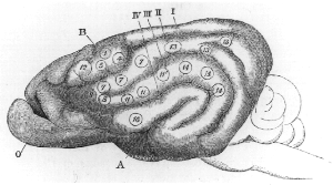
Cortical remapping, also referred to as cortical reorganization, is the process by which an existing cortical map is affected by a stimulus resulting in the creating of a 'new' cortical map. Every part of the body is connected to a corresponding area in the brain which creates a cortical map. When something happens to disrupt the cortical maps such as an amputation or a change in neuronal characteristics, the map is no longer relevant. The part of the brain that is in charge of the amputated limb or neuronal change will be dominated by adjacent cortical regions that are still receiving input, thus creating a remapped area. Remapping can occur in the sensory or motor system. The mechanism for each system may be quite different. Cortical remapping in the somatosensory system happens when there has been a decrease in sensory input to the brain due to deafferentation or amputation, as well as a sensory input increase to an area of the brain. Motor system remapping receives more limited feedback that can be difficult to interpret.

<span class="mw-page-title-main">Tactile hallucination</span> Hallucination involving perception of tactile input

Tactile hallucination is the false perception of tactile sensory input that creates a hallucinatory sensation of physical contact with an imaginary object. It is caused by the faulty integration of the tactile sensory neural signals generated in the spinal cord and the thalamus and sent to the primary somatosensory cortex (SI) and secondary somatosensory cortex (SII). Tactile hallucinations are recurrent symptoms of neurological diseases such as schizophrenia, Parkinson's disease, Ekbom's syndrome and delirium tremens. Patients who experience phantom limb pains also experience a type of tactile hallucination. Tactile hallucinations are also caused by drugs such as cocaine and alcohol.

During every moment of an organism's life, sensory information is being taken in by sensory receptors and processed by the nervous system. Sensory information is stored in sensory memory just long enough to be transferred to short-term memory. Humans have five traditional senses: sight, hearing, taste, smell, touch. Sensory memory (SM) allows individuals to retain impressions of sensory information after the original stimulus has ceased. A common demonstration of SM is a child's ability to write letters and make circles by twirling a sparkler at night. When the sparkler is spun fast enough, it appears to leave a trail which forms a continuous image. This "light trail" is the image that is represented in the visual sensory store known as iconic memory. The other two types of SM that have been most extensively studied are echoic memory, and haptic memory; however, it is reasonable to assume that each physiological sense has a corresponding memory store. For example, children have been shown to remember specific "sweet" tastes during incidental learning trials but the nature of this gustatory store is still unclear. However, sensory memories might be related to a region of the thalamus, which serves as a source of signals encoding past experiences in the neocortex.

<span class="mw-page-title-main">Limb telescoping</span>

Limb telescoping is the progressive shortening of a phantom limb as the cortical regions are reorganized following an amputation. During this reorganization, proximal portions of the residual limb are perceived as more distal parts of the phantom limb. Such effect is responsible for increased phantom pain due to the discrepancy between the patient’s body perception and their actual body. This effect may last from weeks up to years after post-amputation.

References

  1. 1 2 3 Sörös P, Vo O, Husstedt IW, Evers S, Gerding H (May 2003). "Phantom eye syndrome: Its prevalence, phenomenology, and putative mechanisms". Neurology. 60 (9): 1542–1543. doi:10.1212/01.wnl.0000059547.68899.f5. PMID   12743251. S2CID   27474612.
  2. 1 2 3 Shah S.I.A (1994). "Painful Phantom Eye" (PDF). Pak J Ophthalmol. 10 (4 (Index Issue)): 77–78.
  3. Quaranta-Leoni FM, Fiorino MG, Quaranta-Leoni F, Di Marino M (2021-08-06). "Anophthalmic Socket Syndrome: Prevalence, Impact and Management Strategies". Clinical Ophthalmology. 15: 3267–3281. doi: 10.2147/OPTH.S325652 . PMC   8354770 . PMID   34393477.
  4. 1 2 3 4 5 6 7 Andreotti AM, Goiato MC, Pellizzer EP, Pesqueira AA, Guiotti AM, Gennari-Filho H, et al. (2014-12-07). "Phantom eye syndrome: a review of the literature". TheScientificWorldJournal. 2014: 686493. doi: 10.1155/2014/686493 . PMC   4273592 . PMID   25548790.
  5. 1 2 3 4 5 Martel A, Baillif S, Thomas P, Almairac F, Galatoire O, Hamedani M, et al. (August 2021). "Phantom eye pain: a multicentric study in 100 patients". Acta Ophthalmologica. 99 (5): e753–e760. doi:10.1111/aos.14657. PMID   33124153.
  6. Ramachandran VS, Hirstein W (September 1998). "The perception of phantom limbs. The D. O. Hebb lecture". Brain. 121 (9): 1603–1630. doi: 10.1093/brain/121.9.1603 . PMID   9762952.
  7. Nikolajsen L, Jensen TS (July 2001). "Phantom limb pain". British Journal of Anaesthesia. 87 (1): 107–116. doi: 10.1093/bja/87.1.107 . PMID   11460799.
  8. Nikolajsen L, Ilkjaer S, Krøner K, Christensen JH, Jensen TS (September 1997). "The influence of preamputation pain on postamputation stump and phantom pain". Pain. 72 (3): 393–405. doi:10.1016/S0304-3959(97)00061-4. PMID   9313280. S2CID   24665497.
  9. Nicolodi M, Frezzotti R, Diadori A, Nuti A, Sicuteri F (June 1997). "Phantom eye: features and prevalence. The predisposing role of headache". Cephalalgia. 17 (4): 501–504. doi:10.1046/j.1468-2982.1997.1704501.x. PMID   9209770. S2CID   34799505.
  10. Sörös P, Knecht S, Bantel C, Imai T, Wüsten R, Pantev C, et al. (February 2001). "Functional reorganization of the human primary somatosensory cortex after acute pain demonstrated by magnetoencephalography". Neuroscience Letters. 298 (3): 195–198. doi:10.1016/S0304-3940(00)01752-3. PMID   11165440. S2CID   30936812.
  11. 1 2 Brown SL, Hope-Stone L, Hussain RN, Heimann H, van der Voort N, Cherry MG (March 2024). "Prevalence, temporal course and risk factors for phantom eye symptoms in uveal melanoma". Eye. 38 (4): 707–713. doi:10.1038/s41433-023-02756-w. PMC   10920882 . PMID   37752341.
  12. Eysel UT, Schweigart G, Mittmann T, Eyding D, Qu Y, Vandesande F, et al. (1999). "Reorganization in the visual cortex after retinal and cortical damage". Restorative Neurology and Neuroscience. 15 (2–3): 153–164. PMID   12671230. Archived from the original on 2013-01-29.
  13. 1 2 Erlenwein J, Diers M, Ernst J, Schulz F, Petzke F (2021-04-01). "Clinical updates on phantom limb pain". PAIN Reports. 6 (1): e888. doi:10.1097/PR9.0000000000000888. ISSN   2471-2531. PMC   7813551 . PMID   33490849.
  14. Roed Rasmussen ML, Prause JU, Ocularist MJ, Toft PB (September–October 2009). "Phantom Eye Syndrome: Types of Visual Hallucinations and Related Phenomena". Ophthalmic Plastic & Reconstructive Surgery. 25 (5): 390–393. doi:10.1097/IOP.0b013e3181b54b06. ISSN   0740-9303. PMID   19966655.
  15. Moura VL, Faurot KR, Gaylord SA, Mann JD, Sill M, Lynch C, et al. (August 2012). "Mind-Body Interventions for Treatment of Phantom Limb Pain in Persons with Amputation". American Journal of Physical Medicine & Rehabilitation. 91 (8): 701–714. doi:10.1097/PHM.0b013e3182466034. ISSN   0894-9115. PMC   6457458 . PMID   22286895.