Phlebovirus

Last updated
Phlebovirus
Viruses-08-00174-g001-A.jpg
Viruses-08-00174-g001-B.jpg
Prototypic phlebovirus virion and genome organization.
Virus classification OOjs UI icon edit-ltr.svg
(unranked): Virus
Realm: Riboviria
Kingdom: Orthornavirae
Phylum: Negarnaviricota
Class: Ellioviricetes
Order: Bunyavirales
Family: Phenuiviridae
Genus:Phlebovirus
Species

See text

Phlebovirus is one of twenty genera of the family Phenuiviridae in the order Bunyavirales . The genus contains 66 species. [1] It derives its name from Phlebotominae, the vectors of member species Naples phlebovirus , which is said to be ultimately from the Greek phlebos, meaning "vein". [2] The proper word for "vein" in ancient Greek is however phleps (φλέψ). [3]

Contents

Virology

Replication cycle of phleboviruses. Viruses-08-00202-g001.webp
Replication cycle of phleboviruses.

Phleboviruses are viruses with a negative-sense RNA genome consisting of three segments. The small segment (S) codes for the viral N protein and a non structural protein, NSs via an ambisense coding strategy. The medium-sized segment (M) codes for a precursor of the viral glycoproteins and non-structural components. The product of the largest segment (L) is the viral RNA-dependent RNA polymerase. [4]

Replication

The Phlebovirus replicates in a 7 step process. First, the cellular attachment is driven through the glycoprotein interactions with host cells. Examples of this are Dendritic Cell-Specific Intercellular Adhesion Molecule-3-Grabbing Non-Integrin (DC-SIGN), heparan sulfate (HS), or Non-Muscle Myosin Heavy Chain (NMMHC-IIA). Second, in the late endosome, the low pH causes fusion activity in the membrane of the Gc protein. Uukuniemi virus (UUKV) penetration is promoted by the expression of vesicle-associated membrane protein 3 (VAMP3). Additionally, the fusion of Rift Valley fever virus (RVFV) in late endosomes is inhibited by the interferon-induced transmembrane proteins 2 and 3 (IFITIM2 and IFITIM3). Third, the viral and endosomal membranes are fused to allow for the release of the viral ribonucleoprotein complexes into the cytoplasm (Also the site of viral transcription and replication). Fourth, the precursor protein, Gn/Gc, is translated at the rough endoplasmic reticulum (ER). This precursor protein is cleaved by signal peptidase. Synthesis of the viral nucleoprotein and viral polymerase in the cytoplasm combines with the newly formed genomic RNA (gRNA) ribonucleic protein complexes (RNP). Fifth, two ER chaperones, binding immunoglobulin protein (BiP) and calnexin, are required to ensure proper folding of GN/Gc. Gn/Gc are similarly catalyzed by protein-disulfide-isomerase through the formation of disulfide bonds. At the same time, calreticulin prevents any misfolded Gn/Gc from being exported to the Golgi. Sixth, The correctly folded Gn/Gc heterodimers are transported to the Golgi apparatus. The cytoplasmic tails of Gn in the budding process associate with RNPs during this time. Seventh, once the budding of the new virus particles is completed, vesicles that contain the virus are transported to the plasma membrane to be released by exocytosis. [5]

Role of Gn and Gc in phlebovirus entry

A study of the family Bunyaviridae showed that bunyavirus particles are pleomorphic. This known fact cased some surprise when studies showed that UUKKV and RVFV particles are spherically shaped and highly ordered. The configuration of Gn and Gc proteins in the viral envelope imposes the order of the particle. The viral envelope forms an icosahedral lattice with a triangulation number of 12. Also included in the lattice composition are 110 hexametric and 12 pentameric capsomeres. For RVFV in particular, 720 Gn/Gc heterodimers are included in the capsomeres. In these cases, Gn forms the spikes of the capsomere while Gc is closer to the lipid membrane, thus placing it underneath. The pH surrounding the capsomere ultimately determines its shape. This is largely due to protonation triggering conformational changes in Gc, commonly included with membrane fusion. An assembly model for the RVFV envelope has been proposed which consists of Gc dimers positioned horizontally with respect to the viral membrane. This is known because the RVFV Gc ectodomain is crystallized as a dimer. This is opposed to the virion interior of bunyaviruses which has no matrix protein, and thus, has no defining organization. This means that on the virion surface, the Gc and Gn proteins must be present in a highly ordered placement.

To begin entry, phlebovirus particles bind to various components of the plasma membrane. These components interact with the viral glycoproteins of phlebovirus and regulate entry efficiency. While these components are not crucial to the actual entry of the virus itself, receptors are components of the plasma membrane that bind to the glycoproteins and are critical for entry. In Phleboviruses, it was determined that glycan-protein interactions play a crucial role in phlebovirus entry.

Heparan Sulfate (HS) is another crucial component aspect in Phlebovirus attachment. It is a glycosaminoglycan (GAG), which is an unbranched polysaccharide made of disaccharide repeats, that results in the creation of a proteoglycan. Cell lines with defined glycosylation defects were analyzed and showed that HS is necessary for the entry of RVFV. This was confirmed by the removal of HS using an enzyme. HS is charge-dependent in their interactions with virus particles, and studies showed that there are basic amino acid clusters on the P78 protein that interact with negative sulfate clusters on HS. In comparison, there were no identified HS binding sites on Gn and Gc. The P78 protein is plentiful in RVFV-infected cells in insects, while infected cells in mammals produce significantly less P78 proteins. The P78 protein is much more efficiently produced in RVFV in mosquito cells as it is required for viral spread in mosquitos. Overall, the cell line is heavily dependent on HS in RVFV entry.

A computational study provided evidence that phlebovirus Gc proteins might be class II membrane fusion proteins. Final proof for this theory was given by the elucidation of the ectodomain's structure of RVFV Gc in its pre-fusion status. Gc has three domains with characteristics that resembled other class II proteins. An internal fusion loop was discovered, which is a critical aspect of all class II proteins. The location of a histidine in Gc resembled a pH sensing feature, which matches class II characteristics. Although there were many similarities within the structure of the RVFV Gc and class II proteins, the interface between domains I and II in RVFV Gc is more rigid and bigger than other class II proteins. Additionally, RVFV Gc has more disulfide bridges centralized in different locations than the other compared proteins. However, its overall structure and functionality is most closely resembling a class II membrane fusion protein compared to any other class.

The pH sensing feature in the Gc protein is of note. The membrane fusion activity of the phlebovirus Gc proteins is very dependent on pH, as a low pH triggers the transport of virions into endolysosomes. Elevating intravesicular pH inhibits phlebovirus entry. However, it is still unclear whether Gc proteins must bind to a receptor before being triggered by pH or not.

After the viral and endosomal membranes have been fused together, the L,M, and S genomic segments (associated with viral polymerase) are released into the cytoplasm. This initiates the transcription to genomic RNA into mRNA. Viral proteins begin undergoing translation before the transcription of mRNA has finished. The Gn and Gc phlebovirus proteins are encoded on the M-segment and undergo synthesis. The precursor Gn/Gc protein cannot be detected in a cell already infected with phlebovirus. It only is visible after the expression of the M-segment. If microsomal membranes are present, the precursor is cleaved, indicating cleaving by a host factor during the synthesis of the viral protein. The signal peptidase complex in the ER membrane is responsible for the cleaving of the precursor. This precursor is then translocated into the ER lumen, in which two hydrophobic domains are inserted with a third, cleaved hydrophobic domain in between. [6]

An emerging group of arthropod-transmitted pathogens

Phleboviruses are arboviruses taxonomically split into tick- and dipteran-borne viruses. Phlebotomus sandflies are the primary sources for dipteran-borne phleboviruses, with Rift Valley fever virus being the exception (RVFV is associated with mosquitos and has a greater variety in its vector range). Maintenance of the viruses is mainly completed through the vector species by means of vertical (transovarial) transmission. Concerns over the potential introduction of RVFV into susceptible areas has grown due to the increasing spread of vector species. The potential ramifications of this spread could cause massive economic loss through the harming of livestock.

Two new members of phlebovirus as causative agents of traumatic human disease have been identified. In rural China, SFTSV, which is transmitted by ticks, was identified as the result of increased cases of a febrile illness combined with thrombocytopenia, leukenocytopenia, multiple organ dysfunction, and a high case-fatality rate. After this discovery, SFTSV was reported in Japan and Korea as well. North America had a similar case, which was found to be a result of the Heartland virus, which is transmitted by ticks. These two discoveries changed the perception of the effect of tick-borne phleboviruses with regards to public health. These discoveries caused the re-classification of the Bhanja virus (BHAV) into the tick-borne phlebovirus group. Novel associations of phlebovirus diseases have been reported in the Mediterranean area. Examples include the sandfly fever Turkey virus, Adria virus, Granada virus, Adana virus, and Medjerda virus, among others.

The Toscana virus has a high rate of vertical transmission, as demonstrated in sandflies through experimental infection. This suggests that there is an amplified role for vertebrate hosts despite the maintenance in nature coming mainly from sandflies. A sandfly is a name for members of any species or genus of flying, blood-sucking dipteran in sandy areas. For example, "sandfly" in the United States refers to horse flies or members of the family Ceratopogonidae. In other parts of the world, "sandfly" refers to members of the subfamily Phlebotominae within Psychodidae. Two of the three main genera were found in the Old World, Phlebotomus and Sergentomyia, and contain the prominent species that transmit the viral pathogens. The third genera was found in the New World and is called Lutzomyia. Other examples are biting midges, or Austrosimulium, a black fly found in New Zealand.

The result of many of these viruses are some sort of fever. Pappataci fevers, or Phlebotomus fevers, are mild 3-day fevers that are similar to influenza and have a rapid onset. They are most common in endemic areas during the summer months, particularly August, which is when sandflies are active. Some more extreme cases are the Toscana virus, which is associated with meningitis in humans, and the Rift Valley fever virus which has caused wide-spread epidemics in livestock in Africa. [7]

Clinical significance

The following twelve viruses have been linked to disease in humans: Alenquer phlebovirus, [8] Bhanja virus, [9] Candiru phlebovirus, [10] Chagres virus, Sandfly fever Naples phlebovirus, Punta Toro phlebovirus, Rift Valley fever virus, Sicilian phlebovirus, Toscana phlebovirus, Uukuniemi virus, Heartland bandavirus [11] (the first tick-borne phlebovirus known to cause disease in the Western Hemisphere, discovered in 2009), and the Sandfly Turkey virus (discovered in China in 2011). [12] They cause symptoms ranging from short self-limiting fevers, such as pappataci fever, to encephalitis and fatal viral hemorrhagic fever.[ citation needed ]

Taxonomy

Phlebovirus is derived from Phlebotominae, which is the taxon of vectors of member species sandfly fever Naples phlebovirus. The name comes from the Greek root phlebos, which means "vein". Serological cross reactivity previosuls defined species in the genus. A change in classification rules was prompted due to the difficulty in detecting new phlebovirus in serological assays. As a result, viral species are now defined by 95% or greater identity in the amino acid sequences of their respective RNA-dependent RNA plymerase (RdRp). Currently, the genus consists of 67 species. Some of the viruses have other hematophagous arthropods as their main vectors. Examples of this include mosquitos for the Rift Valley fever virus, and the Mukawa virus, which has been isolated from ticks, but remains in Phlebovirus despite the common shift of tick-borne viruses from Phlebovirus to Uukuvirus. In addition to ticks and mosquitos, some Phleboviruses have been isolated from vertebrates like rodents in America and opossums or sloths in Africa. This wide variety of sources shows the possible presence of diversified epidemiological cycles. [13]

The following species are recognized: [1]

As of 2015, within the phlebovirus there are four genetic groups of tick-borne phleboviruses: the SFTS group, the Bhanja group, the Uukuniemi group, [14] and the Kaisodi group. [15]

See also

Related Research Articles

<i>Orthohantavirus</i> Genus of viruses

Orthohantavirus is a genus of single-stranded, enveloped, negative-sense RNA viruses in the family Hantaviridae within the order Bunyavirales. Members of this genus may be called orthohantaviruses or simply hantaviruses.

<i>Bunyavirales</i> Order of RNA viruses

Bunyavirales is an order of segmented negative-strand RNA viruses with mainly tripartite genomes. Member viruses infect arthropods, plants, protozoans, and vertebrates. It is the only order in the class Ellioviricetes. The name Bunyavirales derives from Bunyamwera, where the original type species Bunyamwera orthobunyavirus was first discovered. Ellioviricetes is named in honor of late virologist Richard M. Elliott for his early work on bunyaviruses.

<span class="mw-page-title-main">Viral protein</span>

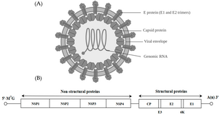
The term viral protein refers to both the products of the genome of a virus and any host proteins incorporated into the viral particle. Viral proteins are grouped according to their functions, and groups of viral proteins include structural proteins, nonstructural proteins, regulatory proteins, and accessory proteins. Viruses are non-living and do not have the means to reproduce on their own, instead depending on their host cell's machinery to do this. Thus, viruses do not code for most of the proteins required for their replication and the translation of their mRNA into viral proteins, but use proteins encoded by the host cell for this purpose.

<i>Tick-borne encephalitis virus</i> Species of virus

Tick-borne encephalitis virus (TBEV) is a positive-strand RNA virus associated with tick-borne encephalitis in the genus Flavivirus.

<i>Alphavirus</i> Genus of viruses

Alphavirus is a genus of RNA viruses, the sole genus in the Togaviridae family. Alphaviruses belong to group IV of the Baltimore classification of viruses, with a positive-sense, single-stranded RNA genome. There are 32 alphaviruses, which infect various vertebrates such as humans, rodents, fish, birds, and larger mammals such as horses, as well as invertebrates. Alphaviruses that could infect both vertebrates and arthropods are referred dual-host alphaviruses, while insect-specific alphaviruses such as Eilat virus and Yada yada virus are restricted to their competent arthropod vector. Transmission between species and individuals occurs mainly via mosquitoes, making the alphaviruses a member of the collection of arboviruses – or arthropod-borne viruses. Alphavirus particles are enveloped, have a 70 nm diameter, tend to be spherical, and have a 40 nm isometric nucleocapsid.

<i>Thogotovirus</i> Genus of viruses

Thogotovirus is a genus of enveloped RNA viruses, one of seven genera in the virus family Orthomyxoviridae. Their single-stranded, negative-sense RNA genome has six or seven segments. Thogotoviruses are distinguished from most other orthomyxoviruses by being arboviruses – viruses that are transmitted by arthropods, in this case usually ticks. Thogotoviruses can replicate in both tick cells and vertebrate cells; one subtype has also been isolated from mosquitoes. A consequence of being transmitted by blood-sucking vectors is that the virus must spread systemically in the vertebrate host – unlike influenza viruses, which are transmitted by respiratory droplets and are usually confined to the respiratory system.

<span class="mw-page-title-main">Pappataci fever</span> Medical condition

Pappataci fever is a vector-borne febrile arboviral infection caused by three serotypes of Phlebovirus. It occurs in subtropical regions of the Eastern Hemisphere. The name, pappataci fever, comes from the Italian word for sandfly; it is the union of the words pappa and taci (silent), distinguishing these insects from blood-feeding mosquitoes, which produce a typical noise while flying.

<i>Orthonairovirus</i> Genus of viruses

Orthonairovirus is a genus of viruses in the family Nairoviridae of the order Bunyavirales that include viruses with circular, negative-sense single stranded RNA. It got its name from the Nairobi sheep disease that affects the gastrointestinal tracts of sheep and goats. The vast majority, and perhaps all viruses in this genus are tick-borne viruses that can have human or other vertebrate hosts.

<i>Orthobunyavirus</i> Genus of viruses

Orthobunyavirus is a genus of the Peribunyaviridae family in the order Bunyavirales. There are currently ~170 viruses recognised in this genus. These have been assembled into 103 species and 20 serogroups.

Toscana phlebovirus (TOSV) is an arbovirus belonging to Bunyavirales, an order of negative-stranded, enveloped RNA viruses. The virus can be transmitted to humans by the bite of an infected sandfly of the genus Phlebotomus. Toscana is not normally associated with disease, as indicated by high seroprevalence rates in endemic areas, but in common with other sandfly transmitted viruses such as Naples virus and Sicilian virus, infection may result in Pappataci fever, an illness with mild fever, headache and myalgia. In serious cases that go undiagnosed, acute meningitis, meningoencephalitis and encephalitis may occur. There is no specific treatment for infection, so treatment is supportive, reducing the severity of symptoms until the immune system has cleared the infection.

Dabie bandavirus, also called SFTS virus, is a tick-borne virus in the genus Bandavirus in the family Phenuiviridae, order Bunyavirales. The clinical condition it caused is known as severe fever with thrombocytopenia syndrome (SFTS). SFTS is an emerging infectious disease that was first described in northeast and central China 2009 and now has also been discovered in Japan, South Korea, Vietnam and Taiwan in 2015. SFTS has a fatality rate of 12% and as high as over 30% in some areas. The major clinical symptoms of SFTS are fever, vomiting, diarrhea, multiple organ failure, thrombocytopenia, leukopenia and elevated liver enzyme levels. Another outbreak occurred in East China in the early half of 2020.

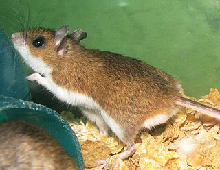
Sangassou orthohantavirus(SANGV) is single-stranded, negative-sense RNA virus species of the genus Orthohantavirus in the Bunyavirales order. It was first isolated in an African wood mouse (Hylomyscus simus) in the forest in Guinea, West Africa in 2010. It is named for the village near where the mouse was trapped. It is the first indigenous Murinae-associated African hantavirus to be discovered.

Naples phlebovirus is an antigenic species of genus Phlebovirus within the family Phenuiviridae of the order Bunyavirales. It is an enveloped RNA virus with a tripartite genome e Uukuniemi (UUK) serogroup. The Sandfly group's natural reservoir are sandflies, while the natural reservoir for Uukuniemi is ticks. The SFNV serogroup consists of two main serocomplexes associated with disease in humans, the Naples and Sicilian serocomplexes. Sandfly fever induces myalgia, fever, and elevated liver enzymes in humans. It is difficult to diagnose outside endemic areas.

In 1954 the Hazara orthonairovirus, one of the 34 tick-borne viruses of the genus Orthonairovirus, was discovered in Pakistan in the Ixodes tick native to that region. Today this virus is studied in mice in an attempt to develop treatments for the highly pathogenic Crimean-Congo Hemorrhagic Fever virus.

The Bhanja virus is a tick-borne virus first discovered in a tick taken from a paralyzed goat in Bhanjanagar, India in 1954. Bhanja virus in humans was first documented in 1974 when Charles Calisher was working with the virus in a lab and contracted it himself. His experience with the virus was mild and included symptoms of mild aching in muscles and joints, moderate headache, slight photophobia. The Bhanja virus is a member of the Bhanja virus serocomplex and is a member of the Bunyavirales order.

Batai orthobunyavirus (BATV) is a RNA virus belonging to order Bunyavirales, genus Orthobunyavirus.

Cache Valley orthobunyavirus (CVV) is a member of the order Bunyavirales, genus Orthobunyavirus, and serogroup Bunyamwera, which was first isolated in 1956 from Culiseta inornata mosquitos collected in Utah's Cache Valley. CVV is an enveloped arbovirus, nominally 80–120 nm in diameter, whose genome is composed of three single-stranded, negative-sense RNA segments. The large segment of related bunyaviruses is approximately 6800 bases in length and encodes a probable viral polymerase. The middle CVV segment has a 4463-nucleotide sequence and the smallest segment encodes for the nucleocapsid, and a second non-structural protein. CVV has been known to cause outbreaks of spontaneous abortion and congenital malformations in ruminants such as sheep and cattle. CVV rarely infects humans, but when they are infected it has caused encephalitis and multiorgan failure.

The Punta Toro virus is a member of the genus Phlebovirus of the order Bunyavirales. It was initially isolated from patients in Colombia and two key patients in Panama. Two individual serotypes of PTV were isolated from these patients, PTV-Adames (A) and PTV-Balliet (B), with PTV-A appearing to be more virulent. PTV is considered to be relatively contained to the Americas with no cases being reported outside of this region. Along with a few other human pathogenic Phleboviruses, PTV is considered to be a significant virus in terms of public health as little information is known about its clinical effects and with further research underway, PTV could have unforeseen impacts on health and virology.

<i>Sepik virus</i> Mosquito transmitted virus endemic to Papua New Guinea

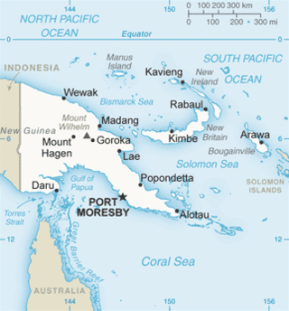
Sepik virus (SEPV) is an arthropod-borne virus (arbovirus) of the genus Flavivirus and family Flaviviridae. Flaviviridae is one of the most well characterized viral families, as it contains many well-known viruses that cause diseases that have become very prevalent in the world, like Dengue virus. The genus Flavivirus is one of the largest viral genera and encompasses over 50 viral species, including tick and mosquito borne viruses like Yellow fever virus and West Nile virus. Sepik virus is much less well known and has not been as well-classified as other viruses because it has not been known of for very long. Sepik virus was first isolated in 1966 from the mosquito Mansoniaseptempunctata, and it derives its name from the Sepik River area in Papua New Guinea, where it was first found. The geographic range of Sepik virus is limited to Papua New Guinea, due to its isolation.

<i>Modoc virus</i> Species of virus

Modoc virus (MODV) is a rodent-associated flavivirus. Small and enveloped, MODV contains positive single-stranded RNA. Taxonomically, MODV is part of the Flavivirus genus and Flaviviridae family. The Flavivirus genus includes nearly 80 viruses, both vector-borne and no known vector (NKV) species. Known flavivirus vector-borne viruses include Dengue virus, Yellow Fever virus, tick-borne encephalitis virus, and West Nile virus.

References

  1. 1 2 "Virus Taxonomy: 2020 Release". International Committee on Taxonomy of Viruses (ICTV). March 2021. Retrieved 19 May 2021.
  2. "ICTV 9th Report (2011) Bunyaviridae". International Committee on Taxonomy of Viruses (ICTV). Retrieved 31 January 2019. Phlebo: refers to phlebotomine vectors of sandfly fever group viruses; Greek phlebos, "vein".
  3. Liddell, H.G. & Scott, R. (1940). A Greek-English Lexicon. Revised and augmented throughout by Sir Henry Stuart Jones. With the assistance of Roderick McKenzie. Oxford: Clarendon Press.
  4. Modrow, Susanne; Falke, Dietrich; Truyen, Uwe; Schätzl, Hermann. Molecular Virology. Springer. p. 460. ISBN   978-3-642-20718-1.
  5. https://www.researchgate.net/figure/Replication-cycle-of-phleboviruses-A-Cellular-attachment-of-phleboviruses-is-driven-by_fig2_305522171.{{cite web}}: Missing or empty |title= (help)
  6. Spiegel, Martin; Plegge, Teresa; Pöhlmann, Stefan (July 2016). "The Role of Phlebovirus Glycoproteins in Viral Entry, Assembly and Release". Viruses. 8 (7): 202. doi: 10.3390/v8070202 . ISSN   1999-4915. PMC   4974537 . PMID   27455305.
  7. Tchouassi, David P.; Marklewitz, Marco; Chepkorir, Edith; Zirkel, Florian; Agha, Sheila B.; Tigoi, Caroline C.; Koskei, Edith; Drosten, Christian; Borgemeister, Christian; Torto, Baldwyn; Junglen, Sandra; Sang, Rosemary (April 2019). "Sand Fly–Associated Phlebovirus with Evidence of Neutralizing Antibodies in Humans, Kenya". Emerging Infectious Diseases. 25 (4): 681–690. doi:10.3201/eid2504.180750. ISSN   1080-6040. PMC   6433041 . PMID   30882303.
  8. Travassos da Rosa AP, Tesh RB, Pinheiro FP, Travassos da Rosa JF, Peterson NE (1983). "Characterization of eight new phlebotomus fever serogroup arboviruses (Bunyaviridae: Phlebovirus) from the Amazon region of Brazil". Am. J. Trop. Med. Hyg. 32 (5): 1164–71. doi:10.4269/ajtmh.1983.32.1164. PMID   6312820.
  9. Vesenjak-Hirjan J, Calisher CH, Beus I. Marton E. First natural clinical human Bhanja virus infection, p 297–301. 1980. In Vesenjak-Hirjan J, Porterfield JS, Arslanagí, c E (ed), Arboviruses in the Mediterranean countries: 6th FEMS Symposium. Fischer, Stuttgart, Germany.
  10. Palacios, Gustavo; Tesh, Robert; Travassos da Rosa, Amelia; Savji, Nazir; Sze, Wilson; Jain, Komal; Serge, Robert; Guzman, Hilda; Guevara, Carolina; Nunes, Marcio; Nunes-Neto, Joaquim; Kochel, Tadeusz; Hutchinson, Stephen; Vasconcelos, Pedro; Lipkin, Ian (2011). "Characterization of the Candiru antigenic complex (Bunyaviridae: Phlebovirus), a highly diverse and reassorting group of viruses affecting humans in tropical America". Journal of Virology. 85 (8): 3811–20. doi:10.1128/JVI.02275-10. PMC   3126144 . PMID   21289119.
  11. Savage, HM; Godsey, MS; Lambert, A; Panella, NA; Burkhalter, KL; Harmon, JR; Lash, RR; Ashley, DC; Nicholson, WL (2013). "First detection of heartland virus (Bunyaviridae: Phlebovirus) from field collected arthropods". Am. J. Trop. Med. Hyg. 89 (3): 445–52. doi:10.4269/ajtmh.13-0209. PMC   3771279 . PMID   23878186.
  12. Yu, X. J.; Liang, M. F.; Zhang, S. Y.; Liu, Y.; Li, J. D.; Sun, Y. L.; Zhang, L.; Zhang, Q. F.; Popov, V. L.; Li, C.; Qu, J.; Li, Q.; Zhang, Y. P.; Hai, R.; Wu, W.; Wang, Q.; Zhan, F. X.; Wang, X. J.; Kan, B.; Wang, S. W.; Wan, K. L.; Jing, H. Q.; Lu, J. X.; Yin, W. W.; Zhou, H.; Guan, X. H.; Liu, J. F.; Bi, Z. Q.; Liu, G. H.; Ren, J. (2011). "Fever with Thrombocytopenia Associated with a Novel Bunyavirus in China". New England Journal of Medicine. 364 (16): 1523–1532. doi:10.1056/NEJMoa1010095. PMC   3113718 . PMID   21410387.
  13. Calisher, Charles H.; Calzolari, Mattia (May 2021). "Taxonomy of Phleboviruses, Emphasizing Those That Are Sandfly-Borne". Viruses. 13 (5): 918. doi: 10.3390/v13050918 . ISSN   1999-4915. PMC   8156068 . PMID   34063467.
  14. Matsuno, K; Weisend, C; Kajihara, M; Matysiak, C; Williamson, BN; Simuunza, M; Mweene, AS; Takada, A; Tesh, RB; Ebihara, H (January 2015). "Comprehensive molecular detection of tick-borne phleboviruses leads to the retrospective identification of taxonomically unassigned bunyaviruses and the discovery of a novel member of the genus phlebovirus". J Virol. 89 (1): 594–604. doi:10.1128/JVI.02704-14. PMC   4301164 . PMID   25339769.
  15. Matsuno, K; Weisend, C; Kajihara, M; Matysiak, C; Williamson, BN; Simuunza, M; Mweene, AS; Takada, A; Tesh, RB; Ebihara, H (2015). "Comprehensive molecular detection of tick-borne phleboviruses leads to the retrospective identification of taxonomically unassigned bunyaviruses and the discovery of a novel member of the genus phlebovirus". J Virol. 89 (1): 594–604. doi: 10.1128/JVI.02704-14 . PMC   4301164 . PMID   25339769.