Phospholipid-derived fatty acids

Last updated

Phospholipid-derived fatty acids (PLFAs) are widely used in microbial ecology as chemotaxonomic markers of bacteria and other organisms. Phospholipids are the primary lipids composing cellular membranes. Phospholipids can be saponified, which releases the fatty acids contained in their diglyceride tail. Once the phospholipids of an unknown sample are saponified, the composition of the resulting PLFA can be compared to the PLFA of known organisms to determine the identity of the sample organism. PLFA analysis may be combined with other techniques, such as stable isotope probing to determine which microbes are metabolically active in a sample. PLFA analysis was pioneered by D.C. White at the University of Tennessee, in the early to mid 1980s. [1]

Contents

Phospholipid bilayer. Each phospholipid consists of a polar hydrophilic head (red) and two hydrophobic fatty acid tails. The fatty acid structure affects the bilayer structure. Fatty acids with an unsaturated tail (blue) disrupt the packing of those with only saturated tails (black). The resulting bilayer has more free space and is consequently more permeable to water and other small molecules. Lipid unsaturation effect.svg
Phospholipid bilayer. Each phospholipid consists of a polar hydrophilic head (red) and two hydrophobic fatty acid tails. The fatty acid structure affects the bilayer structure. Fatty acids with an unsaturated tail (blue) disrupt the packing of those with only saturated tails (black). The resulting bilayer has more free space and is consequently more permeable to water and other small molecules.

Phospholipid fatty acid analysis

PLFAs are an essential structural component of all microbial cellular membranes. PLFA analysis is a technique widely used for estimation of the total biomass and to observe broad changes in the community composition of the living microbiota of soil and aqueous environments. There has been a surge of interest in PLFAs in recent years, evident from the large increase in peer-reviewed journal references on the subject. [2] However, there is increasing concern that some researchers are assigning PLFAs to specific microbial classes when in fact those PLFAs are present in a broad range of life forms. [2] Phospholipids can occur in many biological classes (such as in plant roots, fungi, as well as in soil bacteria), so care has to be taken in over-assigning PLFA biomarkers to the wrong class. Even though phospholipids occur in many different life forms, the fatty acid side chains between differing life forms can be quite unique. Polyunsaturated fatty acids (e.g. 18:3 ω3c) are found in plants, algae and cyanobacteria, but are often not present in bacteria. Monounsaturated fatty acids (particularly at the omega-7 position), odd-chain saturated fatty acids (e.g. 15:0), branched-chain fatty acids (mainly iso or anetiso and 10-methyl) and cyclopropane fatty acids (e.g. 19:0 cyclo ω7c) are mostly synthesized by bacteria. The polyunsaturated fatty acid, 18:2 ω6c (linoleic acid), is found in soil fungi, whereas the monounsaturated fatty acid, 16:1 ω5c, is predominant in Arbuscular mycorrhizal fungi (AMF).

The basic premise is that as individual organisms (especially bacteria and fungi) die, phospholipids are rapidly degraded and the remaining phospholipid content of the sample is assumed to be from living organisms. As the phospholipids of different groups of bacteria and fungi contain a variety of somewhat unusual fatty acids, they can serve as useful biomarkers for such groups. PLFA profiles and composition can be determined by purifying the phospholipids and then cleaving the fatty acids for further analysis. Knowledge of the composition and metabolic activity of the microbiota in soils, water and waste materials is useful in optimizing crop production, in bioremediation and in understanding microbial ecosystems. Soil microbial community analysis by PLFA has been a widely used technique due to the sensitive, reproducible measurement of the dominant portions of the soil microbiota and the fact that PLFA does not require cultivation of the organisms. [3] Sampling of soil populations by culturing has proven not cost effective and results in biased results due to the differing ease of culturing of some organisms. The main drawback of PLFA has been that the extraction time is very long and cumbersome. A new 96-well plate PLFA extraction procedure has been developed which represents a 4-to-5 fold increase in throughput over traditional PLFA extraction methods. This new method, coupled to new software tools for analyzing the PLFA data, will be useful to laboratories performing large numbers of PLFA analyses, or for laboratories wanting to begin PLFA research. [4]

Tuberculostearic acid, a PLFA biomarker for Actinomycetes species. Tuberculostearic acid.png
Tuberculostearic acid, a PLFA biomarker for Actinomycetes species.
Palmitoleic acid, a PLFA biomarker for Gram-negative species. Palmitoleic acid.svg
Palmitoleic acid, a PLFA biomarker for Gram-negative species.
Linoleic acid, a PLFA biomarker for Fungal species. Linoleic acid.png
Linoleic acid, a PLFA biomarker for Fungal species.

Phospholipid fatty acid biomarkers (common case)

  15:0 (Pentadecanoic acid) – Bacteria
  Other straight chain (e.g. 16:0, Palmitic acid) – Prokaryotes and Eukaryotes
 iso-branched (e.g. 17:0 iso, 15-Methylpalmitic acid) – Gram-positive bacteria
 anteiso-branched (e.g. 17:0 anteiso, 14-Methylpalmitic acid) – Gram-positive bacteria
  10-methyl branched (e.g. 19:0 10-methyl, Tuberculostearic acid) – Actinomycetota
  16:1 ω5c (11-Hexadecenoic acid) – Arbuscular mycorrhizal fungi (AMF), hyphae (AMF spores are found in the Neutral Lipid Fraction using NLFA Analysis)
  Omega-5 and 7 positions (e.g. 16:1 ω7c, Palmitoleic acid) – Gram-negative bacteria
  16:1 ω8c (8-Hexadecenoic acid) – Methane-oxidizing bacteria Type I
  18:1 ω8c (10-Octadecenoic acid) – Methane-oxidizing bacteria Type II
  Omega-9 position (e.g. 16:1 ω9c, cis-7-Palmitoleic acid) – Fungi and Gram-positive bacteria
  18:2 ω6c, (Linoleic acid) – Fungi
  20:2 ω6c, 20:3 ω6c, 20:4 ω6c – Protozoa
  Other PUFAs – Eukaryotes

Background of PLFA analysis

Early studies of the living soil microbial communities were largely based on attempts at culturing bacteria and fungi of soil. However, due to difficulty in culturing many of the organisms, the differential growth rates of the organisms, and labor involved, this proved to be not satisfactory. A 1965 article proposed using molecules produced by the organisms as biomarkers for the microbial communities. [5] In the following two decades, rapid progress was made in development of gas chromatographs (GC) and of fused silica capillary columns for the GC instruments, enabling better analysis of biological materials, including fatty acid methyl esters (FAMEs). PLFA analysis can be used for microbial community structure and activity through the use of “signature” fatty acids. [6] The basic concept is that the phospholipid content represents living organisms as these compounds are rapidly decomposed in aerobic mixed communities and that some of the neutral lipid components such as the lipopolysaccharides of Gram-negative bacteria do not reflect organisms alive at the time of sampling.

PLFA sample preparation

Although the method of sample collection is different for soil, water samples, etc., the extraction-derivatization is generally similar to the following protocol from an article on soil microbial communities. [7] The lipids were extracted from the dried soil sample by use of a chloroform-methanol-phosphate buffer mixture by use of a brief sonication followed by shaking for 2 hours and centrifugation to pellet the soil material. The liquid above the soil had additional chloroform and water added to cause separation of the lipid-containing chloroform from the buffer/methanol phase. The lipids were fractionated on a solid-phase extraction column and the neutral lipids, free fatty acids and other materials discarded and the phospholipid phase then dried, prior esterification to form the fatty acid methyl esters (FAMEs) [7] to make them suitable for analysis.

Analysis of FAMEs

Gas chromatography (GC) analysis of fatty acid methyl esters (FAMEs) is the method of choice for analysis of PLFAs from soil. The GC is coupled with either a mass spectrometer detector (MSD) or a flame ionization detector (FID). The GC-MS system is more expensive to purchase and maintain, requires considerable skill to operate, and is typically used for qualitative analysis only. Identification of fatty acids using the GC-FID system is typically use for both qualitative and quantitative analysis of FAMEs, and is usually dependent on comparison of retention times of the unknown fatty acid compounds in comparison to purchased FAME standards. A commercially available, fatty-acid based microbial identification system (using GC-FID), which reproducibly names and quantitates the FAMEs, has been widely adopted for PLFA analysis. [8]

The PLFA components of soil microbiota

Actinomycetes are Gram-positive bacteria that are some of the most common bacteria in soil, freshwater and marine environments. Actinomycetes are active in decomposition of organic matter and give rise to the rich "earthy" smell of freshly tilled soils. This group of bacteria produce distinctive biomarker fatty acids having a methyl branch at the 10th carbon, such as 16:0 10-methyl and 18:0 10-methyl. [9] Some common species of soil actinomycetes include Rhodococcus , Nocardia , Corynebacterium , and Streptomyces .

Gram-positive bacteria include aerobic Bacillus species especially those related to B. cereus and to B. subtilis. These bacteria are common in the bulk soil and increase in numbers in the rhizosphere. The PLFA profiles of these Gram-positive species have high percentages of biomarker branched-chain fatty acids such as 15:0 iso and 15:0 anteiso. Thus, the sum of the iso and anteiso fatty acids in a PLFA analysis may provide an estimate of the abundance of the Gram-positive bacteria (other than actinomycetes) in the sample.

Gram-negative bacteria are a major component of the plant rhizosphere and improve plant growth by increasing solubility of phosphate, producing ionophore compounds that increase uptake of iron or other minerals and may produce antifungal compounds. [10] Gram-negative bacteria produce high levels of monounsaturated fatty acids (e.g. 16:1 omega-7 and 18:1 omega-9) during active metabolism but convert much of the unsaturated fatty acid composition to cyclopropane fatty acids (e.g. 17:0 cyclopropane and 19:0 cyclopropane) when metabolism and cell division slow due to shortage of nutrition or other stress. Thus, in PLFA analysis, the sum of monounsaturated and cyclopropane fatty acids may provide an estimate of the abundance of Gram-negative bacteria. A high ratio of cyclopropane to monounsaturated fatty acid indicates stress conditions. [3]

Anaerobic bacteria in agriculture are primarily a factor in soils of low-oxygen levels such as occur in greater depths or of wet conditions such as in rice paddies. Using PLFA analysis in early sampling, the bacteria- archaea consortia in rice paddy soil was about 44% aerobic bacteria, 32% facultatively anaerobic- bacteria and 24% archaea. Under longer term flooding, the levels were 27%, 36% and 37% respectively and with total biomass being significantly lower. [11] Dimethyl acetals (DMA) formed during derivatization are considered to be biomarkers of anaerobic bacteria.[ citation needed ]

Archaea are universally distributed in soils and have been shown to control nitrification in acidic conditions [12] and to contribute to ammonia oxidation in agricultural and forest soils. [13] However, as the phospholipids of archaea are not ester linked as in bacteria, but are ether linked, they are not significantly present in routine PLFA sample preparation which is designed to cleave ester-linked fatty acids.

Arbuscular mycorrhizae fungi (AMF) penetrate the walls of cortical cells of about 80% of all vascular plant families, generating a symbiotic relationship. The fungi form membrane structures adjacent to the plant cell membrane allowing exchange of phosphorus, nitrogen compounds and minerals from the fungus and the plant provides the fungus primarily with photosynthesis-derived sugars. As the AMF are obligate symbiotic fungi, they are not free-living in the soil. The AMF hyphae in the root form lipid materials which then are transported to the hyphae that extend into the soil from the root and thus may occur in a soil sample. [14] Vesicles are lipid storage organs of AMF and these and the hyphae in the soil contain the fatty acids 18:2 w6c (often used as an indicator of fungal content of the PLFA analysis) as well as containing the fatty acid 16:1 w5c which has been recommended as a biomarker for AMF (PLFA fraction:AMF hyphae and NLFA fraction:AMF spores). [15]

Applications of PLFA analysis

Sampling of agricultural soils for analysis of chemical composition (e.g. pH, N, P, K, Ca, Mg, etc.) has long been practiced in crop production and while there has been recognition of the importance of the soil microbiota, tools for studying the microbiota have been developed relatively recently.

High value vegetable crops

Many high-value vegetable crops easily justify soil testing both for chemical content and the soil microbiota. [16] Conventional, low-input and organic farming systems showed a rapid response of the soil microbial communities to wet/dry cycles and that increases in bacterial cyclopropyl fatty acids were useful to detect periods of stress. [17] Lines of transgenic corn (maize) expressing Bacillus thuringiensis endotoxins were found to have small effect on soil microbial communities when compared by PLFA analysis to their non-transgenic isolines. [7] Successful exotic invasive plant species can have profound effects on the microbial communities of the soil [18] perhaps thus improving their competitiveness. Grassland restoration practices of tillage, weeding and herbicide use showed an impact on microbial communities of the upper soil but very small changes on the microbiota of lower soil layers and that after 4 years of recovery the communities were very similar to untreated plots. [19]

Bioremediation

Bioremediation has been studied using PLFA analysis of soil microbiota from sites contaminated by diesel fuel, [20] crude oil, [21] explosives, [22] olive mill waste, [23] pentachlorophenol, [24] coal tar [25] and PCBs. [26] There are reports of the effects on PLFAs of heavy metals on arbuscular fungi [27] and on bacteria, [9] of polycyclic aromatic hydrocarbons on rice paddy bacteria [28] and of methylene chloride on bacteria. [29]

Phytoplankton

Phytoplankton (eukaryotic algae) are microscopic photosynthesizing plants that inhabit the sunlit layers of oceans and bodies of freshwater. As the primary source of elaborated carbon compounds, they are vital to the aquatic food web. Phytoplankton produce considerable amounts of polyunsaturated fatty acids (PUFA), including Eicosapentaenoic acid (EPA, 20:5 w3c), with microalgae being the origin of omega-3 fatty acids in fish oil. [30] The diverse taxonomic groups in algae vary in abundance dependent on environmental conditions such as temperature, salinity, sunlight, and nutrient availability. The PLFA biomarker compositions were found to enable determination of the prevalence of the major groups in several marine environments. [31] In a study of reservoir sedimentary deposits, an assumption was made that the community PUFA content constituted ca. 50% of the total microeukaryotic PLFAs. [32] It was also assumed that "The ratio of omega-3 to omega-6 fatty acids describes the relative contribution of phototrophic to heterotrophic members of the microeukaryotic community…." [32]

Aquatic environments

In contrast to the considerable microbial diversity in soils, free-living microbes distributed by marine currents and exposed to algal exudates exhibit global distributions for a few dominant microbial groups of relatively few species. [33] Streambed sediments displayed a variation in microbial community structure (as measured by PLFA) related to the forest environment and geographic location of the stream, with much of the variation determined by use of the algal biomarker fatty acid 18:3 w3. [34] By PLFA analysis, considerable spatial and seasonal variations were determined in a freshwater reservoir sedimentary microbial community. [32]

Forestry

Coniferous forests are dependent on available nutrients in soil rather than agricultural fertilizers and thus are routinely colonized by symbiotic mycorrhizal fungi. The mycorrhizae may be ectomycorrhizae (EMF) and/or arbuscular (AMF) in type in the forest. [35] The amount of total PLFA in soil provides an estimate of the total soil fungi (not including AMF). The AMF can be estimated by the amount of 16:1 w5c fatty acid in the PLFA. [35] Water stress was indicated by an increase in [PLFA ratios of saturated, monounsaturated and (cyclo 17:0 + cyclo 19:0) / (16:1 w7c + 18:1 w7c)] in a Douglas fir forest. [36] Boreal forests with low soil pH values had elevated EM PLFAs and raising the pH of the soil increased bacterial PLFAs. [37] The introduction of photosynthates through tree roots is a major source of carbon for soil microbiota and influences the composition of fungal and bacterial communities. [38] Forest areas without tree roots had less fungal biomarkers and more actinobacterial biomarkers than areas with tree roots. [39] Addition of nitrogen fertilizer to an oak forest reduced the ectomycorrhizal fungal content of the soil microbiota. [40]

Composting

Composting of organic materials is the microbial degradation of heterogeneous organic material under moist, self-heating, aerobic conditions. Initially, activity by mesophilic organisms leads to a rapid rise in temperature, followed by thermophilic organisms dominating the degradation process leading to a cooling period in which mesophilic bacteria again dominate populations. A commercial FAME extraction protocol developed for identification of bacteria, a mild alkaline methanolysis protocol and PLFA-extraction/derivatization were compared for effectiveness. [41] The PLFA protocol gave the most detailed information about community succession, however, the other two protocols were much simpler and appeared suitable for analysis of microbial FAME profiles in compost. [41]

Wastewater treatment

Activated sludge technology is the most widely used method for wastewater treatment. Complex microbial communities in activated sludge processes are needed for the stable removal efficiency of organic pollutants. PLFA analysis can be used to monitor the microbial community composition of activated sludge reactors, which microbial groups are predominant, and the efficiency of such systems. [42] [43]

High mountain soil microbial studies

Soil microbial community composition in high mountain areas is less known when compared to other plant communities. The microbial community PLFA can be characterized by composition and its relation with soil N availability, N mineralization and nitrification potential. [44]

Related Research Articles

<span class="mw-page-title-main">Human microbiome</span> Microorganisms in or on human skin and biofluids

The human microbiome is the aggregate of all microbiota that reside on or within human tissues and biofluids along with the corresponding anatomical sites in which they reside, including the gastrointestinal tract, skin, mammary glands, seminal fluid, uterus, ovarian follicles, lung, saliva, oral mucosa, conjunctiva, and the biliary tract. Types of human microbiota include bacteria, archaea, fungi, protists, and viruses. Though micro-animals can also live on the human body, they are typically excluded from this definition. In the context of genomics, the term human microbiome is sometimes used to refer to the collective genomes of resident microorganisms; however, the term human metagenome has the same meaning.

<span class="mw-page-title-main">Exoenzyme</span> Exoenzyme

An exoenzyme, or extracellular enzyme, is an enzyme that is secreted by a cell and functions outside that cell. Exoenzymes are produced by both prokaryotic and eukaryotic cells and have been shown to be a crucial component of many biological processes. Most often these enzymes are involved in the breakdown of larger macromolecules. The breakdown of these larger macromolecules is critical for allowing their constituents to pass through the cell membrane and enter into the cell. For humans and other complex organisms, this process is best characterized by the digestive system which breaks down solid food via exoenzymes. The small molecules, generated by the exoenzyme activity, enter into cells and are utilized for various cellular functions. Bacteria and fungi also produce exoenzymes to digest nutrients in their environment, and these organisms can be used to conduct laboratory assays to identify the presence and function of such exoenzymes. Some pathogenic species also use exoenzymes as virulence factors to assist in the spread of these disease-causing microorganisms. In addition to the integral roles in biological systems, different classes of microbial exoenzymes have been used by humans since pre-historic times for such diverse purposes as food production, biofuels, textile production and in the paper industry. Another important role that microbial exoenzymes serve is in the natural ecology and bioremediation of terrestrial and marine environments.

<span class="mw-page-title-main">Psychrophile</span> Organism capable of growing and reproducing in the cold

Psychrophiles or cryophiles are extremophilic organisms that are capable of growth and reproduction in low temperatures, ranging from −20 °C (−4 °F) to 20 °C (68 °F). They are found in places that are permanently cold, such as the polar regions and the deep sea. They can be contrasted with thermophiles, which are organisms that thrive at unusually high temperatures, and mesophiles at intermediate temperatures. Psychrophile is Greek for 'cold-loving', from Ancient Greek ψυχρός (psukhrós) 'cold, frozen'.

<i>Sphingomonas</i> Genus of bacteria

Sphingomonas was defined in 1990 as a group of Gram-negative, rod-shaped, chemoheterotrophic, strictly aerobic bacteria. They possess ubiquinone 10 as their major respiratory quinone, contain glycosphingolipids (GSLs), specifically ceramide, instead of lipopolysaccharide (LPS) in their cell envelopes, and typically produce yellow-pigmented colonies. The GSL serves to protect the bacteria from antibacterial substances. Unlike most Gram-negative bacteria, Sphingomonas cannot carry endotoxins due to the lack of lipopolysaccharides, and has a hydrophobic surface characterized by the short nature of the GSL's carbohydrate portion.

<span class="mw-page-title-main">Arbuscular mycorrhiza</span> Symbiotic penetrative association between a fungus and the roots of a vascular plant

An arbuscular mycorrhiza (AM) is a type of mycorrhiza in which the symbiont fungus penetrates the cortical cells of the roots of a vascular plant forming arbuscules. Arbuscular mycorrhiza is a type of endomycorrhiza along with ericoid mycorrhiza and orchid mycorrhiza .They are characterized by the formation of unique tree-like structures, the arbuscules. In addition, globular storage structures called vesicles are often encountered.

<span class="mw-page-title-main">Mycoremediation</span> Process of using fungi to degrade or sequester contaminants in the environment

Mycoremediation is a form of bioremediation in which fungi-based remediation methods are used to decontaminate the environment. Fungi have been proven to be a cheap, effective and environmentally sound way for removing a wide array of contaminants from damaged environments or wastewater. These contaminants include heavy metals, organic pollutants, textile dyes, leather tanning chemicals and wastewater, petroleum fuels, polycyclic aromatic hydrocarbons, pharmaceuticals and personal care products, pesticides and herbicides in land, fresh water, and marine environments.

<span class="mw-page-title-main">Rhizosphere</span> Region of soil or substrate comprising the root microbiome

The rhizosphere is the narrow region of soil or substrate that is directly influenced by root secretions and associated soil microorganisms known as the root microbiome. Soil pores in the rhizosphere can contain many bacteria and other microorganisms that feed on sloughed-off plant cells, termed rhizodeposition, and the proteins and sugars released by roots, termed root exudates. This symbiosis leads to more complex interactions, influencing plant growth and competition for resources. Much of the nutrient cycling and disease suppression by antibiotics required by plants occurs immediately adjacent to roots due to root exudates and metabolic products of symbiotic and pathogenic communities of microorganisms. The rhizosphere also provides space to produce allelochemicals to control neighbours and relatives.

Mycolic acids are long fatty acids found in the cell walls of Mycobacteriales taxon, a group of bacteria that includes Mycobacterium tuberculosis, the causative agent of the disease tuberculosis. They form the major component of the cell wall of many Mycobacteriales species. Despite their name, mycolic acids have no biological link to fungi; the name arises from the filamentous appearance their presence gives Mycobacteriales under high magnification. The presence of mycolic acids in the cell wall also gives Mycobacteriales a distinct gross morphological trait known as "cording". Mycolic acids were first isolated by Stodola et al. in 1938 from an extract of M. tuberculosis.

<span class="mw-page-title-main">Soil biology</span> Study of living things in soil

Soil biology is the study of microbial and faunal activity and ecology in soil. Soil life, soil biota, soil fauna, or edaphon is a collective term that encompasses all organisms that spend a significant portion of their life cycle within a soil profile, or at the soil-litter interface. These organisms include earthworms, nematodes, protozoa, fungi, bacteria, different arthropods, as well as some reptiles, and species of burrowing mammals like gophers, moles and prairie dogs. Soil biology plays a vital role in determining many soil characteristics. The decomposition of organic matter by soil organisms has an immense influence on soil fertility, plant growth, soil structure, and carbon storage. As a relatively new science, much remains unknown about soil biology and its effect on soil ecosystems.

<span class="mw-page-title-main">Surfactin</span> Chemical compound

Surfactin is a cyclic lipopeptide, commonly used as an antibiotic for its capacity as a surfactant. It is an amphiphile capable of withstanding hydrophilic and hydrophobic environments. The Gram-positive bacterial species Bacillus subtilis produces surfactin for its antibiotic effects against competitors. Surfactin showcases antibacterial, antiviral, antifungal, and hemolytic effects.

<span class="mw-page-title-main">Archaea</span> Domain of single-celled organisms

Archaea is a domain of single-celled organisms. These microorganisms lack cell nuclei and are therefore prokaryotic. Archaea were initially classified as bacteria, receiving the name archaebacteria, but this term has fallen out of use.

<span class="mw-page-title-main">Microbiota</span> Community of microorganisms

Microbiota are the range of microorganisms that may be commensal, mutualistic, or pathogenic found in and on all multicellular organisms, including plants. Microbiota include bacteria, archaea, protists, fungi, and viruses, and have been found to be crucial for immunologic, hormonal, and metabolic homeostasis of their host.

Decomposition in animals is a process that begins immediately after death and involves the destruction of soft tissue, leaving behind skeletonized remains. The chemical process of decomposition is complex and involves the breakdown of soft tissue, as the body passes through the sequential stages of decomposition. Autolysis and putrefaction also play major roles in the disintegration of cells and tissues.

Soil microbiology is the study of microorganisms in soil, their functions, and how they affect soil properties. It is believed that between two and four billion years ago, the first ancient bacteria and microorganisms came about on Earth's oceans. These bacteria could fix nitrogen, in time multiplied, and as a result released oxygen into the atmosphere. This led to more advanced microorganisms, which are important because they affect soil structure and fertility. Soil microorganisms can be classified as bacteria, actinomycetes, fungi, algae and protozoa. Each of these groups has characteristics that define them and their functions in soil.

Syntrophococcus sucromutans is a Gram-negative strictly anaerobic chemoorganotrophic Bacillota. These bacteria can be found forming small chains in the habitat where it was first isolated, the rumen of cows. It is the type strain of genus Syntrophococcus and it has an uncommon one-carbon metabolic pathway, forming acetate from formate as a product of sugar oxidation.

A phytobiome consists of a plant (phyto) situated in its specific ecological area (biome), including its environment and the associated communities of organisms which inhabit it. These organisms include all macro- and micro-organisms living in, on, or around the plant including bacteria, archaea, fungi, protists, insects, animals, and other plants. The environment includes the soil, air, and climate. Examples of ecological areas are fields, rangelands, forests. Knowledge of the interactions within a phytobiome can be used to create tools for agriculture, crop management, increased health, preservation, productivity, and sustainability of cropping and forest systems.

<span class="mw-page-title-main">Mycorrhiza helper bacteria</span> Group of organisms

Mycorrhiza helper bacteria (MHB) are a group of organisms that form symbiotic associations with both ectomycorrhiza and arbuscular mycorrhiza. MHBs are diverse and belong to a wide variety of bacterial phyla including both Gram-negative and Gram-positive bacteria. Some of the most common MHBs observed in studies belong to the phylas Pseudomonas and Streptomyces. MHBs have been seen to have extremely specific interactions with their fungal hosts at times, but this specificity is lost with plants. MHBs enhance mycorrhizal function, growth, nutrient uptake to the fungus and plant, improve soil conductance, aid against certain pathogens, and help promote defense mechanisms. These bacteria are naturally present in the soil, and form these complex interactions with fungi as plant root development starts to take shape. The mechanisms through which these interactions take shape are not well-understood and needs further study.

<span class="mw-page-title-main">Plant microbiome</span> Assembly of microorganisms near plants

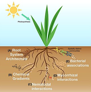
The plant microbiome, also known as the phytomicrobiome, plays roles in plant health and productivity and has received significant attention in recent years. The microbiome has been defined as "a characteristic microbial community occupying a reasonably well-defined habitat which has distinct physio-chemical properties. The term thus not only refers to the microorganisms involved but also encompasses their theatre of activity".

Hydrocarbonoclastic bacteria are a heterogeneous group of prokaryotes which can degrade and utilize hydrocarbon compounds as source of carbon and energy. Despite being present in most of environments around the world, several of these specialized bacteria live in the sea and have been isolated from polluted seawater.

An oleaginous microorganism is a type of microbe that accumulates lipid as a normal part of its metabolism. Oleaginous microbes may accumulate an array of different lipid compounds, including polyhydroxyalkanoates, triacylglycerols, and wax esters. Various microorganisms, including bacteria, fungi, and yeast, are known to accumulate lipids. These organisms are often researched for their potential use in producing fuels from waste products.

References

  1. Morris, Brandon EL; Crable, Bryan R; Suflita, Joseph M (2008). "On the contributions of David Cleaveland White, MD, PhD to microbial ecology: Celebrating the life of a pioneer". The ISME Journal. 2 (8): 797–804. doi: 10.1038/ismej.2008.65 . PMID   18563187. S2CID   40933979.
  2. 1 2 Frostegård, Åsa; Tunlid, Anders; Bååth, Erland (2011). "Use and misuse of PLFA measurements in soils". Soil Biology and Biochemistry. 43 (8): 1621–1625. doi:10.1016/j.soilbio.2010.11.021.
  3. 1 2 Kaur A, Chaudhary A, Kaur A, Choudhary R, Kaushik R (2005). "Phospholipid fatty acid - A bioindicator of environmental monitoring and assessment in soil ecosystem" (PDF). Current Science. 89 (7): 1103–1112. Retrieved 23 July 2016.
  4. Buyer, Jeffrey S.; Sasser, Myron (2012). "High throughput phospholipid fatty acid analysis of soils". Applied Soil Ecology. 61: 127–130. doi: 10.1016/j.apsoil.2012.06.005 .
  5. Zuckerkandl, E; Pauling, L (1965). "Molecules as documents of evolutionary history". Journal of Theoretical Biology. 8 (2): 357–66. doi:10.1016/0022-5193(65)90083-4. PMID   5876245.
  6. White DC (1983). "Analysis of microorganisms in terms of quantity and activity in natural environments". In Slater JHW; Wimpenny R (eds.). Microbes in Their Natural Environment. Cambridge University Press.
  7. 1 2 3 Blackwood, C. B.; Buyer, J. S. (2004). "Soil microbial communities associated with Bt and non-Bt corn in three soils". Journal of Environmental Quality. 33 (3): 832–6. doi:10.2134/jeq2004.0832. PMID   15224917.
  8. Piotrowska-Seget Z, Mrozik A (2003). "Signature lipid biomarker (SLB) analysis in determining changes in community structure of soil microorganisms". Polish J Environ Stud. 12 (6): 669–75. Retrieved 23 July 2016.
  9. 1 2 Frostegard A, Tunlid A, Baath E (1993). "Phospholipid Fatty Acid composition, biomass, and activity of microbial communities from two soil types experimentally exposed to different heavy metals". Appl Environ Microbiol. 59 (11): 3605–17. doi:10.1128/AEM.59.11.3605-3617.1993. PMC   182506 . PMID   16349080.
  10. Pandey, A; Trivedi, P; Kumar, B; Palni, L. M. (2006). "Characterization of a phosphate solubilizing and antagonistic strain of Pseudomonas putida (B0) isolated from a sub-alpine location in the Indian Central Himalaya". Current Microbiology. 53 (2): 102–7. doi:10.1007/s00284-006-4590-5. PMID   16832725. S2CID   23129843.
  11. Bai, Q; Gattinger, A; Zelles, L (2000). "Characterization of Microbial Consortia in Paddy Rice Soil by Phospholipid Analysis". Microbial Ecology. 39 (4): 273–281. doi:10.1007/s002480000020. PMID   10882432. S2CID   25374515.
  12. Gubry-Rangin, C; Nicol, G. W.; Prosser, J. I. (2010). "Archaea rather than bacteria control nitrification in two agricultural acidic soils". FEMS Microbiology Ecology. 74 (3): 566–74. doi: 10.1111/j.1574-6941.2010.00971.x . PMID   21039653.
  13. Szukics, U; Hackl, E; Zechmeister-Boltenstern, S; Sessitsch, A (2012). "Rapid and dissimilar response of ammonia oxidizing archaea and bacteria to nitrogen and water amendment in two temperate forest soils". Microbiological Research. 167 (2): 103–9. doi:10.1016/j.micres.2011.04.002. PMC   4339039 . PMID   21632226.
  14. Pfeffer, P. E.; Douds Jr, D. D.; Becard, G; Shachar-Hill, Y (1999). "Carbon uptake and the metabolism and transport of lipids in an arbuscular mycorrhiza". Plant Physiology. 120 (2): 587–98. doi:10.1104/pp.120.2.587. PMC   59298 . PMID   10364411.
  15. Van Aarle, I. M.; Olsson, P. A. (2003). "Fungal lipid accumulation and development of mycelial structures by two arbuscular mycorrhizal fungi". Applied and Environmental Microbiology. 69 (11): 6762–7. doi:10.1128/aem.69.11.6762-6767.2003. PMC   262256 . PMID   14602638.
  16. Buyer, Jeffrey S.; Teasdale, John R.; Roberts, Daniel P.; Zasada, Inga A.; Maul, Jude E. (2010). "Factors affecting soil microbial community structure in tomato cropping systems". Soil Biology and Biochemistry. 42 (5): 831–841. doi:10.1016/j.soilbio.2010.01.020.
  17. Lundquist, E.J; Scow, K.M; Jackson, L.E; Uesugi, S.L; Johnson, C.R (1999). "Rapid response of soil microbial communities from conventional, low input, and organic farming systems to a wet/dry cycle". Soil Biology and Biochemistry. 31 (12): 1661–1675. doi:10.1016/S0038-0717(99)00080-2.
  18. Kourtev, Peter S.; Ehrenfeld, Joan G.; Häggblom, Max (2002). "Exotic Plant Species Alter the Microbial Community Structure and Function in the Soil". Ecology. 83 (11): 3152–3166. doi:10.1890/0012-9658(2002)083[3152:EPSATM]2.0.CO;2.
  19. Potthoff, Martin; Steenwerth, Kerri L.; Jackson, Louise E.; Drenovsky, Rebecca E.; Scow, Kate M.; Joergensen, Rainer G. (2006). "Soil microbial community composition as affected by restoration practices in California grassland". Soil Biology and Biochemistry. 38 (7): 1851–1860. doi:10.1016/j.soilbio.2005.12.009.
  20. Ringelberg, D; Richmond, M; Foley, K; Reynolds, C (2008). "Utility of lipid biomarkers in support of bioremediation efforts at army sites". Journal of Microbiological Methods. 74 (1): 17–25. doi:10.1016/j.mimet.2007.07.007. PMID   17714813.
  21. MacNaughton, S. J.; Stephen, J. R.; Venosa, A. D.; Davis, G. A.; Chang, Y. J.; White, D. C. (1999). "Microbial population changes during bioremediation of an experimental oil spill". Applied and Environmental Microbiology. 65 (8): 3566–74. doi:10.1128/AEM.65.8.3566-3574.1999. PMC   91535 . PMID   10427050.
  22. Fuller, M. E.; Manning Jr, J. F. (2004). "Microbiological changes during bioremediation of explosives-contaminated soils in laboratory and pilot-scale bioslurry reactors". Bioresource Technology. 91 (2): 123–33. doi:10.1016/s0960-8524(03)00180-9. PMID   14592740.
  23. Morillo, J. A.; Aguilera, M; Antízar-Ladislao, B; Fuentes, S; Ramos-Cormenzana, A; Russell, N. J.; Monteoliva-Sánchez, M (2008). "Molecular microbial and chemical investigation of the bioremediation of two-phase olive mill waste using laboratory-scale bioreactors". Applied Microbiology and Biotechnology. 79 (2): 309–17. doi:10.1007/s00253-008-1422-5. PMID   18347793. S2CID   25268355.
  24. Hansen, L. D.; Nestler, C; Ringelberg, D; Bajpai, R (2004). "Extended bioremediation of PAH/PCP contaminated soils from the POPILE wood treatment facility". Chemosphere. 54 (10): 1481–93. Bibcode:2004Chmsp..54.1481H. doi:10.1016/j.chemosphere.2003.09.046. PMID   14659950.
  25. Antizar-Ladislao, B; Beck, A. J.; Spanova, K; Lopez-Real, J; Russell, N. J. (2007). "The influence of different temperature programmes on the bioremediation of polycyclic aromatic hydrocarbons (PAHs) in a coal-tar contaminated soil by in-vessel composting". Journal of Hazardous Materials. 144 (1–2): 340–7. doi:10.1016/j.jhazmat.2006.10.031. PMID   17113229.
  26. Slater, H; Gouin, T; Leigh, M. B. (2011). "Assessing the potential for rhizoremediation of PCB contaminated soils in northern regions using native tree species". Chemosphere. 84 (2): 199–206. Bibcode:2011Chmsp..84..199S. doi:10.1016/j.chemosphere.2011.04.058. PMC   3502615 . PMID   21596420.
  27. Hassan Sel, D; Boon, E; St-Arnaud, M; Hijri, M (2011). "Molecular biodiversity of arbuscular mycorrhizal fungi in trace metal-polluted soils". Molecular Ecology. 20 (16): 3469–83. doi:10.1111/j.1365-294X.2011.05142.x. PMID   21668808. S2CID   5483017.
  28. Su, Y. H.; Yang, X. Y. (2009). "Interactions between selected PAHs and the microbial community in rhizosphere of a paddy soil". Science of the Total Environment. 407 (3): 1027–34. Bibcode:2009ScTEn.407.1027S. doi:10.1016/j.scitotenv.2008.09.026. PMID   19000632.
  29. Schwartz, E; Trinh, S. V.; Scow, K. M. (2002). "Impact of methylene chloride on microorganisms and phenanthrene mineralization in soil". Journal of Environmental Quality. 31 (1): 144–9. doi:10.2134/jeq2002.1440. PMID   11837417.
  30. Wen, Z. Y.; Chen, F (2003). "Heterotrophic production of eicosapentaenoic acid by microalgae". Biotechnology Advances. 21 (4): 273–94. doi:10.1016/s0734-9750(03)00051-x. PMID   14499126.
  31. Dijkman, NA; Kromkamp, JC (2006). "Phospholipid-derived fatty acids as chemotaxonomic markers for phytoplankton: Application for inferring phytoplankton composition". Marine Ecology Progress Series. 324: 113–125. Bibcode:2006MEPS..324..113D. doi: 10.3354/meps324113 .
  32. 1 2 3 Smoot, J. C.; Findlay, R. H. (2001). "Spatial and Seasonal Variation in a Reservoir Sedimentary Microbial Community as Determined by Phospholipid Analysis". Microbial Ecology. 42 (3): 350–358. doi:10.1007/s002480000102. PMID   12024260. S2CID   31982751.
  33. Hagström, A; Pommier, T; Rohwer, F; Simu, K; Stolte, W; Svensson, D; Zweifel, U. L. (2002). "Use of 16S ribosomal DNA for delineation of marine bacterioplankton species". Applied and Environmental Microbiology. 68 (7): 3628–33. doi:10.1128/aem.68.7.3628-3633.2002. PMC   126765 . PMID   12089052.
  34. Findlay, R. H.; Yeates, C; Hullar, M. A.; Stahl, D. A.; Kaplan, L. A. (2008). "Biome-level biogeography of streambed microbiota". Applied and Environmental Microbiology. 74 (10): 3014–21. doi:10.1128/AEM.01809-07. PMC   2394931 . PMID   18378660.
  35. 1 2 Nilsson, L. O.; Giesler, R; Bååth, E; Wallander, H (2005). "Growth and biomass of mycorrhizal mycelia in coniferous forests along short natural nutrient gradients". New Phytologist. 165 (2): 613–22. doi: 10.1111/j.1469-8137.2004.01223.x . PMID   15720671.
  36. Moore-Kucera, J; Dick, R. P. (2008). "PLFA profiling of microbial community structure and seasonal shifts in soils of a Douglas-fir chronosequence". Microbial Ecology. 55 (3): 500–11. doi:10.1007/s00248-007-9295-1. PMID   17786504. S2CID   19270098.
  37. Högberg, M. N.; Högberg, P; Myrold, D. D. (2007). "Is microbial community composition in boreal forest soils determined by pH, C-to-N ratio, the trees, or all three?". Oecologia. 150 (4): 590–601. Bibcode:2006Oecol.150..590H. doi:10.1007/s00442-006-0562-5. PMID   17033802. S2CID   5686327.
  38. Yarwood, S. A.; Myrold, D. D.; Högberg, M. N. (2009). "Termination of belowground C allocation by trees alters soil fungal and bacterial communities in a boreal forest". FEMS Microbiology Ecology. 70 (1): 151–62. doi: 10.1111/j.1574-6941.2009.00733.x . PMID   19656196.
  39. Brant, J. B.; Myrold, D. D.; Sulzman, E. W. (2006). "Root controls on soil microbial community structure in forest soils". Oecologia. 148 (4): 650–9. Bibcode:2006Oecol.148..650B. doi:10.1007/s00442-006-0402-7. PMID   16547734. S2CID   13240195.
  40. Nilsson, L. O.; Bååth, E; Falkengren-Grerup, U; Wallander, H (2007). "Growth of ectomycorrhizal mycelia and composition of soil microbial communities in oak forest soils along a nitrogen deposition gradient". Oecologia. 153 (2): 375–84. Bibcode:2007Oecol.153..375N. doi:10.1007/s00442-007-0735-x. PMID   17453252. S2CID   28446290.
  41. 1 2 Steger, K; Jarvis, A; Smårs, S; Sundh, I (2003). "Comparison of signature lipid methods to determine microbial community structure in compost". Journal of Microbiological Methods. 55 (2): 371–82. doi:10.1016/s0167-7012(03)00187-8. PMID   14529958.
  42. Yi, T; Lee, E. H.; Kang, S; Shin, J; Cho, K. S. (2012). "Structure and dynamics of microbial community in full-scale activated sludge reactors". Journal of Industrial Microbiology & Biotechnology. 39 (1): 19–25. doi: 10.1007/s10295-011-0994-8 . PMID   21633845. S2CID   279624.
  43. Chang, Jun-Jun; Liang, Wei; Xiao, En-Rong; Wu, Zhen-Bin (2011). "Effect of intermittent aeration on the microbial community structure of activated sludge in a submerged membrane bioreactor". Water and Environment Journal. 25 (2): 214–218. doi:10.1111/j.1747-6593.2009.00213.x. S2CID   83881859.
  44. Jiménez, J.J.; Igual, J.M. H.; Villar, L.; Benito-Alonso, J.L.; Abadias-Ullod, J. (2019). "Hierarchical drivers of soil microbial community structure variability in "Monte Perdido" Massif (Central Pyrenees)". Scientific Reports. 9 (8768): 1–17. doi: 10.1038/s41598-019-45372-z . PMC   6584728 . PMID   31217456.