Photoacoustic microscopy

Last updated
Photoacoustic imaging schematic PASchematics v2.png
Photoacoustic imaging schematic

Photoacoustic microscopy is an imaging method based on the photoacoustic effect and is a subset of photoacoustic tomography. Photoacoustic microscopy takes advantage of the local temperature rise that occurs as a result of light absorption in tissue. Using a nanosecond pulsed laser beam, tissues undergo thermoelastic expansion, resulting in the release of a wide-band acoustic wave that can be detected using a high-frequency ultrasound transducer. [1] Since ultrasonic scattering in tissue is weaker than optical scattering, photoacoustic microscopy is capable of achieving high-resolution images at greater depths than conventional microscopy methods. Furthermore, photoacoustic microscopy is especially useful in the field of biomedical imaging due to its scalability. By adjusting the optical and acoustic foci, lateral resolution may be optimized for the desired imaging depth. [2]

Contents

Photoacoustic signal

The goal of photoacoustic microscopy is to find the local pressure rise , which can be used to calculate the absorption coefficient according to the formula:

where is the percentage of light converted to heat, is the local optical fluence (J/cm2), and the dimensionless Gruneisen parameter is defined as:

where is the thermal coefficient of volume expansion (K−1), is the isothermal compressibility (Pa−1), and is the density (kg/m3). [3]

Following the initial pressure rise, a photoacoustic wave propagates at the speed of sound within the medium and can be detected with an ultrasound transducer.

Image reconstruction

One of the major benefits of photoacoustic microscopy is the simplicity of image reconstruction. A laser pulse excites tissue in the axial direction and the resulting photoacoustic waves are detected by an ultrasound transducer. The transducer then converts the mechanical energy into a voltage signal that can be read by an analog-to-digital converter for post-processing. A one-dimensional image, known as an A-line, is formed as a result of each laser pulse. Hilbert transform of an A-line reveals depth-encoded information. A 3D photoacoustic image can then be formed by combining multiple A-lines produced by 2D raster scanning. [3]

Synthetic Aperture Image Reconstruction

Altering delays of the elements on an ultrasound transducer allows one to focus ultrasound waves similar to passing through an acoustic lens. This delay-and-sum method enables one to find the signal at each focal point. However, the lateral resolution is limited by the presence of side lobes, which appear at polar angles and are dependent on the width of each element. [4]

Contrast

In photoacoustic imaging modalities, including photoacoustic microscopy, contrast is based on photon excitation and is thus determined by the optical properties of the tissue. When an electron absorbs a photon, it moves to a higher energy state. Upon returning to a lower energy level, the electron undergoes either radiative or nonradiative relaxation. During radiative relaxation, the electron releases energy in the form of a photon. On the other hand, an electron undergoing nonradiative relaxation releases energy as heat. The heat then induces a pressure rise that propagates as a photoacoustic wave. Due to the fact that almost all molecules are capable of nonradiative relaxation, photoacoustic microscopy has the potential to image a wide range of endogenous and exogenous agents. By contrast, fewer molecules are capable of radiative relaxation, thus limiting fluorescence microscopy techniques such as one-photon and two-photon microscopy. [3] Current research in photoacoustic microscopy takes advantage of both endogenous and exogenous contrast agents to gain functional information about the body, from blood saturation levels to cancer proliferation rate.

Endogenous Contrast Agents

Absorption profile of oxy- and deoxyhemoglobin HbAbs v3.png
Absorption profile of oxy- and deoxyhemoglobin

Endogenous contrast agents, molecules naturally occurring within the body, are useful in photoacoustic microscopy due to the fact that they may be imaged non-invasively. Endogenous agents are also non-toxic and do not affect the properties of the tissue being studied. In particular, endogenous absorbers can be classified based on their absorbing wavelengths. [2]

Ultraviolet Absorbers

Within the ultraviolet light range (λ = 180 to 400 nm), the primary absorber in the body is DNA and RNA. By using ultraviolet photoacoustic microscopy, DNA and RNA can be imaged in the cell nuclei without the use of fluorescence labeling. Since cancer is associated with DNA replication failure, UV photoacoustic microscopy has the potential to be used for early cancer detection. [5]

Visible Light Absorbers

Visible light absorbers (λ = 400 to 700 nm) include oxyhemoglobin, deoxyhemoglobin, melanin, and cytochrome c. Visible light photoacoustic microscopy is particularly useful in determining hemoglobin concentration and oxygen saturation due to the difference in absorption profiles of oxyhemoglobin and deoxyhemoglobin. Real-time analysis can then be used to determine blood flow speed and oxygen metabolism rate. [3] In addition, photoacoustic microscopy is capable of early melanoma detection due to the high concentration of melanin found in skin cancer cells.

Near-Infrared Absorbers

Near-Infrared absorbers (λ = 700 to 1400 nm) include water, lipids, and glucose. Photoacoustic determination of blood glucose levels can be used for treating diabetes, while studying lipid concentrations within blood vessels is important for monitoring the progression of atherosclerosis. [2] It is still feasible to quantify and compare deoxyhemoglobin and hemoglobin concentrations at this wavelength, trading deeper tissue penetration for lower absorption. [6]

Exogenous Contrast Agents

Although endogenous contrasts agents are noninvasive and simpler to use, they are limited by their inherent behavior and concentration, making it difficult to monitor certain processes if optical absorption is weak. On the other hand, exogenous agents can be engineered to specifically bind to certain molecules of interest. In addition, the concentration of exogenous agents can be optimized to produce a greater signal and provide more contrast. Through selective binding, exogenous contrast agents are capable of targeting specific molecules of interest while also enhancing resulting images. [3]

Organic Dyes

Organic dyes, such as ICG-PEG and Evans blue, are used to enhance vasculature as well as to improve tumor imaging. In addition, dyes are easily filtered out of the body due to their small size (≤ 3 nm). [2]

Nanoparticles

Nanoparticles are currently being researched due to their chemical inactivity and ability to target tumor cells. These properties allow for cancer propagation to be monitored and potentially enables intraoperative cancer removal. However, more studies on short-term toxicity effects are necessary to determine if nanoparticles are suitable for clinical research. [2] Gold nanoparticles have shown promise as a contrast agent for image-guided medicine. AuNPs have been widely used as contrast agents due to their strong and tunable optical absorption. [7]

Fluorescent Proteins

Fluorescent proteins have been developed for fluorescence microscopy imaging and are unique in that they can be genetically encoded and therefore do not need to be delivered into the body. Using photoacoustic microscopy, fluorescent proteins can be visualized at depths beyond the limit of typical microscopy methods. [2] Frequency-dependent acoustic attenuation in tissue and dampening of higher frequencies limits the bandwidth of light propagation through deeper regions in tissue. Fluorescent proteins act as light source at the target region, bypassing the limitation of optical attenuation. However, the effectiveness of fluorescent proteins is limited by low fluence changes, as the light diffusion equation predicts lower than 5% increase. [8]

Resolution

Mouse ear vasculature imaged using OR-PAM at 532 nm OR-PAM Mouse Ear Vasculature.jpg
Mouse ear vasculature imaged using OR-PAM at 532 nm
Mouse ear vasculature imaged using AR-PAM at 532 nm AR-PAM Mouse Ear Vasculature.jpg
Mouse ear vasculature imaged using AR-PAM at 532 nm
Photoacoustic micrograph of methanol fixed human red blood cells using 405 nm. HumanRBCsPAM.png
Photoacoustic micrograph of methanol fixed human red blood cells using 405 nm.

Photoacoustic microscopy achieves greater penetration than conventional microscopy due to ultrasonic detection. As a result, axial resolution is defined acoustically and is determined by the formula:

where is the speed of sound in the medium and is the photoacoustic signal bandwidth. The axial resolution of the system can be improved by using a wider bandwidth ultrasound transducer as long as the bandwidth matches that of the photoacoustic signal. The lateral resolution of photoacoustic microscopy depends on the optical and acoustic foci of the system. Optical-resolution photoacoustic microscopy (OR-PAM) uses a tighter optical focus than acoustic focus, while acoustic-resolution photoacoustic microscopy (AR-PAM) uses a tighter acoustic focus than optical focus. [9] [10]

Optical-Resolution Photoacoustic microscopy

Due to a tighter optical focus, OR-PAM is more useful for imaging in the quasi-ballistic range of depths up to 1 mm. [9] The lateral resolution of OR-PAM is determined by the formula:

where is the optical wavelength and is the numerical aperture of the optical objective lens. [2] The lateral resolution of OR-PAM can be improved by using a shorter laser pulse and tighter focusing of the laser spot. OR-PAM systems can typically achieve a lateral resolution of 0.2 to 10 μm, allowing OR-PAM to be classified as a super-resolution imaging method.

Acoustic-Resolution Photoacoustic microscopy

At depths greater than 1 mm and up to 3 mm, acoustic-resolution photoacoustic microscopy (AR-PAM) is more useful due to greater optical scattering. Acoustic scattering is much weaker beyond the optical diffusion limit, making AR-PAM more practical as it provides higher lateral resolution at these depths. The lateral resolution of AR-PAM is determined by the formula:

where is the central wavelength of the photoacoustic wave and is the numerical aperture of the ultrasound transducer. [2] Higher lateral resolution can therefore be achieved by increasing the center frequency of the ultrasound transducer and tighter acoustic focusing. AR-PAM systems can typically achieve a lateral resolution of 15 to 50 μm.

Dark-field Confocal Photoacoustic microscopy

Depiction of PAM raster scanning path Raster-scan.svg
Depiction of PAM raster scanning path

By ignoring ballistic light, dark-field confocal photoacoustic microscopy reduces surface signal. This method uses a dark-field pulsed laser and high-NA ultrasonic detection, with the fiber output end coaxially aligned with the focused ultrasound transducer. Filtration of ballistic light relies on the altered shape of the excitation laser beam instead of an opaque disk, as used in conventional dark-field microscopy. The general reconstruction technique is used to convert the photoacoustic signal into one A-line, and B-line images are produced by raster scanning. [4]

Biomedical applications

Photoacoustic microscopy has a wide range of applications in the biomedical field. Due to its ability to image a variety of molecules based on optical wavelength, photoacoustic microscopy can be used to gain functional information about the body noninvasively. Blood flow dynamics and oxygen metabolic rates can be measured and correlated to studies of atherosclerosis or tumor proliferation. Exogenous agents can be used to bind to cancerous tissue, enhancing image contrast and aiding in surgical removal. On the same note, photoacoustic microscopy is useful in early cancer diagnosis due to the difference in optical absorption properties compared to healthy tissue. [1]

See also

Related Research Articles

<span class="mw-page-title-main">Microscopy</span> Viewing of objects which are too small to be seen with the naked eye

Microscopy is the technical field of using microscopes to view objects and areas of objects that cannot be seen with the naked eye. There are three well-known branches of microscopy: optical, electron, and scanning probe microscopy, along with the emerging field of X-ray microscopy.

The term biophotonics denotes a combination of biology and photonics, with photonics being the science and technology of generation, manipulation, and detection of photons, quantum units of light. Photonics is related to electronics and photons. Photons play a central role in information technologies, such as fiber optics, the way electrons do in electronics.

<span class="mw-page-title-main">Photoacoustic imaging</span> Imaging using the photoacoustic effect

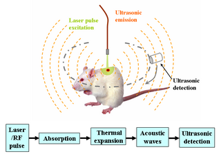
Photoacoustic imaging or optoacoustic imaging is a biomedical imaging modality based on the photoacoustic effect. Non-ionizing laser pulses are delivered into biological tissues and part of the energy will be absorbed and converted into heat, leading to transient thermoelastic expansion and thus wideband ultrasonic emission. The generated ultrasonic waves are detected by ultrasonic transducers and then analyzed to produce images. It is known that optical absorption is closely associated with physiological properties, such as hemoglobin concentration and oxygen saturation. As a result, the magnitude of the ultrasonic emission, which is proportional to the local energy deposition, reveals physiologically specific optical absorption contrast. 2D or 3D images of the targeted areas can then be formed.

<span class="mw-page-title-main">Molecular imaging</span> Imaging molecules within living patients

Molecular imaging is a field of medical imaging that focuses on imaging molecules of medical interest within living patients. This is in contrast to conventional methods for obtaining molecular information from preserved tissue samples, such as histology. Molecules of interest may be either ones produced naturally by the body, or synthetic molecules produced in a laboratory and injected into a patient by a doctor. The most common example of molecular imaging used clinically today is to inject a contrast agent into a patient's bloodstream and to use an imaging modality to track its movement in the body. Molecular imaging originated from the field of radiology from a need to better understand fundamental molecular processes inside organisms in a noninvasive manner.

Functional imaging is a medical imaging technique of detecting or measuring changes in metabolism, blood flow, regional chemical composition, and absorption.

<span class="mw-page-title-main">Monte Carlo method for photon transport</span>

Modeling photon propagation with Monte Carlo methods is a flexible yet rigorous approach to simulate photon transport. In the method, local rules of photon transport are expressed as probability distributions which describe the step size of photon movement between sites of photon-matter interaction and the angles of deflection in a photon's trajectory when a scattering event occurs. This is equivalent to modeling photon transport analytically by the radiative transfer equation (RTE), which describes the motion of photons using a differential equation. However, closed-form solutions of the RTE are often not possible; for some geometries, the diffusion approximation can be used to simplify the RTE, although this, in turn, introduces many inaccuracies, especially near sources and boundaries. In contrast, Monte Carlo simulations can be made arbitrarily accurate by increasing the number of photons traced. For example, see the movie, where a Monte Carlo simulation of a pencil beam incident on a semi-infinite medium models both the initial ballistic photon flow and the later diffuse propagation.

Biological imaging may refer to any imaging technique used in biology. Typical examples include:

Acoustic microscopy is microscopy that employs very high or ultra high frequency ultrasound. Acoustic microscopes operate non-destructively and penetrate most solid materials to make visible images of internal features, including defects such as cracks, delaminations and voids.

<span class="mw-page-title-main">Thermoacoustic imaging</span>

Thermoacoustic imaging was originally proposed by Theodore Bowen in 1981 as a strategy for studying the absorption properties of human tissue using virtually any kind of electromagnetic radiation. But Alexander Graham Bell first reported the physical principle upon which thermoacoustic imaging is based a century earlier. He observed that audible sound could be created by illuminating an intermittent beam of sunlight onto a rubber sheet. Shortly after Bowen's work was published, other researchers proposed methodology for thermoacoustic imaging using microwaves. In 1994 researchers used an infrared laser to produce the first thermoacoustic images of near-infrared optical absorption in a tissue-mimicking phantom, albeit in two dimensions (2D). In 1995 other researchers formulated a general reconstruction algorithm by which 2D thermoacoustic images could be computed from their "projections," i.e. thermoacoustic computed tomography (TCT). By 1998 researchers at Indiana University Medical Center extended TCT to 3D and employed pulsed microwaves to produce the first fully three-dimensional (3D) thermoacoustic images of biologic tissue [an excised lamb kidney ]. The following year they created the first fully 3D thermoacoustic images of cancer in the human breast, again using pulsed microwaves. Since that time, thermoacoustic imaging has gained widespread popularity in research institutions worldwide. As of 2008, three companies were developing commercial thermoacoustic imaging systems – Seno Medical, Endra, Inc. and OptoSonics, Inc.

The photoacoustic Doppler effect is a type of Doppler effect that occurs when an intensity modulated light wave induces a photoacoustic wave on moving particles with a specific frequency. The observed frequency shift is a good indicator of the velocity of the illuminated moving particles. A potential biomedical application is measuring blood flow.

Ultrasound-modulated optical tomography (UOT), also known as Acousto-Optic Tomography (AOT), is a hybrid imaging modality that combines light and sound; it is a form of tomography involving ultrasound. It is used in imaging of biological soft tissues and has potential applications for early cancer detection. As a hybrid modality which uses both light and sound, UOT provides some of the best features of both: the use of light provides strong contrast and sensitivity ; these two features are derived from the optical component of UOT. The use of ultrasound allows for high resolution, as well as a high imaging depth. However, the difficulty of tackling the two fundamental problems with UOT have caused UOT to evolve relatively slowly; most work in the field is limited to theoretical simulations or phantom / sample studies.

Preclinical imaging is the visualization of living animals for research purposes, such as drug development. Imaging modalities have long been crucial to the researcher in observing changes, either at the organ, tissue, cell, or molecular level, in animals responding to physiological or environmental changes. Imaging modalities that are non-invasive and in vivo have become especially important to study animal models longitudinally. Broadly speaking, these imaging systems can be categorized into primarily morphological/anatomical and primarily molecular imaging techniques. Techniques such as high-frequency micro-ultrasound, magnetic resonance imaging (MRI) and computed tomography (CT) are usually used for anatomical imaging, while optical imaging, positron emission tomography (PET), and single photon emission computed tomography (SPECT) are usually used for molecular visualizations.

Photo-activated localization microscopy and stochastic optical reconstruction microscopy (STORM) are widefield fluorescence microscopy imaging methods that allow obtaining images with a resolution beyond the diffraction limit. The methods were proposed in 2006 in the wake of a general emergence of optical super-resolution microscopy methods, and were featured as Methods of the Year for 2008 by the Nature Methods journal. The development of PALM as a targeted biophysical imaging method was largely prompted by the discovery of new species and the engineering of mutants of fluorescent proteins displaying a controllable photochromism, such as photo-activatible GFP. However, the concomitant development of STORM, sharing the same fundamental principle, originally made use of paired cyanine dyes. One molecule of the pair, when excited near its absorption maximum, serves to reactivate the other molecule to the fluorescent state.

Multi-spectral optoacoustic tomography (MSOT), also known as functional photoacoustic tomography (fPAT), is an imaging technology that generates high-resolution optical images in scattering media, including biological tissues. MSOT illuminates tissue with light of transient energy, typically light pulses lasting 1-100 nanoseconds. The tissue absorbs the light pulses, and as a result undergoes thermo-elastic expansion, a phenomenon known as the optoacoustic or photoacoustic effect. This expansion gives rise to ultrasound waves (photoechoes) that are detected and formed into an image. Image formation can be done by means of hardware or computed tomography. Unlike other types of optoacoustic imaging, MSOT involves illuminating the sample with multiple wavelengths, allowing it to detect ultrasound waves emitted by different photoabsorbing molecules in the tissue, whether endogenous or exogenous. Computational techniques such as spectral unmixing deconvolute the ultrasound waves emitted by these different absorbers, allowing each emitter to be visualized separately in the target tissue. In this way, MSOT can allow visualization of hemoglobin concentration and tissue oxygenation or hypoxia. Unlike other optical imaging methods, MSOT is unaffected by photon scattering and thus can provide high-resolution optical images deep inside biological tissues.

Super-resolution photoacoustic imaging is a set of techniques used to enhance spatial resolution in photoacoustic imaging. Specifically, these techniques primarily break the optical diffraction limit of the photoacoustic imaging system. It can be achieved in a variety of mechanisms, such as blind structured illumination, multi-speckle illumination, or photo-imprint photoacoustic microscopy in Figure 1.

Three-photon microscopy (3PEF) is a high-resolution fluorescence microscopy based on nonlinear excitation effect. Different from two-photon excitation microscopy, it uses three exciting photons. It typically uses 1300 nm or longer wavelength lasers to excite the fluorescent dyes with three simultaneously absorbed photons. The fluorescent dyes then emit one photon whose energy is three times the energy of each incident photon. Compared to two-photon microscopy, three-photon microscopy reduces the fluorescence away from the focal plane by , which is much faster than that of two-photon microscopy by . In addition, three-photon microscopy employs near-infrared light with less tissue scattering effect. This causes three-photon microscopy to have higher resolution than conventional microscopy.

Deep learning in photoacoustic imaging

Deep learning in photoacoustic imaging combines the hybrid imaging modality of photoacoustic imaging (PA) with the rapidly evolving field of deep learning. Photoacoustic imaging is based on the photoacoustic effect, in which optical absorption causes a rise in temperature, which causes a subsequent rise in pressure via thermo-elastic expansion. This pressure rise propagates through the tissue and is sensed via ultrasonic transducers. Due to the proportionality between the optical absorption, the rise in temperature, and the rise in pressure, the ultrasound pressure wave signal can be used to quantify the original optical energy deposition within the tissue.

A specific branch of contrast-enhanced ultrasound, acoustic angiography is a minimally invasive and non-ionizing medical imaging technique used to visualize vasculature. Acoustic angiography was first developed by the Dayton Laboratory at North Carolina State University and provides a safe, portable, and inexpensive alternative to the most common methods of angiography such as Magnetic Resonance Angiography and Computed Tomography Angiography. Although ultrasound does not traditionally exhibit the high resolution of MRI or CT, high-frequency ultrasound (HFU) achieves relatively high resolution by sacrificing some penetration depth. HFU typically uses waves between 20 and 100 MHz and achieves resolution of 16-80μm at depths of 3-12mm. Although HFU has exhibited adequate resolution to monitor things like tumor growth in the skin layers, on its own it lacks the depth and contrast necessary for imaging blood vessels. Acoustic angiography overcomes the weaknesses of HFU by combining contrast-enhanced ultrasound with the use of a dual-element ultrasound transducer to achieve high resolution visualization of blood vessels at relatively deep penetration levels.

Photoacoustic flow cytometry or PAFC is a biomedical imaging modality that utilizes photoacoustic imaging to perform flow cytometry. A flow of cells passes a photoacoustic system producing individual signal response. Each signal is counted to produce a quantitative evaluation of the input sample.

Ultrasound-switchable fluorescence (USF) imaging is a deep optics imaging technique. In last few decades, fluorescence microscopy has been highly developed to image biological samples and live tissues. However, due to light scattering, fluorescence microscopy is limited to shallow tissues. Since fluorescence is characterized by high contrast, high sensitivity, and low cost which is crucial to investigate deep tissue information, developing fluorescence imaging technique with high depth-to-resolution ratio would be promising.. Recently, ultrasound-switchable fluorescence imaging has been developed to achieve high signal-to-noise ratio (SNR) and high spatial resolution imaging without sacrificing image depth.

References

  1. 1 2 H.F. Zhang; K. Maslov; G. Stoica; L.V. Wang (2006). "Functional photoacoustic microscopy for high-resolution and noninvasive in vivo imaging" (PDF). Nature Biotechnology. 24 (7): 848–851. doi:10.1038/nbt1220. PMID   16823374. S2CID   912509.
  2. 1 2 3 4 5 6 7 8 L.V. Wang; J. Yao (2013). "Photoacoustic Microscopy". Laser Photonics Rev. 7 (5): 10. Bibcode:2013LPRv....7..758Y. doi:10.1002/lpor.201200060. PMC   3887369 . PMID   24416085.
  3. 1 2 3 4 5 Y. Zhou; J. Yao; L.V. Wang (2016). "Tutorial of photoacoustic tomography". J. Biomed. Opt. 21 (6): 061007. Bibcode:2016JBO....21f1007Z. doi:10.1117/1.JBO.21.6.061007. PMC   4834026 . PMID   27086868.
  4. 1 2 L.V. Wang; H.I. Wu (2007). Biomedical Optics. Wiley. ISBN   978-0-471-74304-0.
  5. L.V. Wang; S. Hu (2012). "Photoacoustic Tomography: In Vivo Imaging from Organelles to Organs". Science. 335 (6075): 1458–1462. Bibcode:2012Sci...335.1458W. doi:10.1126/science.1216210. PMC   3322413 . PMID   22442475.
  6. A. Edwards; C. Richardson (1993). "Measurement of hemoglobin flow and blood flow by near-infrared spectroscopy". Journal of Applied Physiology. 75 (4): 1884–9. doi:10.1152/jappl.1993.75.4.1884. PMID   8282646.
  7. W. Li; X. Chen (2015). "Gold nanoparticles for photoacoustic imaging". Nanomedicine. 10 (2): 299–320. doi:10.2217/nnm.14.169. PMC   4337958 . PMID   25600972.
  8. D. Razansky; M. Distel; C. Vinegoni (2009). "Multispectral opto-acoustic tomography of deep-seated fluorescent proteins in vivo". Nature Photonics. 3 (7): 412–7. Bibcode:2009NaPho...3..412R. doi:10.1038/nphoton.2009.98.
  9. 1 2 L.V. Wang; J. Yao (2016). "A practical guide to photoacoustic tomography in the life sciences". Nature Methods. 13 (8): 627–638. doi:10.1038/NMETH.3925. PMC   4980387 . PMID   27467726.
  10. Wang, Lihong V. (2009-08-28). "Multiscale photoacoustic microscopy and computed tomography". Nature Photonics. 3 (9): 503–509. Bibcode:2009NaPho...3..503W. doi:10.1038/nphoton.2009.157. ISSN   1749-4885. PMC   2802217 . PMID   20161535.